

Start Here
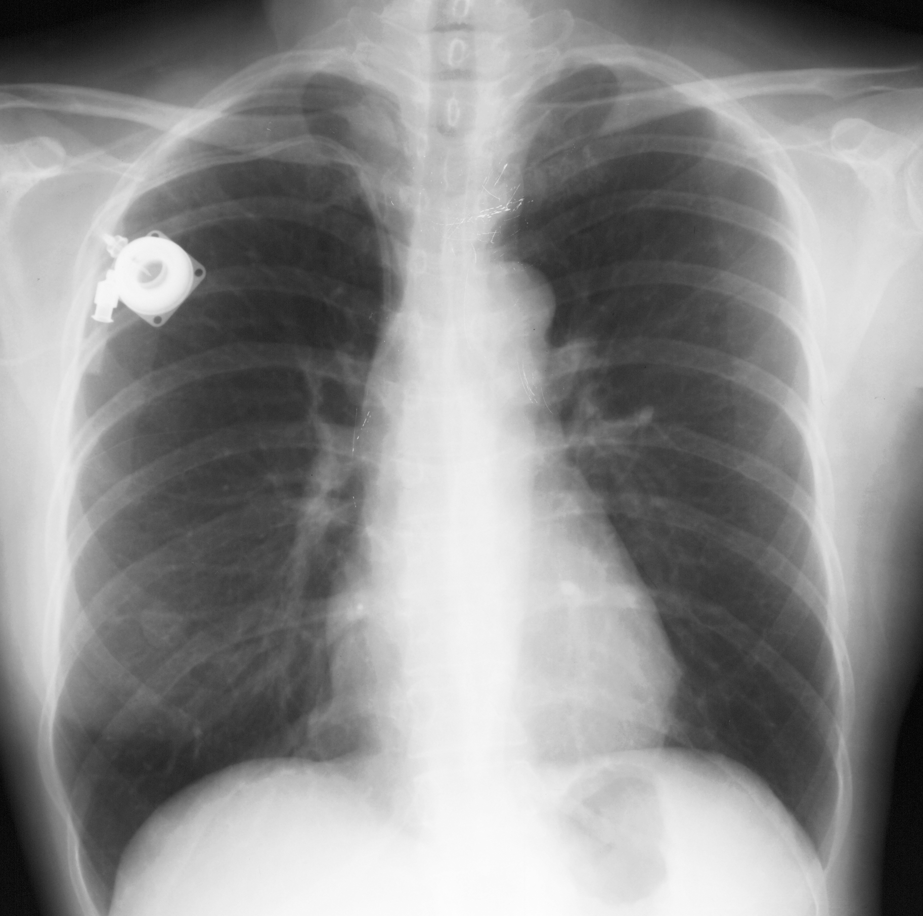
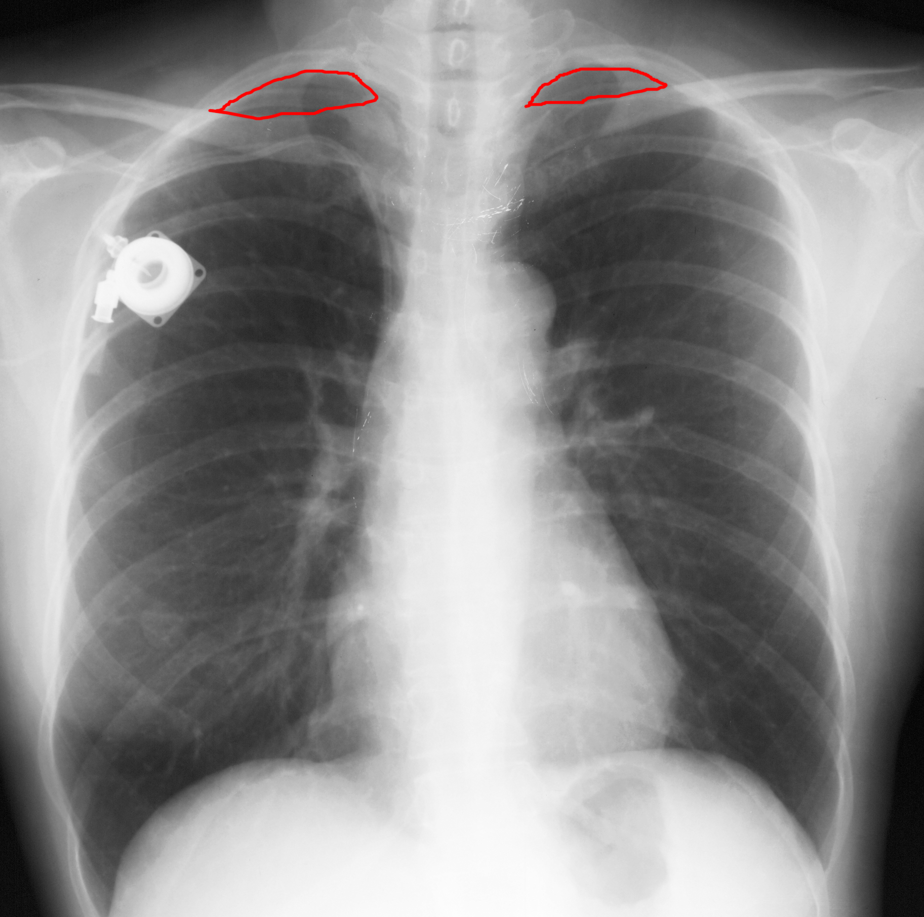
This is the chest radiograph that was shown in class.
Question 1:
a) What do you think of the lungs, comparing one side to the other?
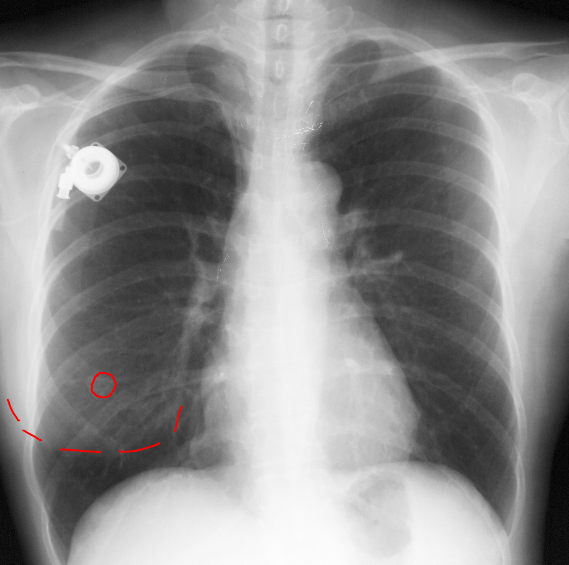
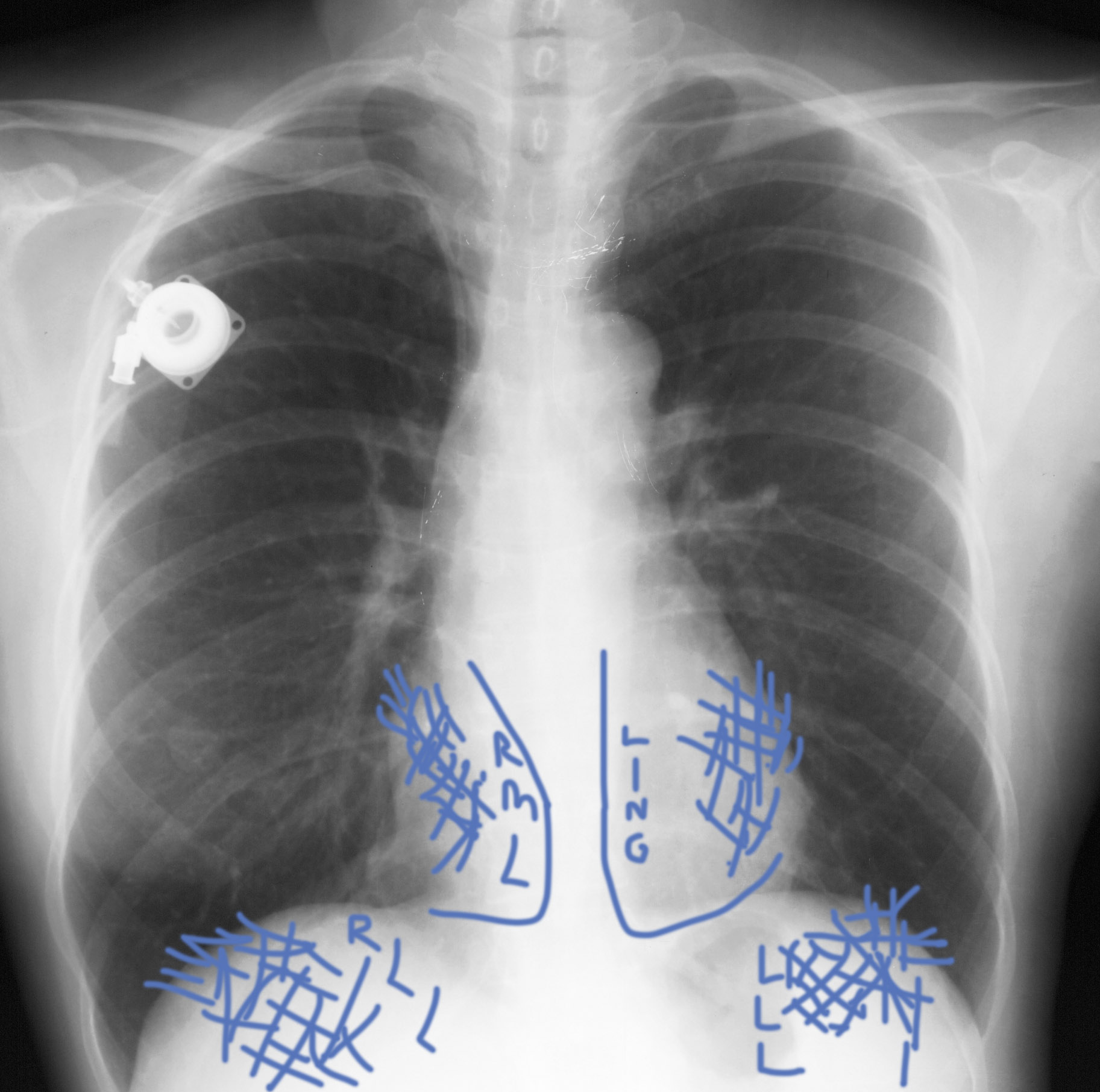
The lung density and distribution of vessels is symmetric from right to left in the upper and middle zones of the chest. In the lower chest, the left lung looks darker than the right, but there is a relatively sharp horizontal transition in the lower right lung, and at the very bottom (in the region of the costophrenic angles), the lung density is again symmetry on the two sides. There are several things to consider to account for this--what do you think is most likely?
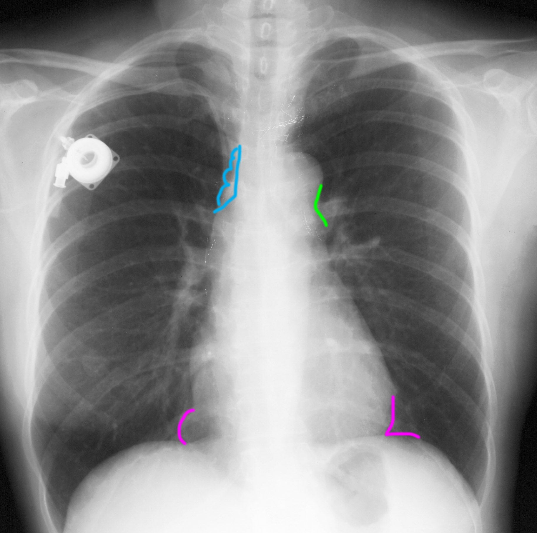
b) In class, we talked about two different potential clinical scenarios for this patient: one with cough and fever (clinical concern for pneumonia) and one with malaise, weight loss and peripheral adenopathy (clinical concern for enlarged nodes from recurrent lymphoma). Neither of these is the actual scenario for this patient. Thinking about the foreign object present and the lung findings, what do you think is a more likely history for this patient?
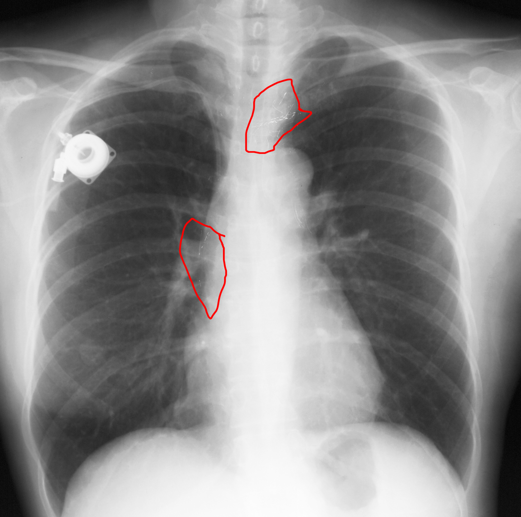
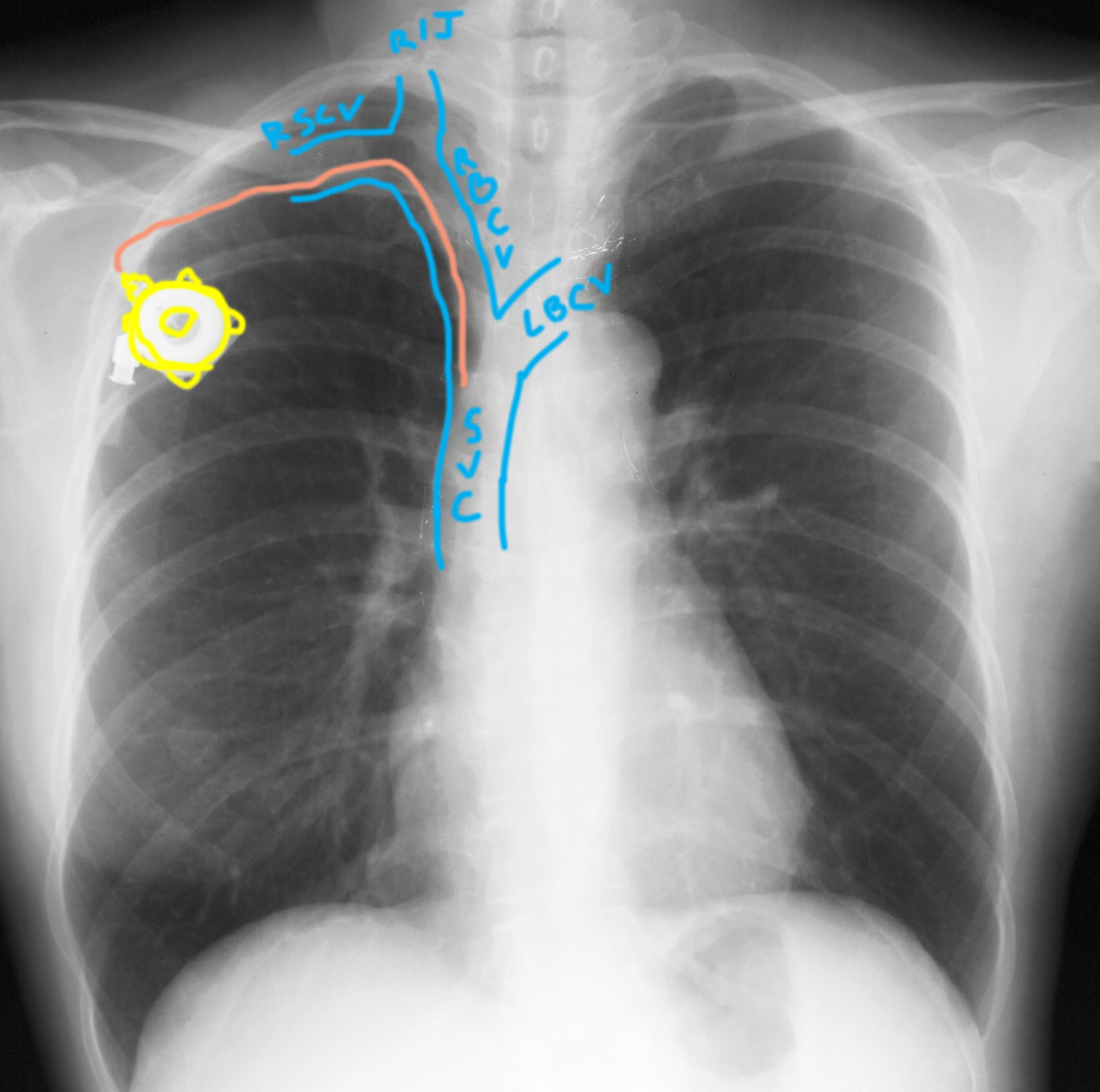
The foreign object (injection port) suggests some serious illness, possibly a malignancy since these ports are most often used to administer chemotherapy. The lung findings could be explained by left mastectomy. The actual history was breast cancer, and the mass in the right cardiophrenic angle was a metastatic node.
Use this chest radiograph to recall the topics covered in class, which include:
1. use of a systematic approach (no more than five items, saving the lungs for last).
2. addition of a targeted search for various common clinical scenarios (in class we discussed pneumonia and enlarged nodes)
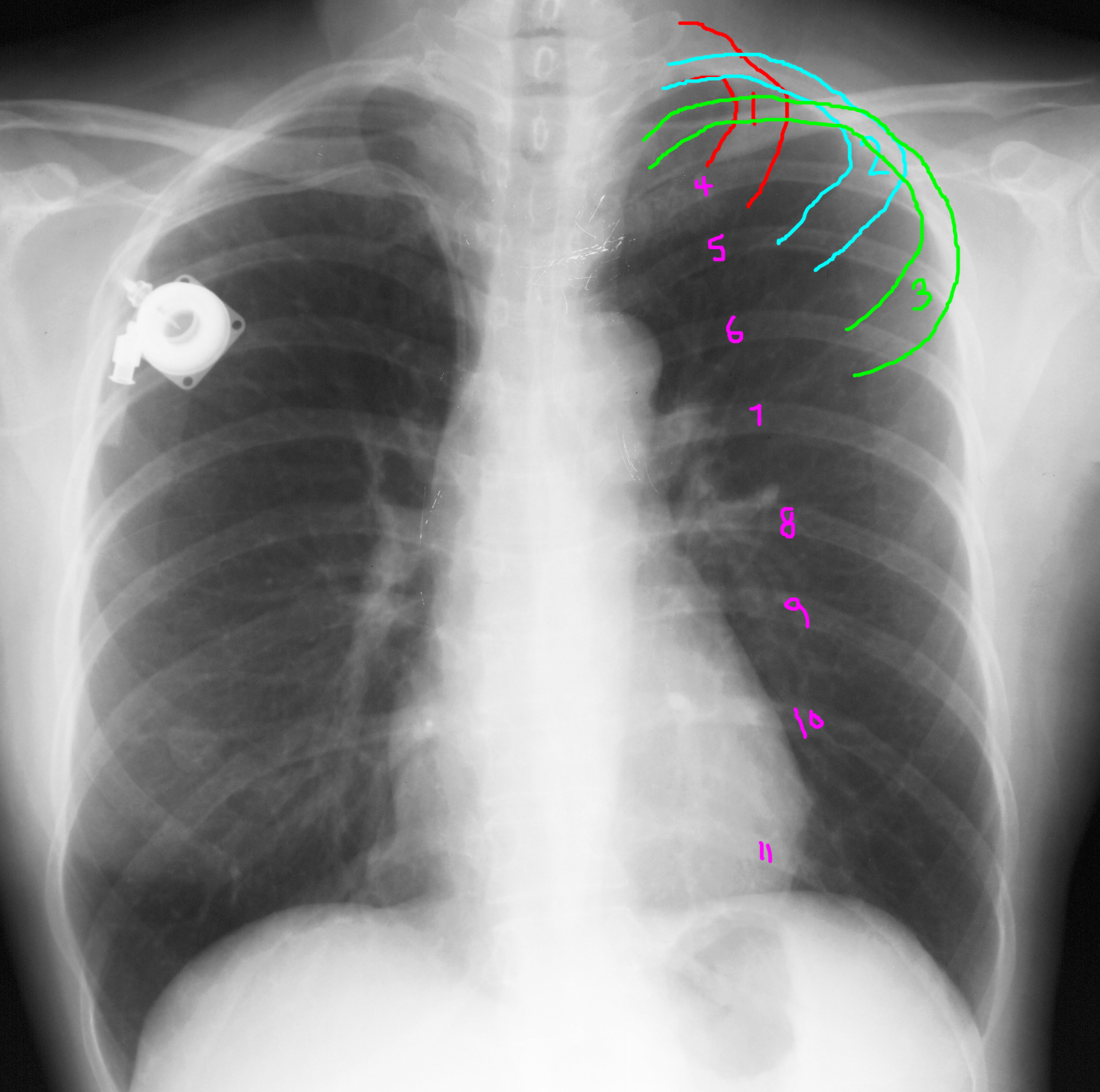
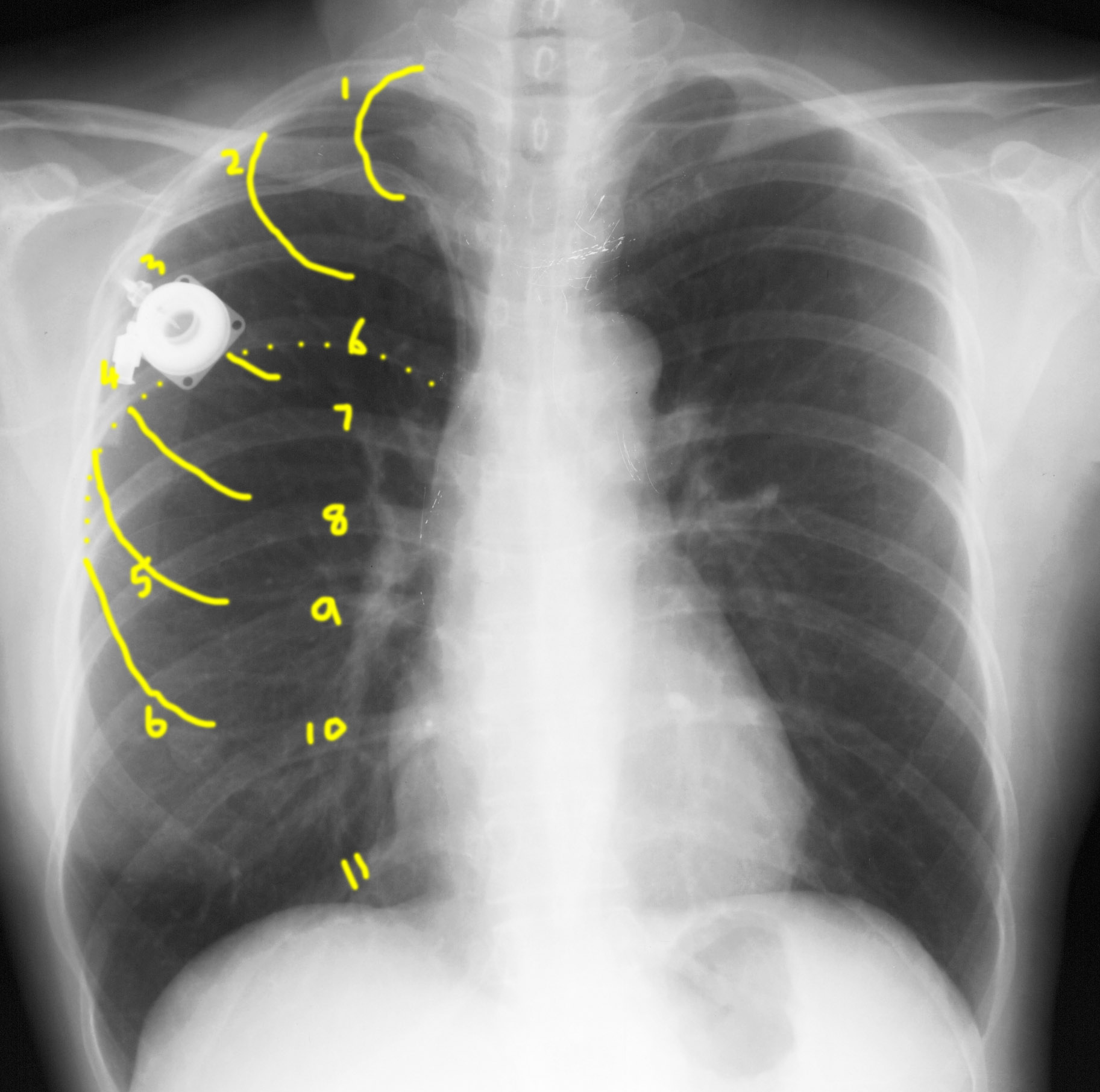
3. how to determine if the patient has taken a deep breath (counting ribs).
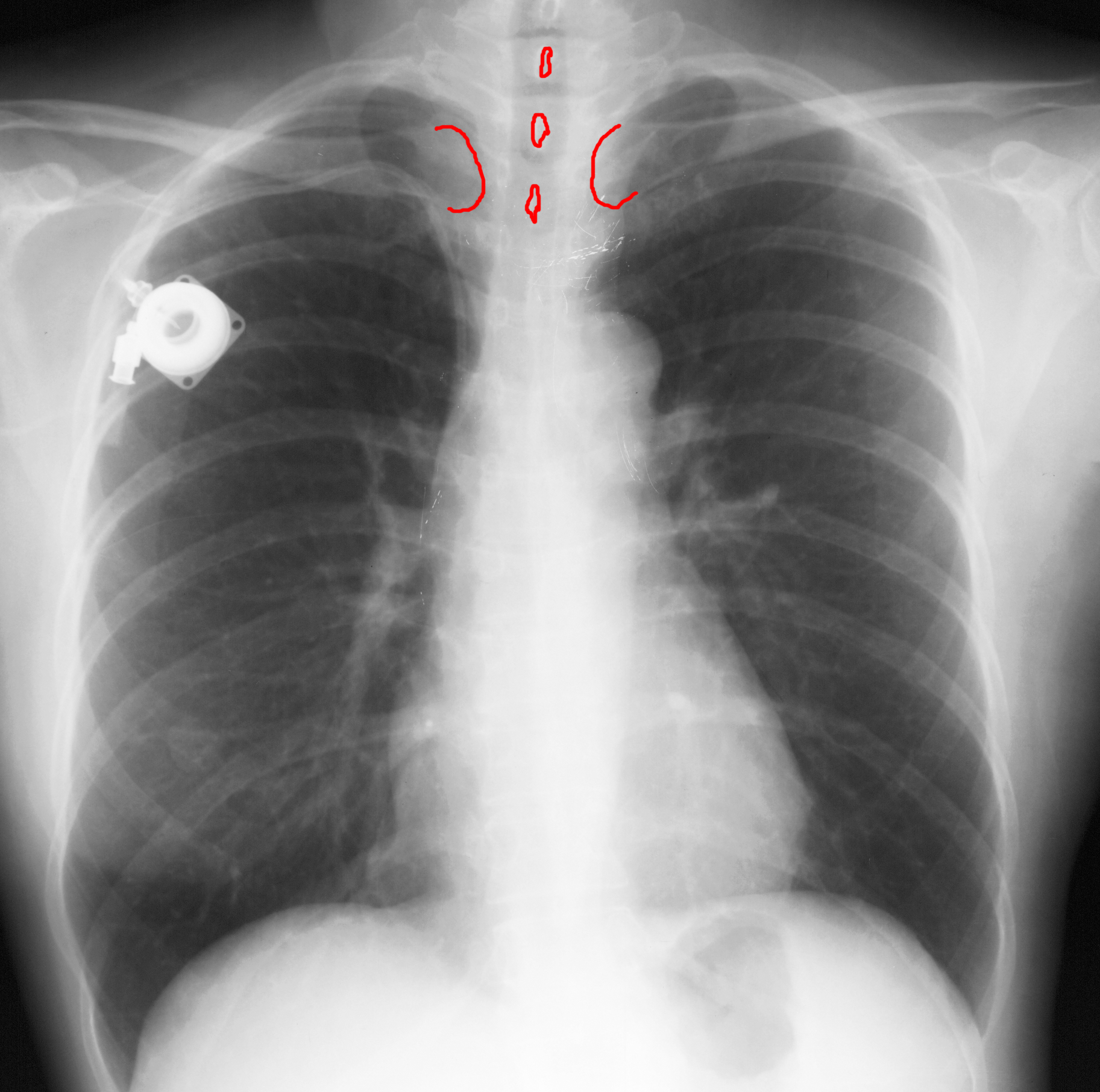
4. how to determine if the patient is rotated.
Click on the labels below to bring up annotations on the image!
