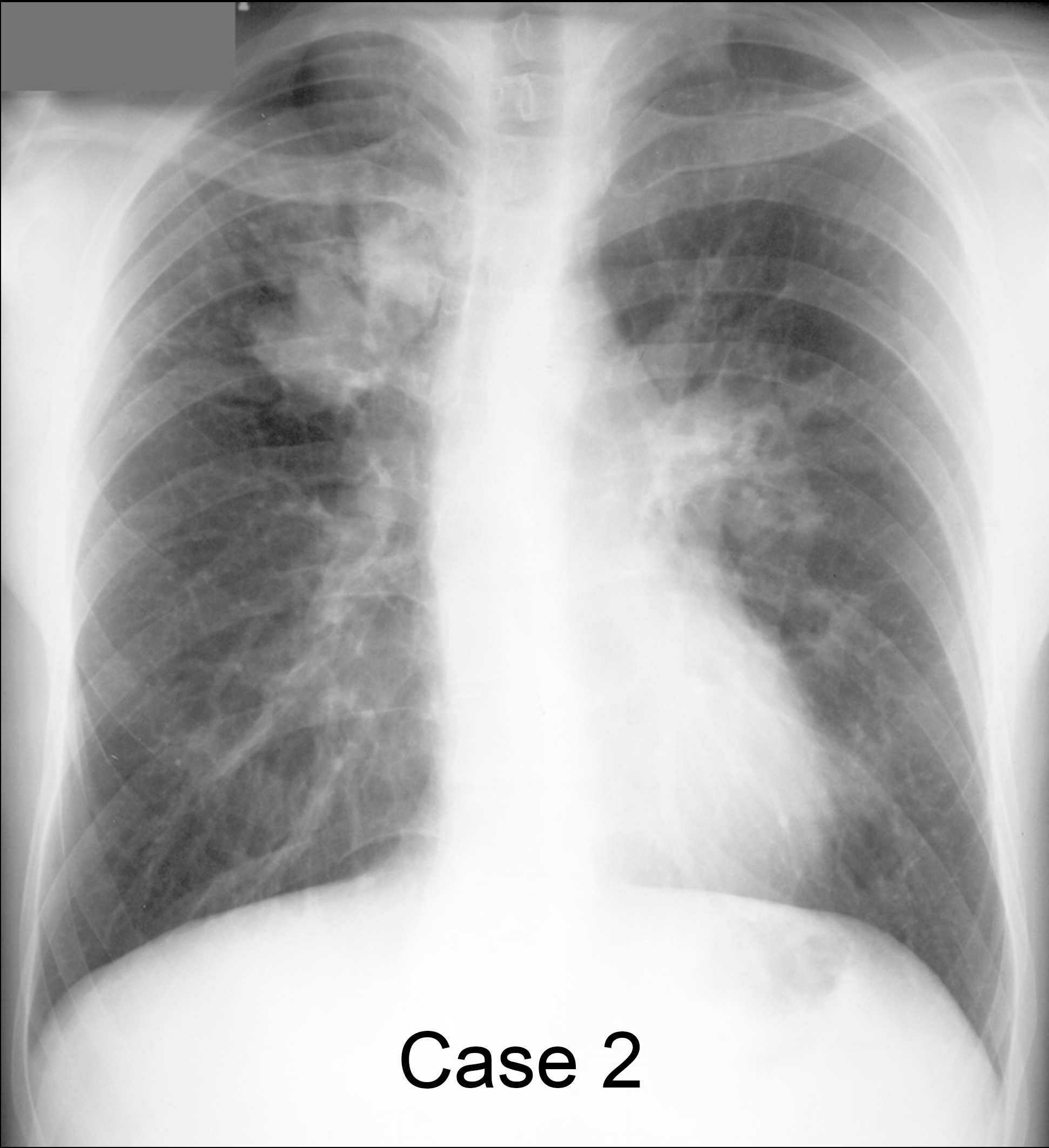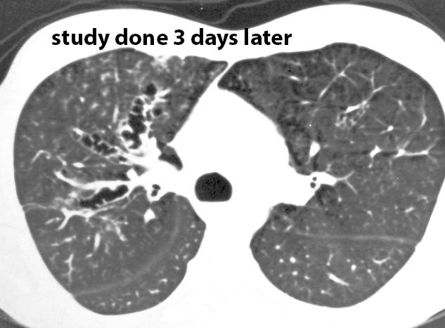


Case 2
Middle aged patient with intermittent shortness of breath and chronic cough.
Further Explanation:
For each case, fill out part of the table, considering the appearance of airways, lung parenchyma, pulmonary vasculature and pleura and enter descriptive adjectives for CXR and CT/other findings. Be sure to evaluate overall lung volume. For DESCRIBING abnormalities, consider their SHAPE, DENSITY and DISTRIBUTION, rather than trying to choose a specific diagnosis from the start.

Case 2
Choose a spokesman from your group to present your findings for this case during class discussion. Pay particular attention to the SHAPE of the abnormalities you see on the radiograph and what this implies about the structures of the lung or pleura that are involved. Think about whether the CT findings and radiographic findings are the same, or if something has changed between the two studies.
Further Explanation:


