

US
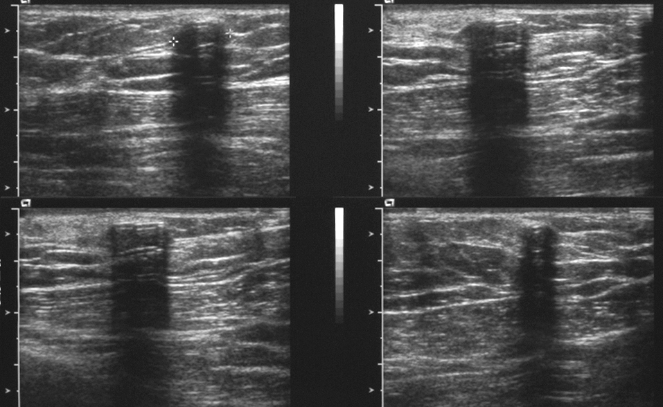
Think about how you identify this type of study, relative radiation dose, field of view, spatial resolution, any patient considerations or preparation, strengths, weaknesses and most frequent uses for this type of study. This is the same patient whose mammogram was shown previously. What artifact is seen on this image, and how can it help you decide what that object might be?
Further Explanation:

US
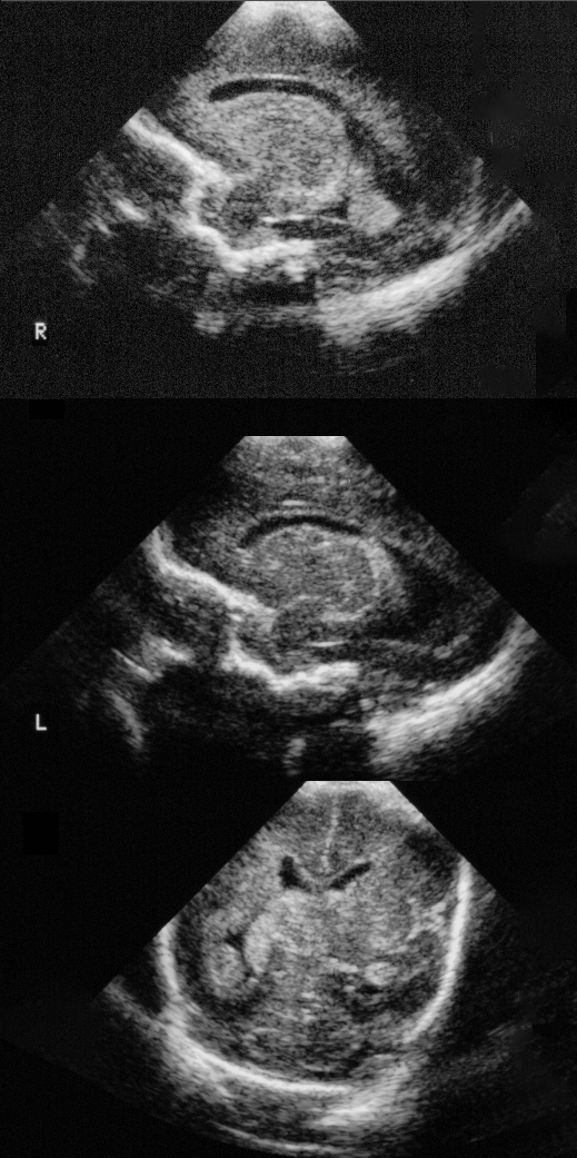
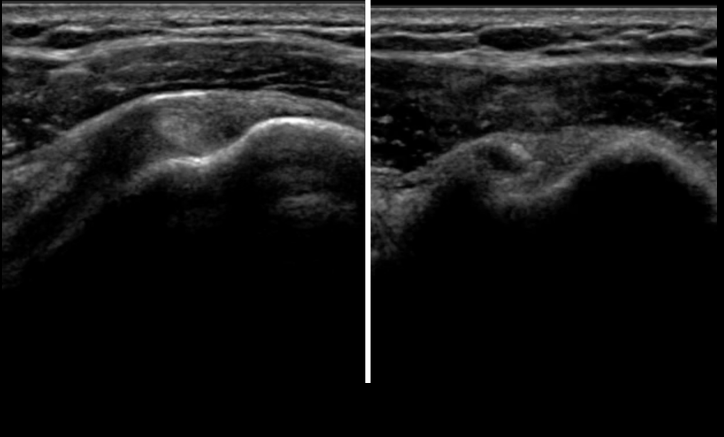
Another ultrasound done in a baby. How does the sound beam enter the body for this type of imaging? What organ are we seeing? Could this be performed in an older child? For the other patient shown, why does the biceps tendon look different in the two views (done within seconds of each other), and what does this tell us about the tissue?
Further Explanation:


