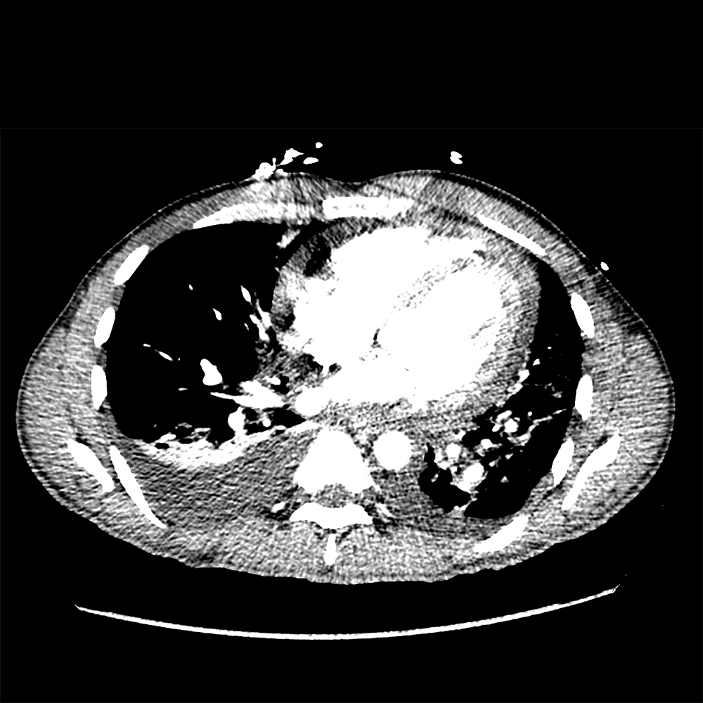


Other CT windows
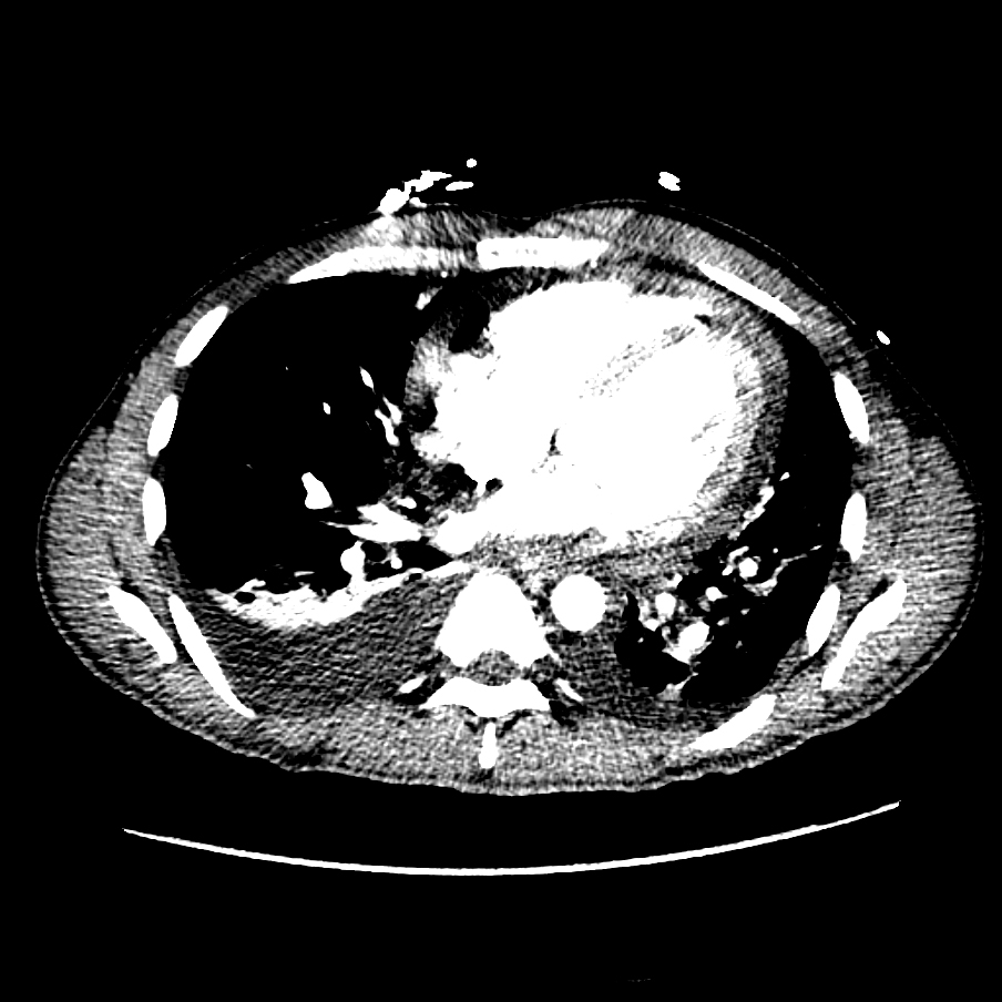
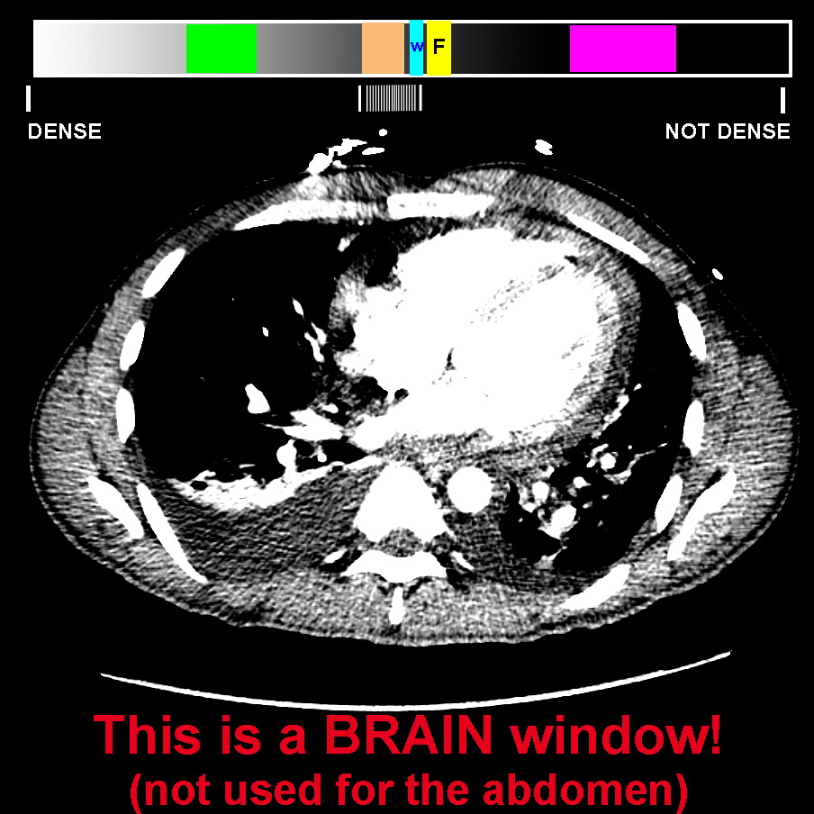
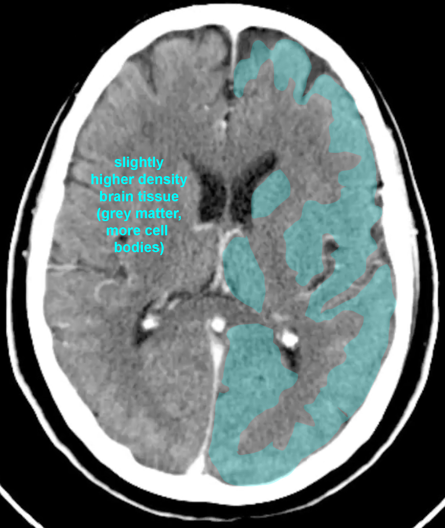
This image shows a different CT window that is specialized for a single tissue in the body.
Question 1:
a) How does this image look different from a soft tissue window?
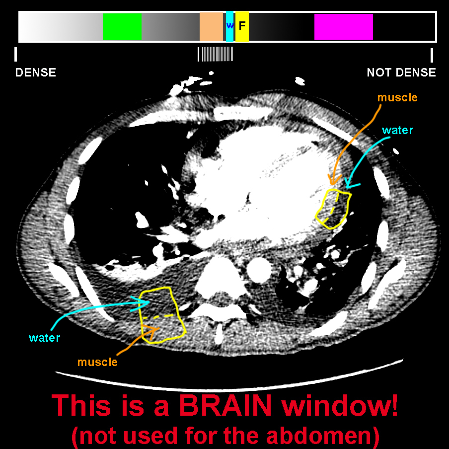
This image looks very HIGH CONTRAST. That meant that all the dark areas are very dark and all the light areas are very bright. If you concentrate on the interface between fluid and muscle (along the pericardial effusion and the pleural effusion), you can see that they look VERY different in terms of shades of grey, because that is the part of the density spectrum that we are focusing on with this window. And the bones are much brighter and fat much darker than on a normal soft tissue window. In fact, the image looks confusing and grainy, not like the usual abdominal CT images.
b) In what part of the body is the difference between water and relatively low density soft tissues of particular importance?
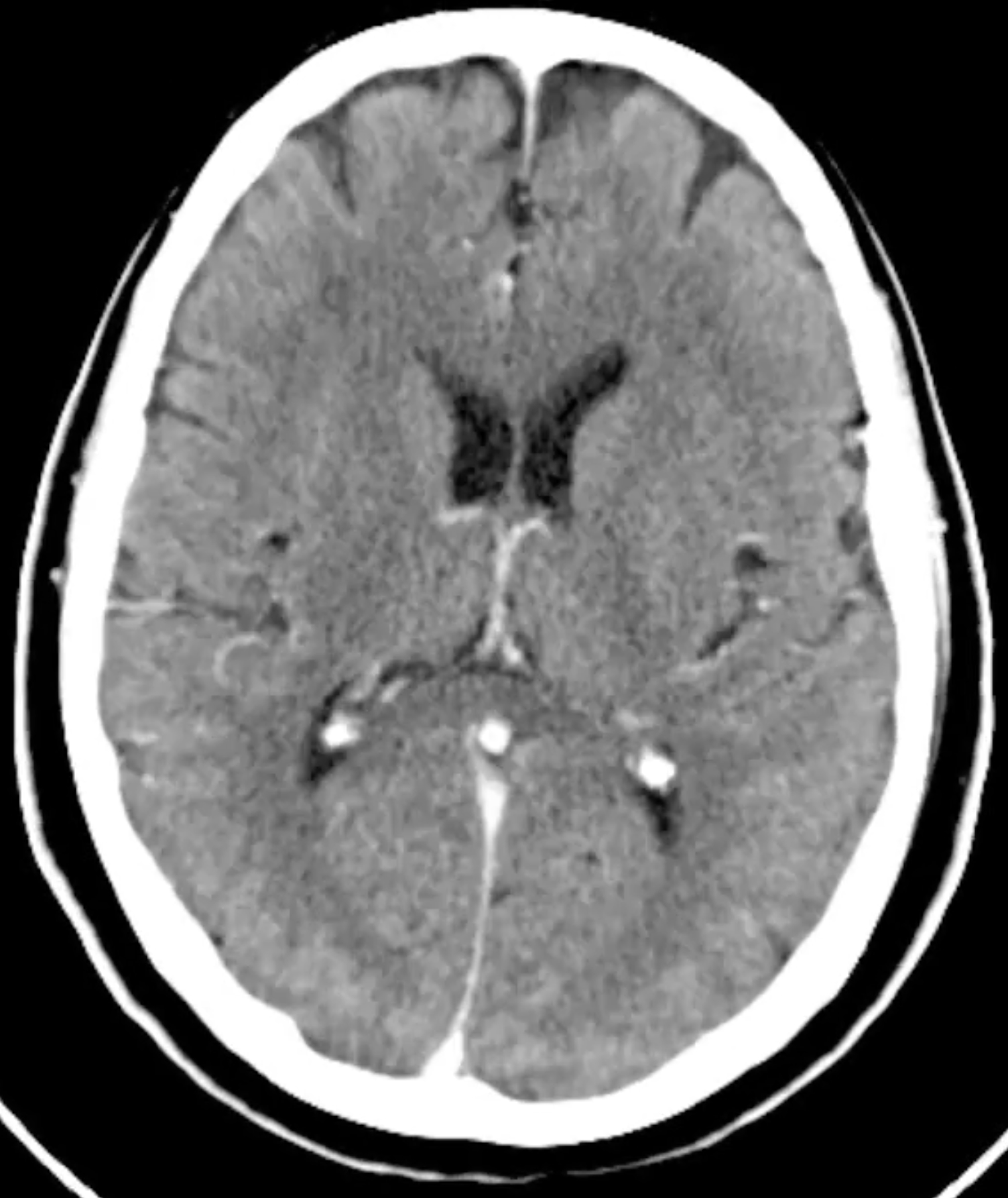
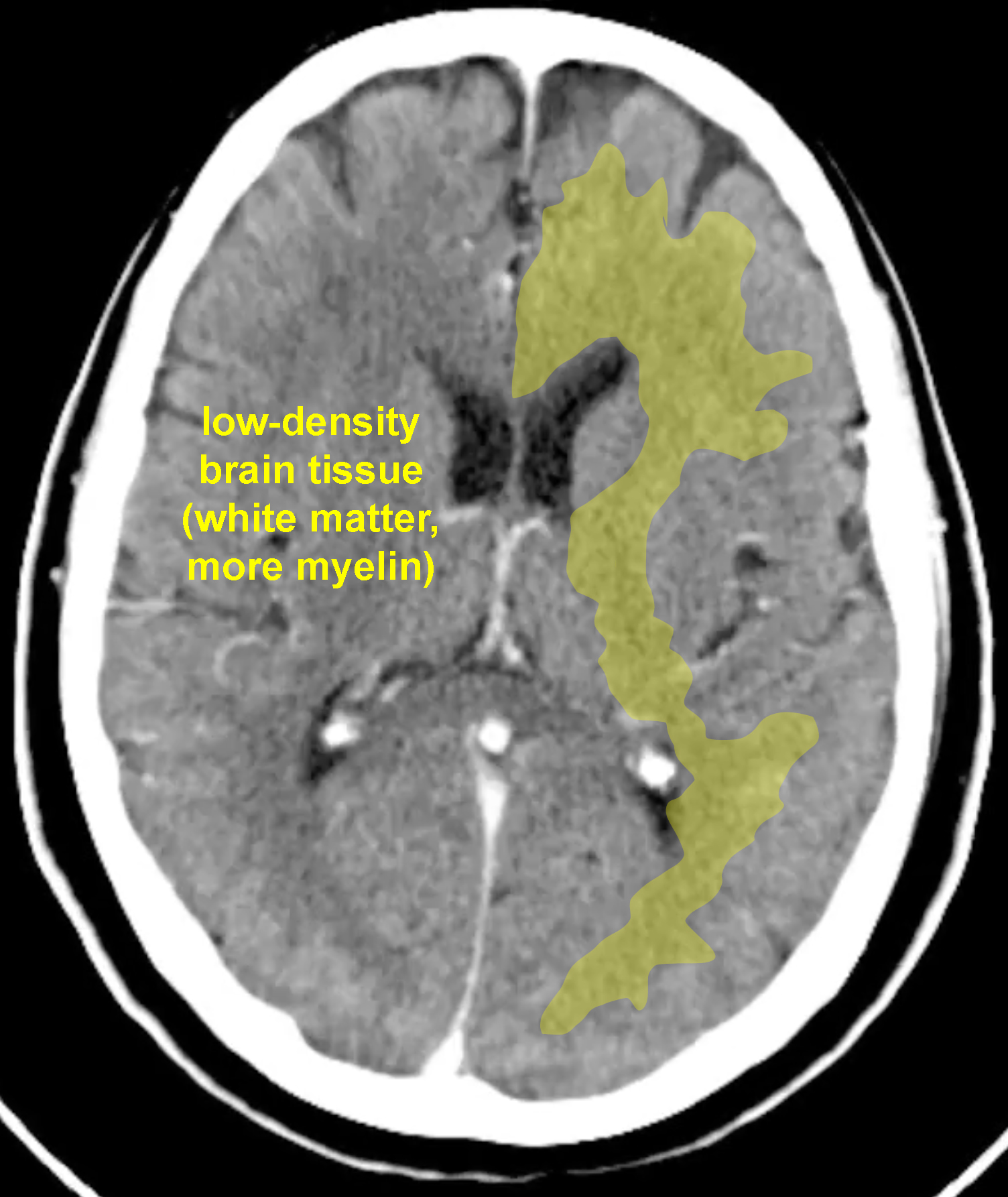
Imaging the brain requires a window that optimizes distinction between soft tissues, that are very close to the density of water. There are not a lot of other densities to see, so the shades of grey in the image can all be very close together, which will increase the contrast between tissues of slightly different densities. The overall brain tissue density is low, just a bit higher than water, and some areas of the brain are slightly less dense than others, due to the abundance of fatty myelin that covers brain cell fibers. This is an example of a BRAIN WINDOW, which would not be used for imaging the abdomen. You can see that white matter (with more myelin) is darker (lower density) than grey matter. The terms 'white' and 'grey' refer to the gross appearance of the tissue, NOT to the shades of grey that you see on a CT scan!
Other CT windows
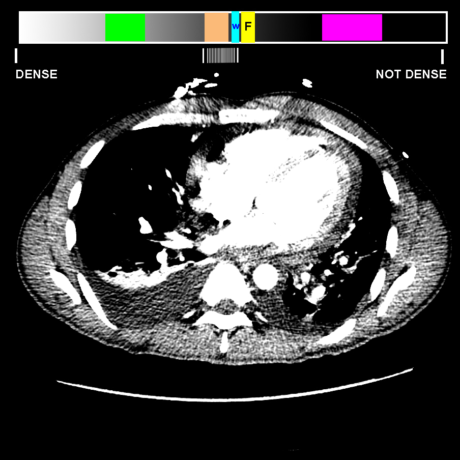
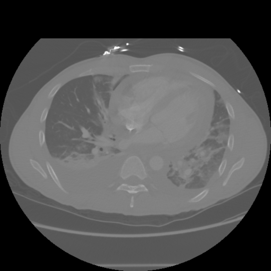
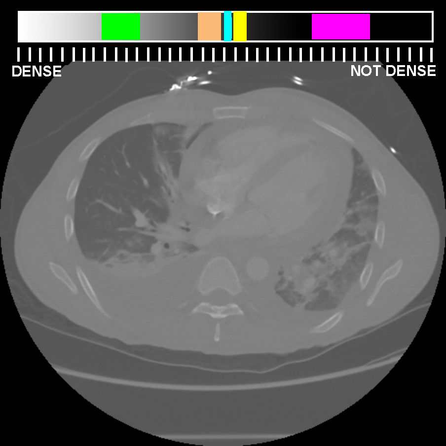
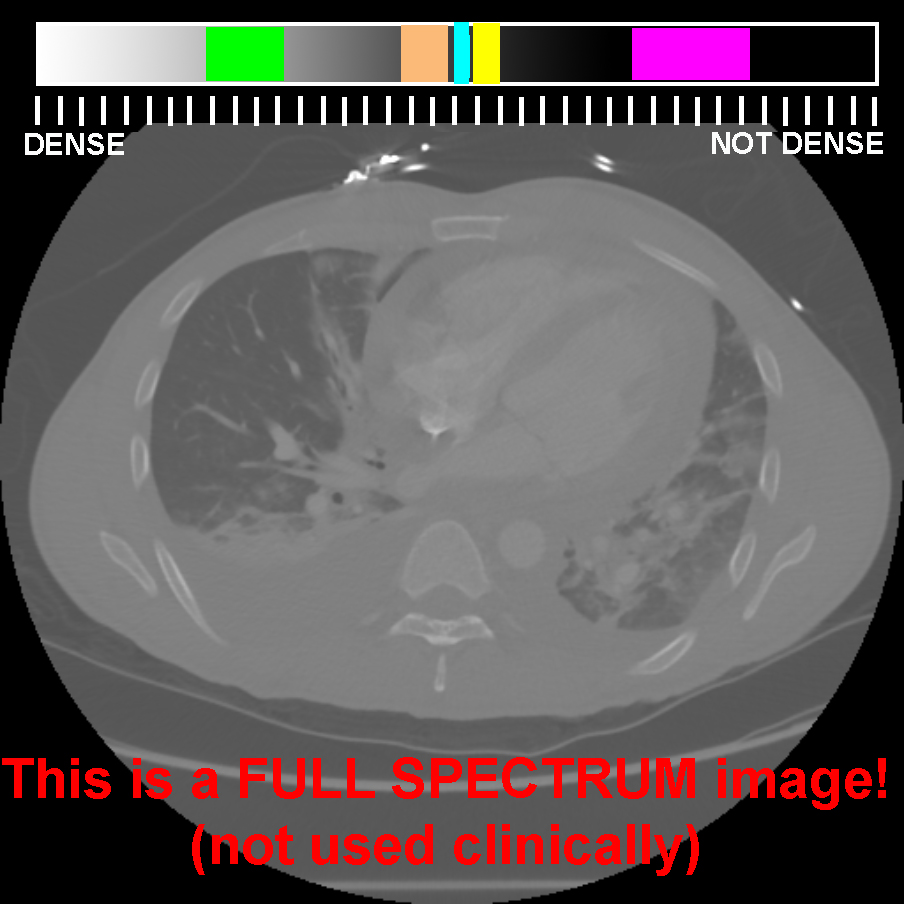
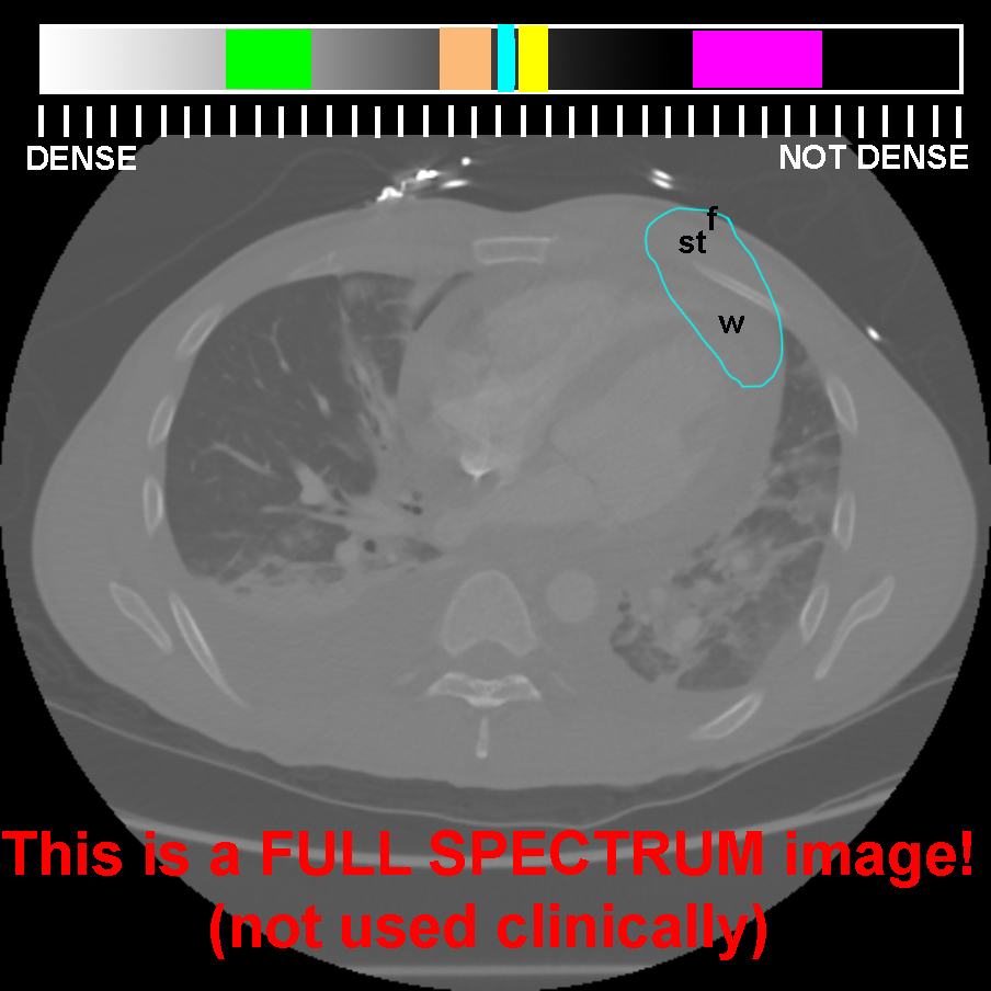
This is an image that shows a window that is not actually used clinically.
Question 2:
a) How does this image differ from a soft tissue window?
This image looks overall washed-out. You can still see structures, but all tissues are more similar terms of shades of grey than on any of the routine windows used in clinical, practice. This is called a 'full spectrum' image. That means that the shades of grey are evenly distributed across the entire spectrum of tissue density, not focusing in on any particular tissue. So it does not give not a very clear view of any of them. If you look at fat, tissue and water, they are very hard to distinguish. Bone and air are still far enough out at the ends of the spectrum that they are visible, but not as clearly as on more appropriate windows.
b) How does this image differ from a brain window?
This image is very LOW in terms of contrast, giving the washed-out look. A brain window of the abdomen appears very HIGH in contrast. Both are not useful for showing anatomy or pathology in the abdomen. Brain windows are specialized to show fat-water-soft tissue differences, just like bone windows are specialized to show all gradations of high density bone, and lung windows are specialized to show all gradations of low density lung or air collections.
Other CT windows
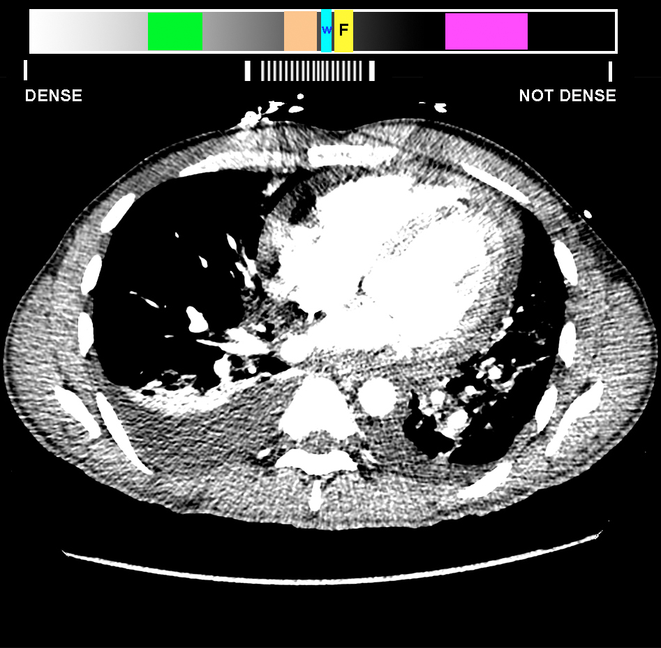
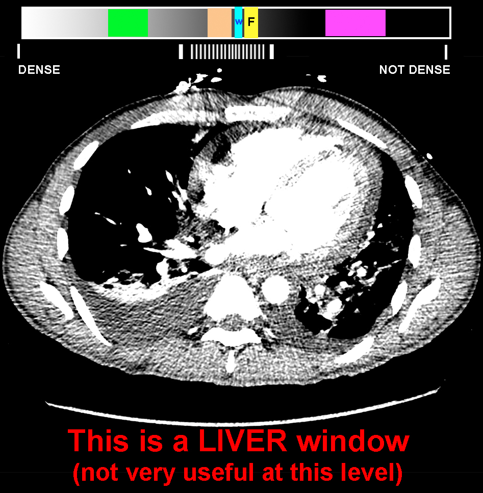
This is another specialized window that is used in the abdomen.
Question 3:
a) What other CT window does this resemble?
This looks a lot like the brain window. It is high in contrast and looks a bit grainy. That is because the organ of interest for this special window is the liver. The range of densities in the liver is a bit wider than for the brain, but we are again interested in focusing in on the central part of the density spectrum, where soft tissue, water and fat are located.
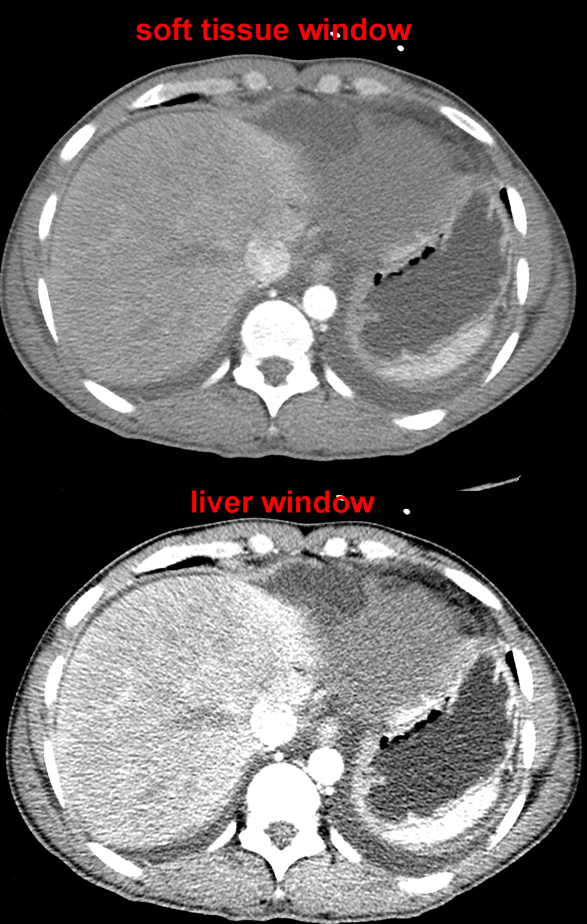
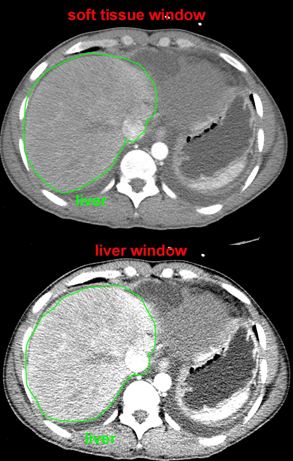
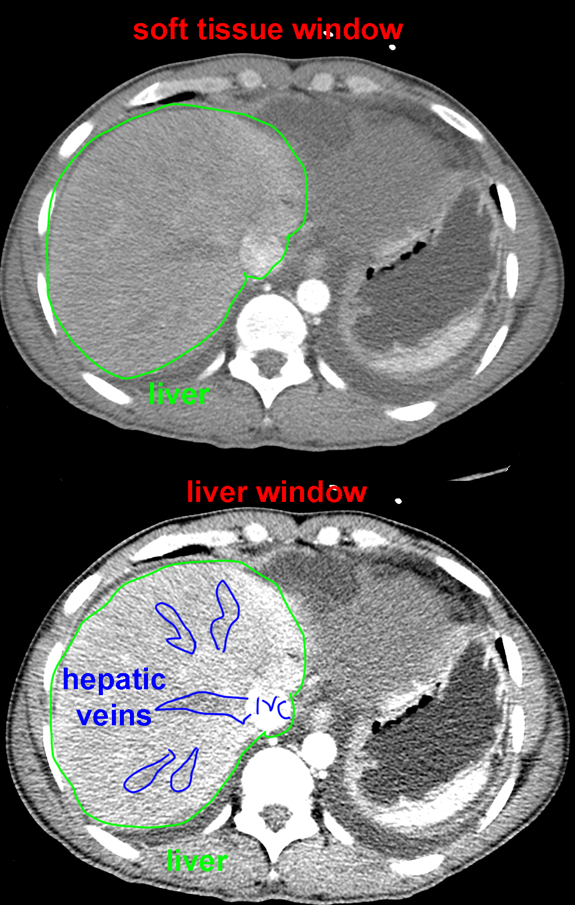
b) You are shown an image from lower in this patient's CT scan below, including the liver. This is the region where a liver window would be helpful. Comparing the top vs bottom image, decide which is a liver window and which is a soft tissue window. Is it easier to see the unenhanced hepatic veins within the liver on the top or the bottom image?
The difference in density between slightly enhancing liver parenchyma and non-enhanced hepatic veins is subtle, and you can see them on both images. The point of the liver window is to make them stand out more clearly by focusing in on the central part of the density spectrum. The hepatic veins have not yet received contrast from the portal system in this scan performed early after injection, so they appear dark, and are slightly easier to see on the bottom image, displayed in liver windows, than on the top soft tissue windowed image. The same is often true for other liver diseases such as spread of tumor or infections--subtle abnormalities are often better seen on liver windows than on routine soft tissue windows.
To return to TOPICS FOR ANATOMY GRADUATE STUDENTS, click HERE.
