


Case 1
This case deals with imaging of the genital organs in the pelvis
Question 1:
a) This patient was being evaluated for vague symptoms. What do you notice on the first slice of her CT scan?
This is an axial CT in soft tissue windows with IV contrast (and oral contrast, but you cannot see this on the first slice of the study--you need to scroll down to the stomach). On the first slice you can see fluid-filled breast implants between the chest wall and subcutaneous fat.
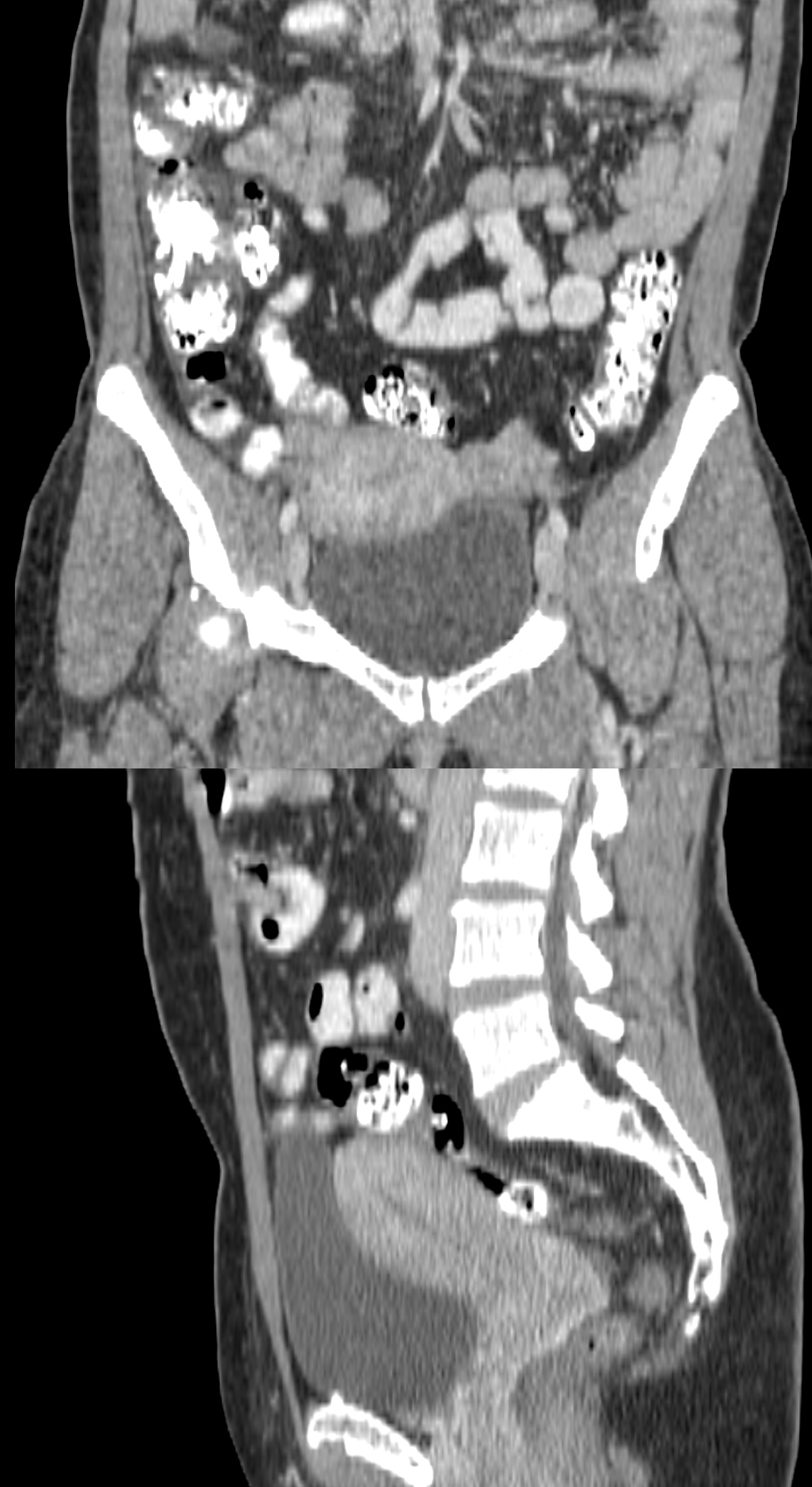
b) What do you notice regarding the renal system in this patient? What do you notice about her uterus on the lower cuts of the study?
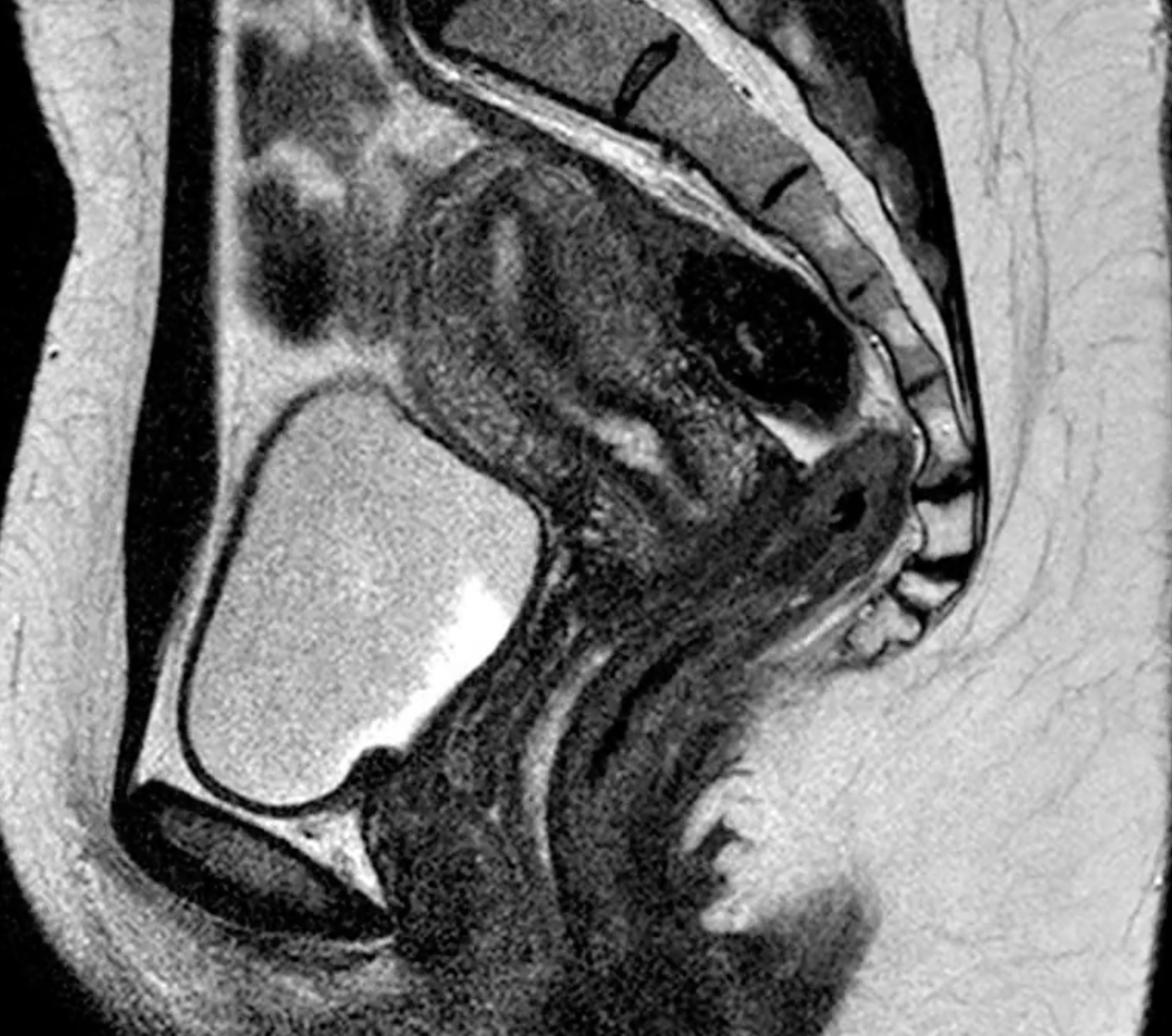
In the middle cuts of the CT, you can see a very large left kidney and no right kidney. In the lower cuts, you can see that her uterus is small and seems to be all on the left. You can never to the 'normal female pelvis' images to see what a normal uterus should look like on CT. There are many ways to image the uterus, including MRI, which you can see in another normal patient by clicking 'another imaging modality'. You can recognize MRI by the fact that cortical bone is very dark (look at the pubic symphysis). Fat is variable in appearance on MR, but if it is white (as in this case) then it is definitely NOT a CT scan. Fat on CT is relatively dark. Fat on MR can be dark or white, depending on how the scan was set up.
c) What is the study shown if you click the 'another way to image the pelvis' link?
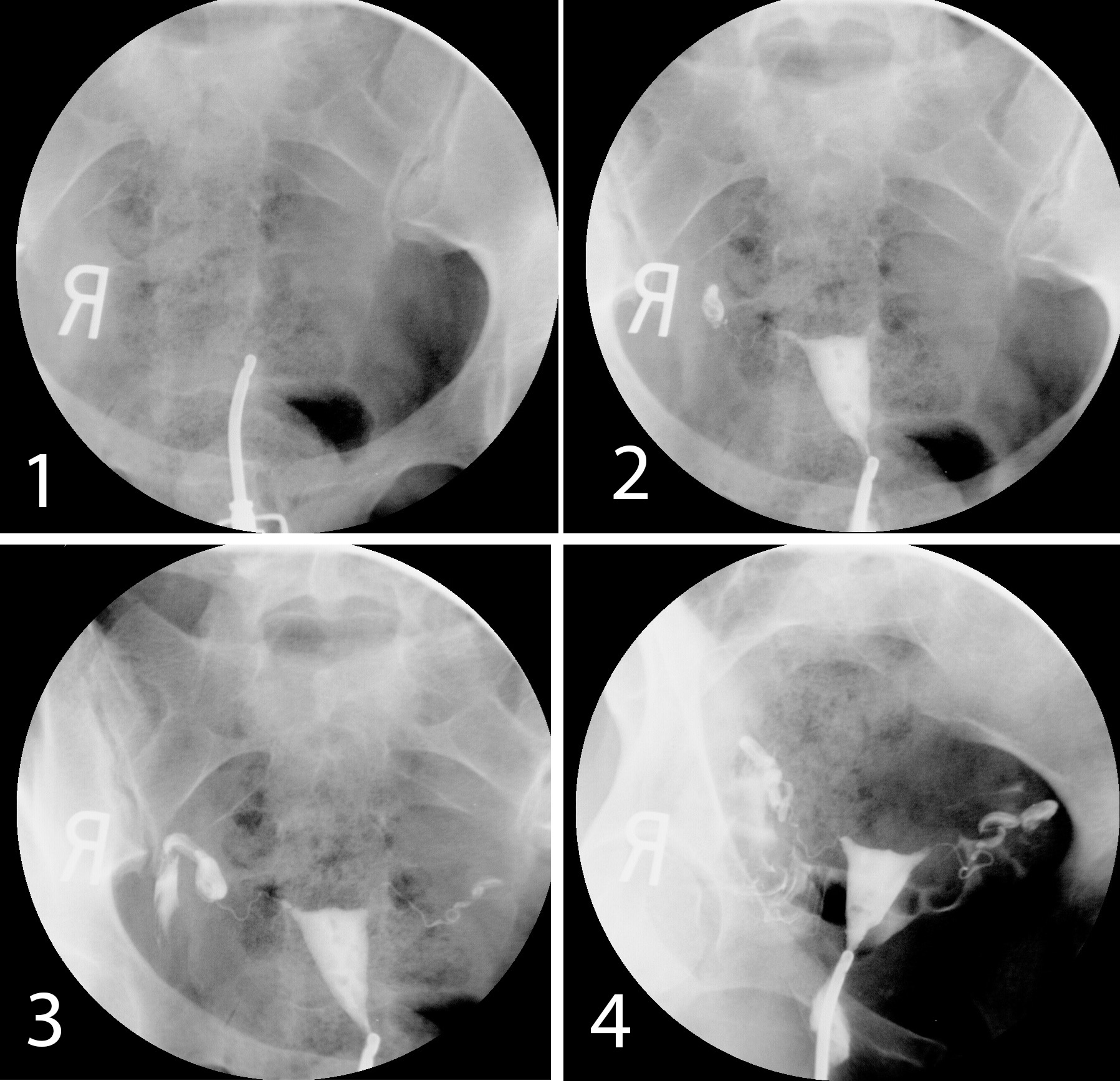
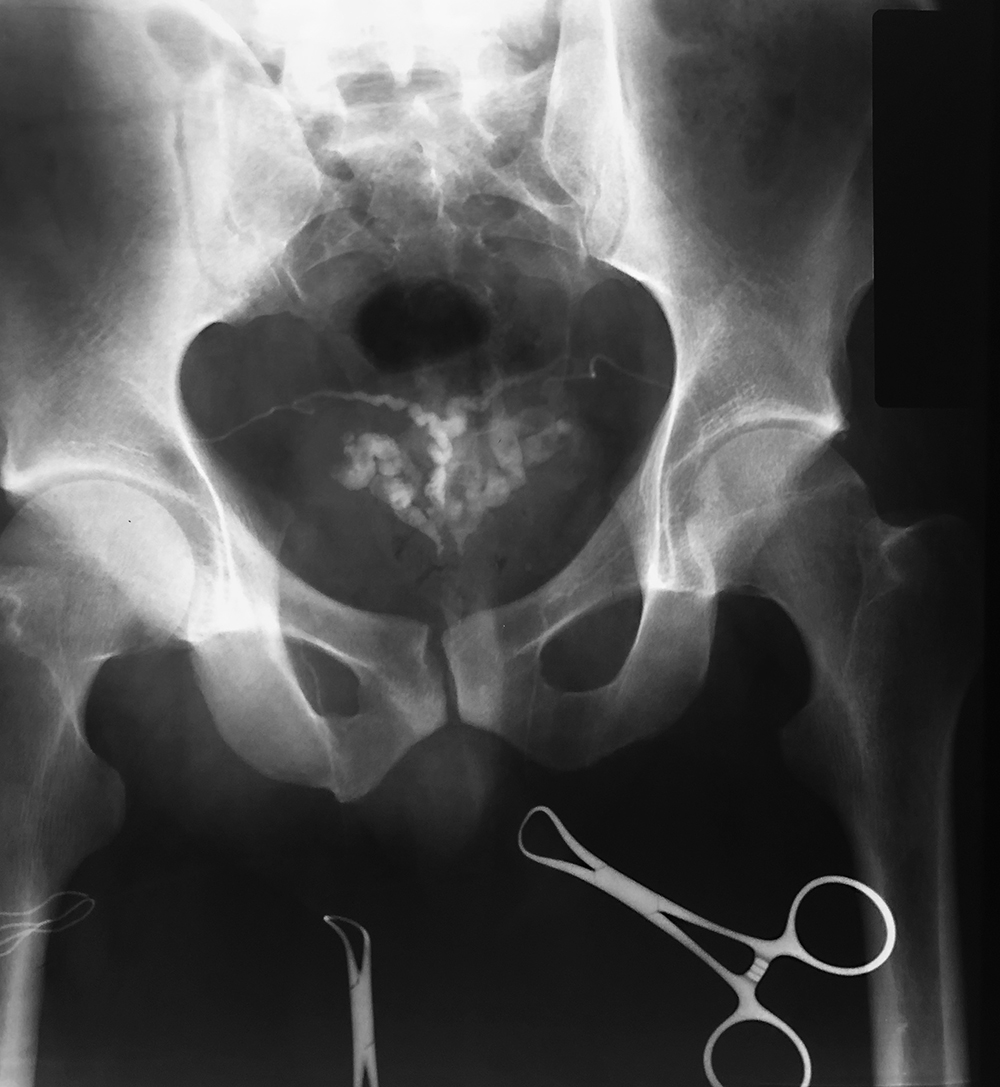
This shows a normal hysterosalpingogram, an interventional study where a cannula (thin tube) is inserted into the cervical os and dense contrast material is injected into the uterine lumen, with sequential radiographic images taken over time. The two thin undulating uterine tubes should fill, and eventually there should be contrast spilling out into the peritoneal cavity, outlining bowel loops.
d) What do you think of the 'same study on the initial patient'?
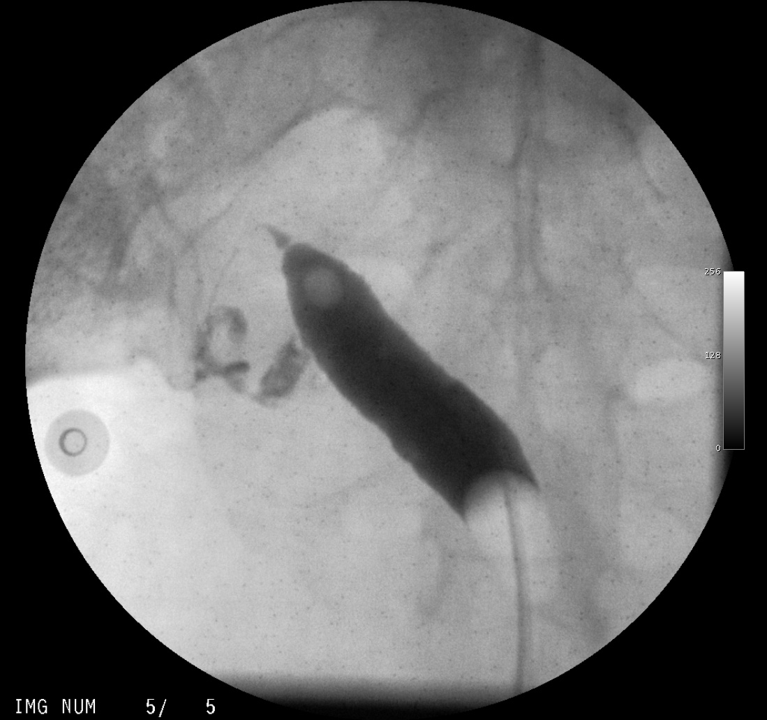
This is the hysterosalpingogram done on the patient with only one kidney. It shows a unicornuate uterine lumen, instead of the usual triangular one seen on the normal example. There is one uterine tube and probable spillage of contrast into the peritoneal space. This case emphasizes the importance of embryology in understanding the importance of a unilateral kidney. Since the kidney and uterus share some embryological origins, it is not surprising that a congenital anomaly in one system might be reflected in the other.
Return to case list for pelvis
Case 1
More pelvic imaging on a different patient
Question 2:
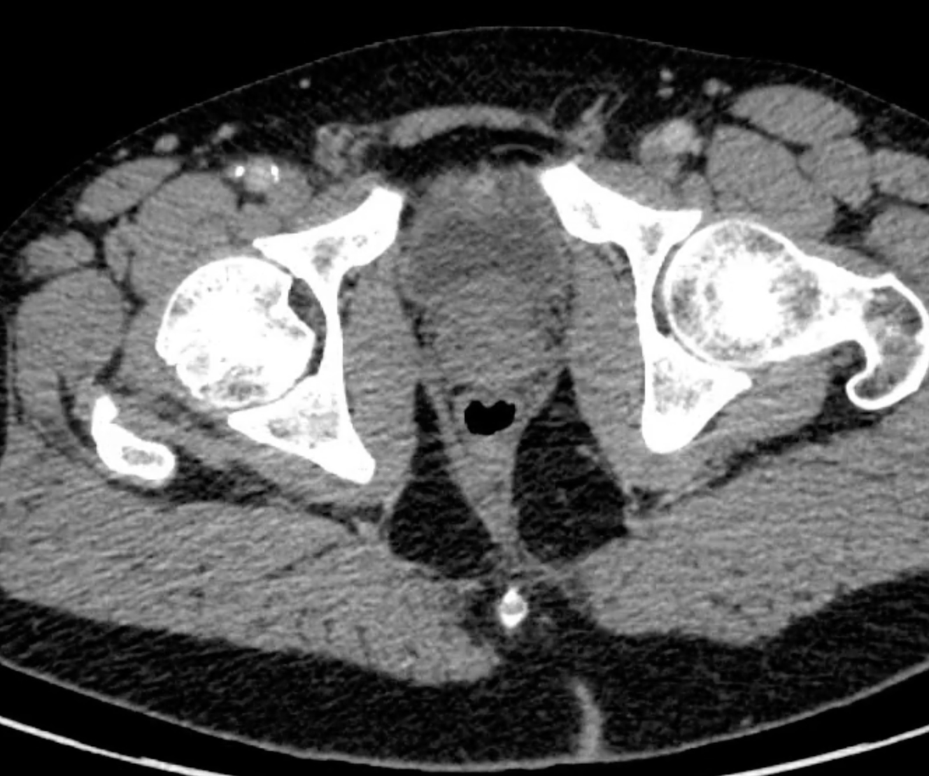
a) What is this study and how can you tell this is not the same patient as the previous page?
This is an axial CT, soft tissue window, with IV contrast. There is no definite GI contrast on this single image, but there might be some on other images showing more proximal parts of the GI tract. You can tell this is not the same patient as the previous page because we are seeing the prostate gland, located between the bladder and the rectum. Like the uterus, the prostate on CT is often a mid-grey, without much internal structure. While CT is very useful for seeing all of the adjacent organs and nodes, it does not tell as much about the internal structure of the prostate as other types of imaging.
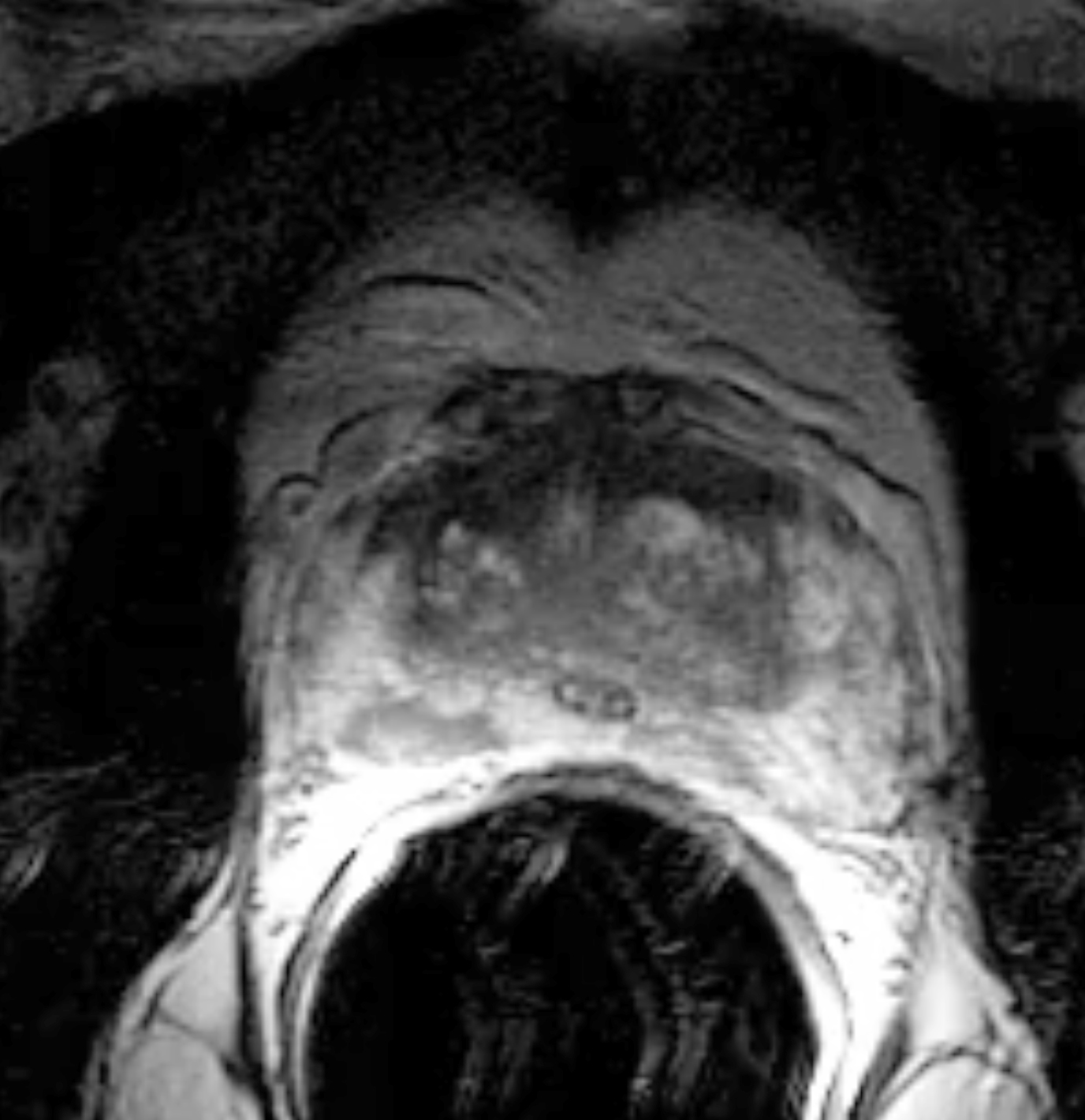
b) What type of imaging is shown when you click 'another type of imaging'?
This is a single image from a prostate MRI. It is oriented the same way as the CT, but the level is not showing us the bladder. The large black circle in the lower part of the image is a balloon filled with air that is positioned in the rectum, to move the prostate into the optimal position for imaging. You can again tell that this is an MR because the fat around the prostate is white. The prostate shows much internal structure, with many layers and heterogeneous tissue signal that might not show up on CT.
c) What about 'and another type of imaging? What part of the body is being imaged, and Is this study normal?
These images are ultrasound. The first image is labeled SAG LT. By convention, sagittal ultrasound images are displayed with the patient's HEAD to the LEFT side of the image. These images show a homogenous oval structure that is mid-grey and is consistent with a normal testis. However all around this testis are tortuous tubular structures that are dark (fluid-filled), and which light up brightly on Doppler imaging, indicating turbulent flow of blood. This is a patient with a varicocele, or dilated veins in the scrotal area. For both prostate and testicles, ultrasound can show internal structure better than CT scanning.
d) What is 'one more type of imaging'?
Somewhat analogous to the hysterosalpingogram, where contrast was injected into the uterine lumen to evaluate the female pelvic organs, one can also inject contrast into the vas deferens, to evaluate the male pelvic organs. The vas on both sides were injected and you can follow the thin line of contrast back to the ejaculatory ducts and the seminal vesicles. This study is called a vasogram. It is rarely performed but can be considered in the workup of infertility.
Return to case list for pelvis
Case 1
Summary of imaging for the abdomen and pelvis
Further Explanation:


