

Back and Limbs Case 1
52 year old patient with vague right arm pain and limitation of motion for many years.
Question 1:
a) What are technical parameters for this image? (Please discuss your answer within your group before clicking 'REVEAL', which will reveal the answer).
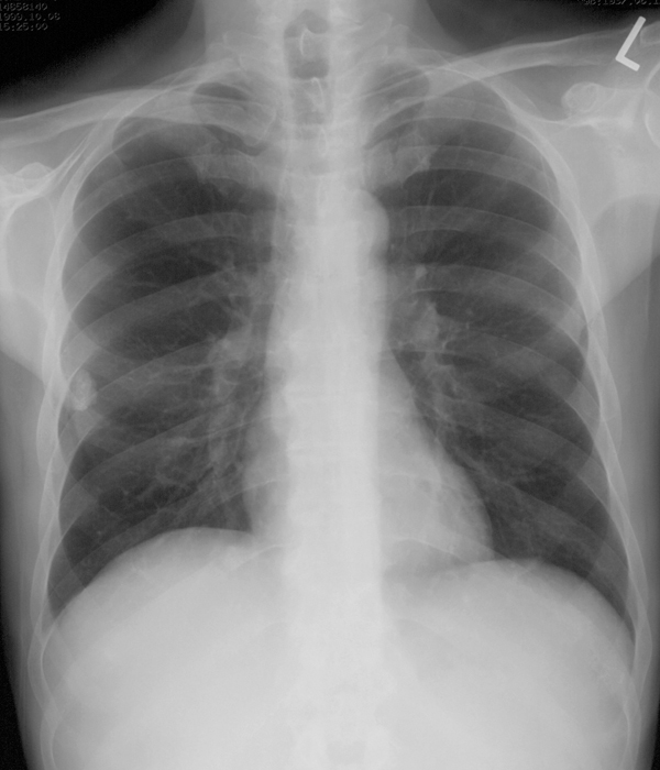
This is a chest radiograph, likely to be in the PA position, which is the preferred position for all patients who are able to stand upright on their own.
b) What do you see on this image that might relate to the reported symptoms?
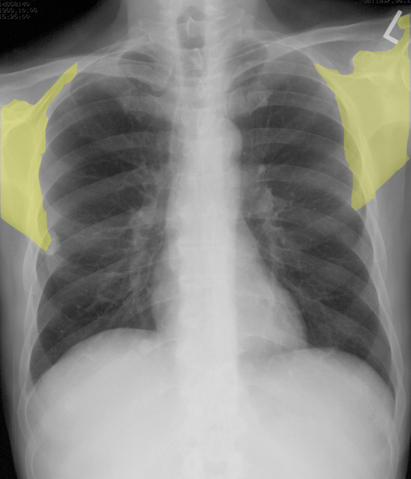
The right shoulder is not held in symmetric position, relative to the left. There are many causes of shoulder pain, some acute and some chronic. The shoulder is prone to dislocation due to the shallow socket of the glenoid, but the history in this case does not sound like a dislocation, which is a more acute problem.
c) Where is metal density on this image?
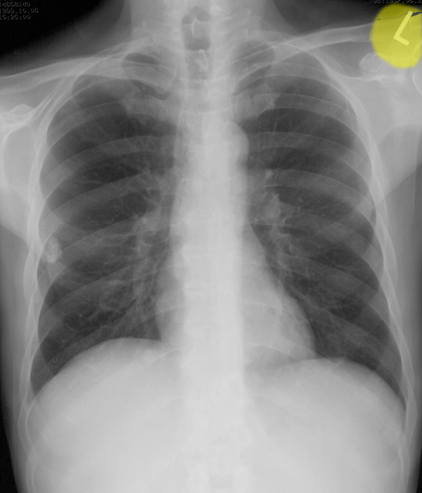
Metal density is not normally present in the body, unless there are foreign objects present (such as bullets or surgical clips or other hardware). But a metallic letter (usually an R or an L) are often still used to indicate the right or left side of the patient. In digital systems, sometimes this indicator is digitally added to the image, but in this case, a metallic object was taped to the receptor. This is the most convincing way to indicate which side of the patient is which, so it is still used in many settings. Metal density will be much whiter than bone or calcification, as is seen in this image.
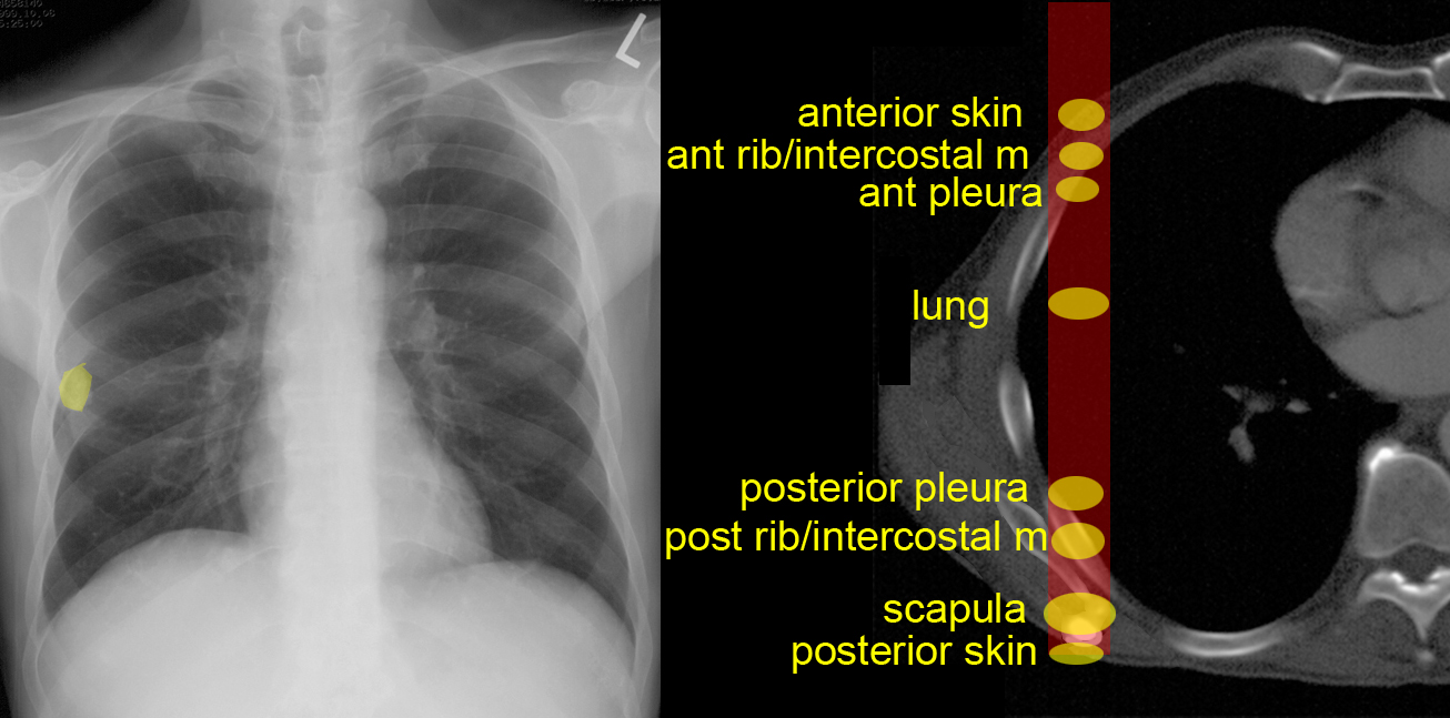
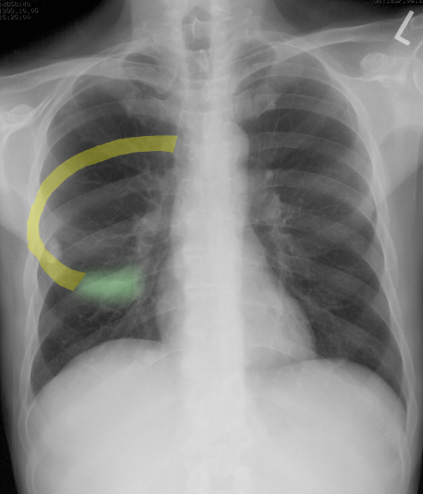
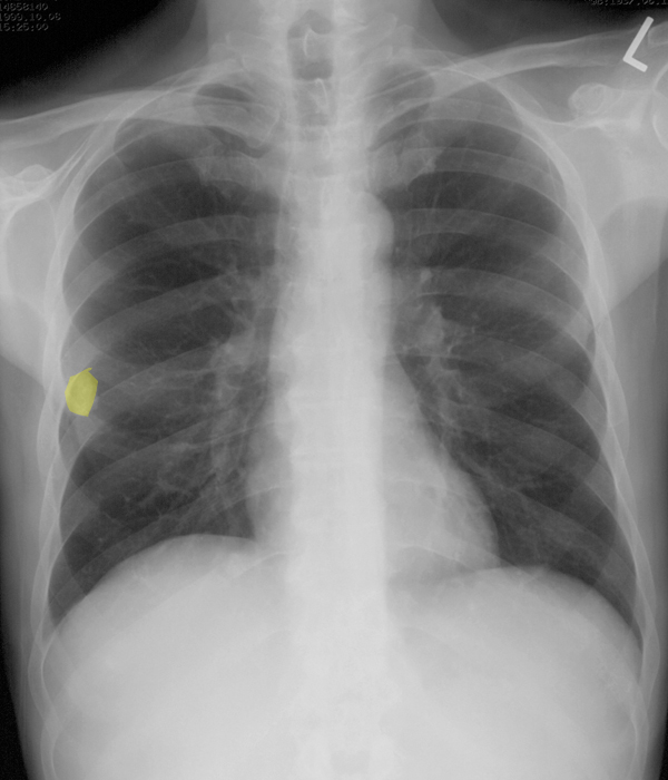
There is another abnormality on the right on this image which you should try to locate before clicking on the label. Once you have identified this oval calcific density object, try to think of where it could be located, from front to back.
Back and Limbs Case 1
This is the next study that was done on this patient.
Question 2:
What are technical parameters for this study?
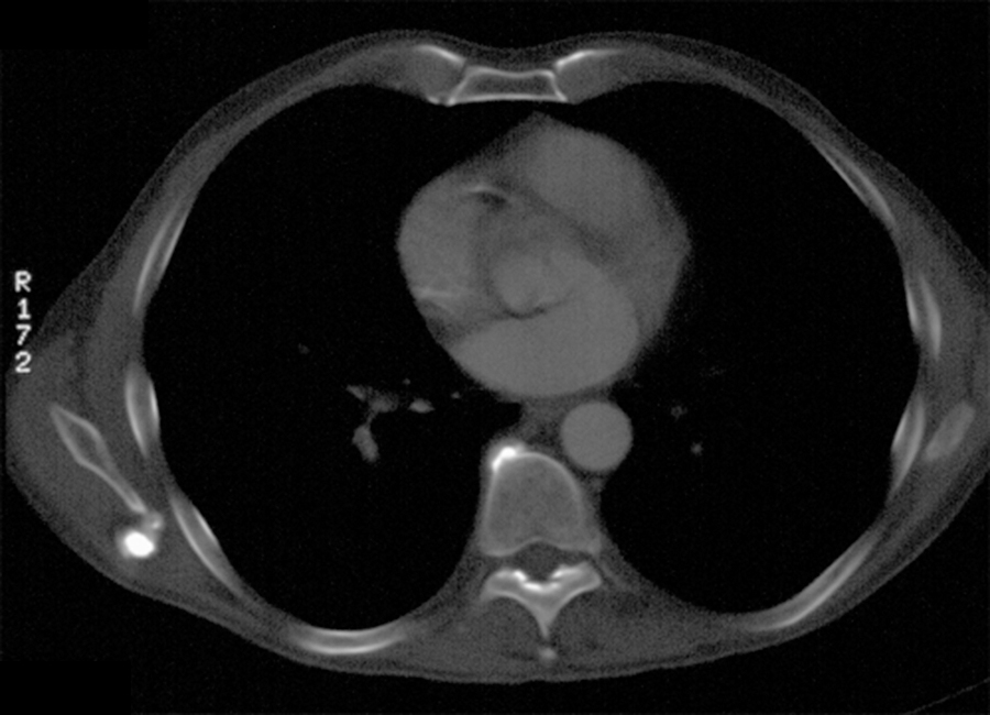
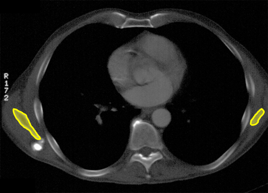
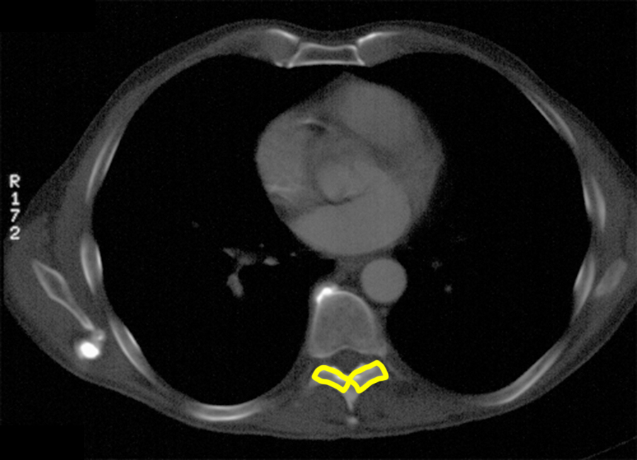
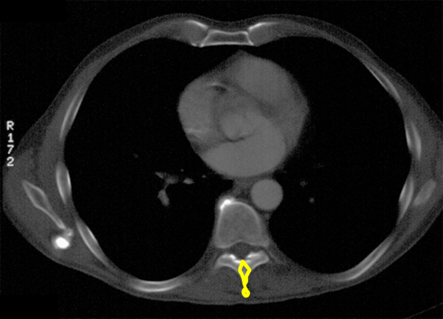
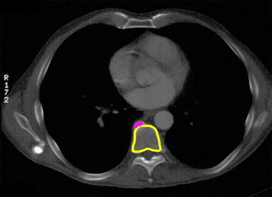
This is an axial CT image. You know it is a CT and not an MR because cortical bone appears white (high attenuation). It is displayed with a bone window, meaning the brightness and contrast have been optimized to show internal details of bones. Notice that you can see the transition from cortical to medullary bone in each bone on the image--every rib and even the small flat bone of the scapula. Some parts of the thoracic spine are visible and labeled below. The spur off the vertebral body is a degenerative change seen due to abnormal forces, often related to loss of the cushioning effect of the intervertebral disc with age.
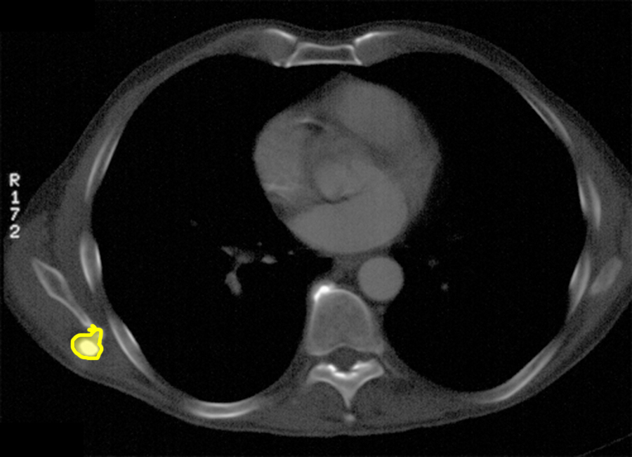
When you see abnormalities on a chest radiograph, particularly when they overly the lungs, then CT is the most common next study to perform. There can be exceptions to this. For heart abnormalities, sometimes ultrasound is useful. For complicated problems related to vessels, MR is often considered. But CT is the most frequently done because it shows the lungs better than ultrasound or MR and it gives a fairly good overview of the heart and vessels as well. Given the location of the rounded calcified lesion of the right scapula on the CT, does this correspond to the position of the lesion on the CXR?
Back and Limbs Case 1
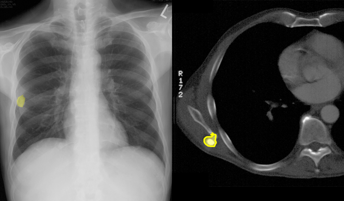
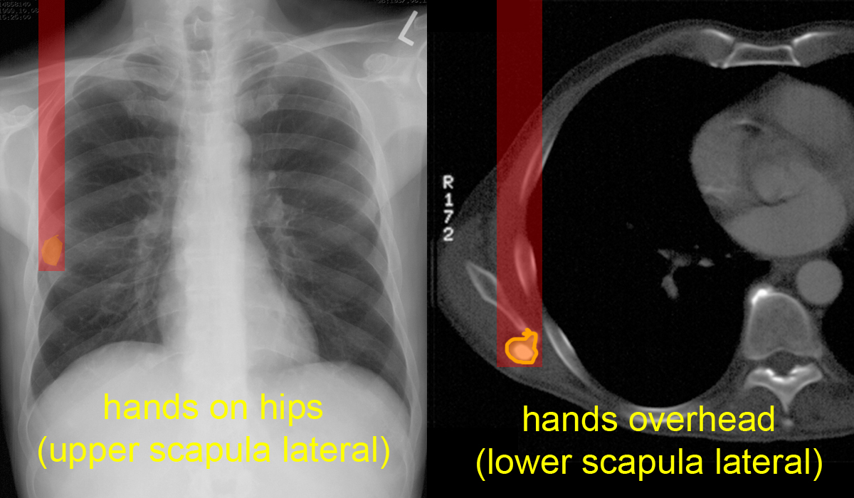
The chest radiograph and CT images are shown side by side
Question 3:
a) How are the arms positioned for a chest radiograph?
Patients are asked to put their hands on their hips and rotate their shoulders forward, which moves the upper scapula laterally. The lower scapula moves laterally also but to a lesser extent. The goal is to get as much of the wing of the scapula as possible off the lung, so that it does not obscure the upper lateral lung and pleural surface.
b) How are the arms positioned for a chest CT scan?
The arms are raised overhead, which rotates the inferior part of the scapula laterally, as well as the rest of the scapula. The purpose of this is to make the entire body more cylindrical in cross-section. With the arms at the sides, patient tissue are significantly thicker from side to side than front to back, and this can produce artifacts as the x-ray tube rotates around the patient. The scapula is a particularly mobile bone, which is mostly attached to the torso via muscles rather than bony articulations.
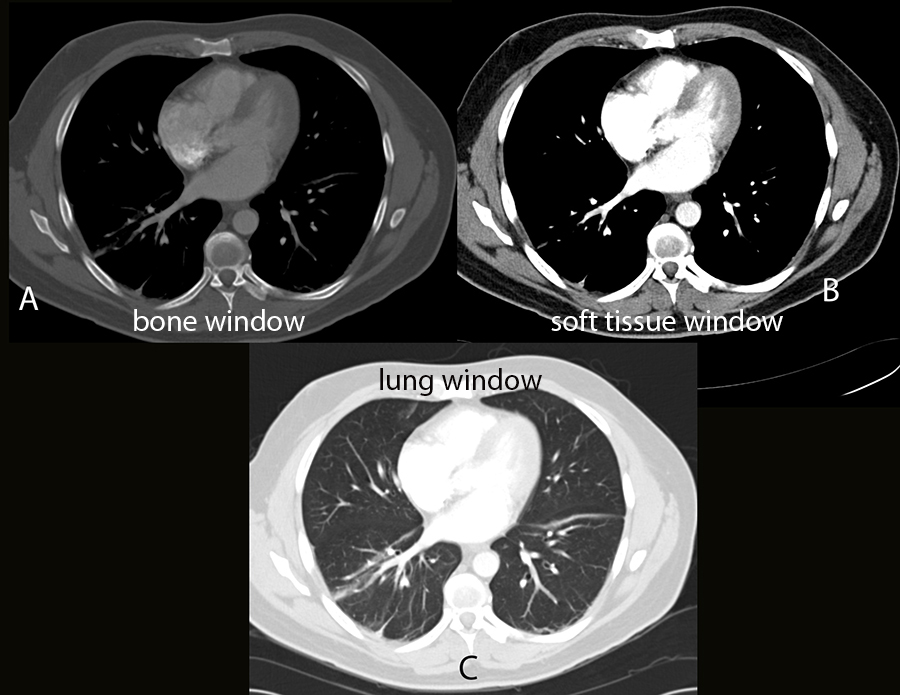
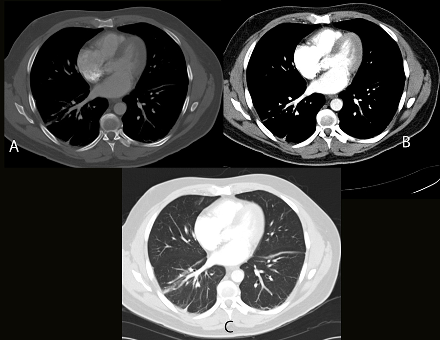
It is important to try to triangulate between the 2D projection image of a radiograph, and the cross-sectional image of a CT scan, to be sure that the finding you see on one modality matches the finding on the other. The link below shows you a CT image at a similar level from another patient, with a normal scapula. Try to decide which body tissues are best shown on each of the images, A-C. CT data can be adjusted in terms of brightness and contrast to highlight different tissues optimally.
Back and Limbs Case 1
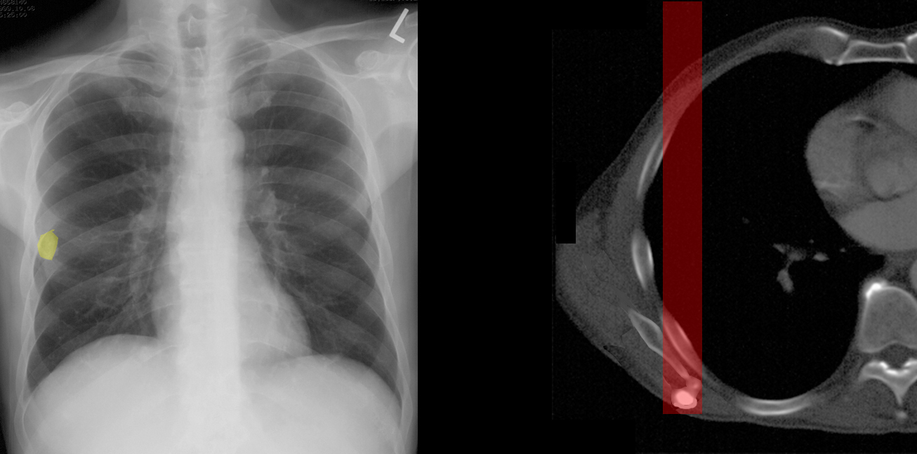
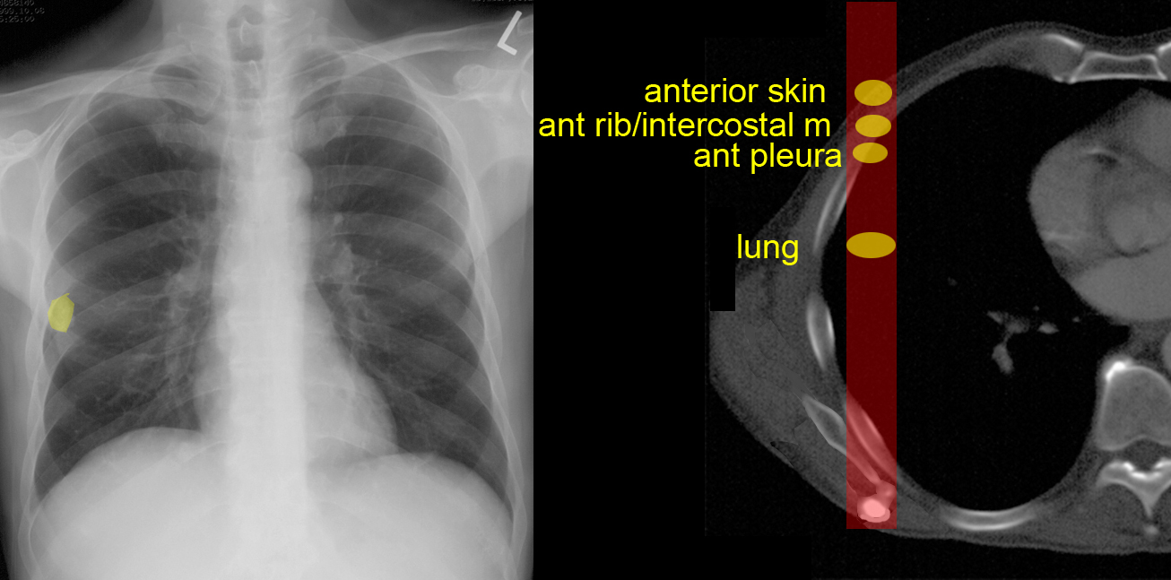
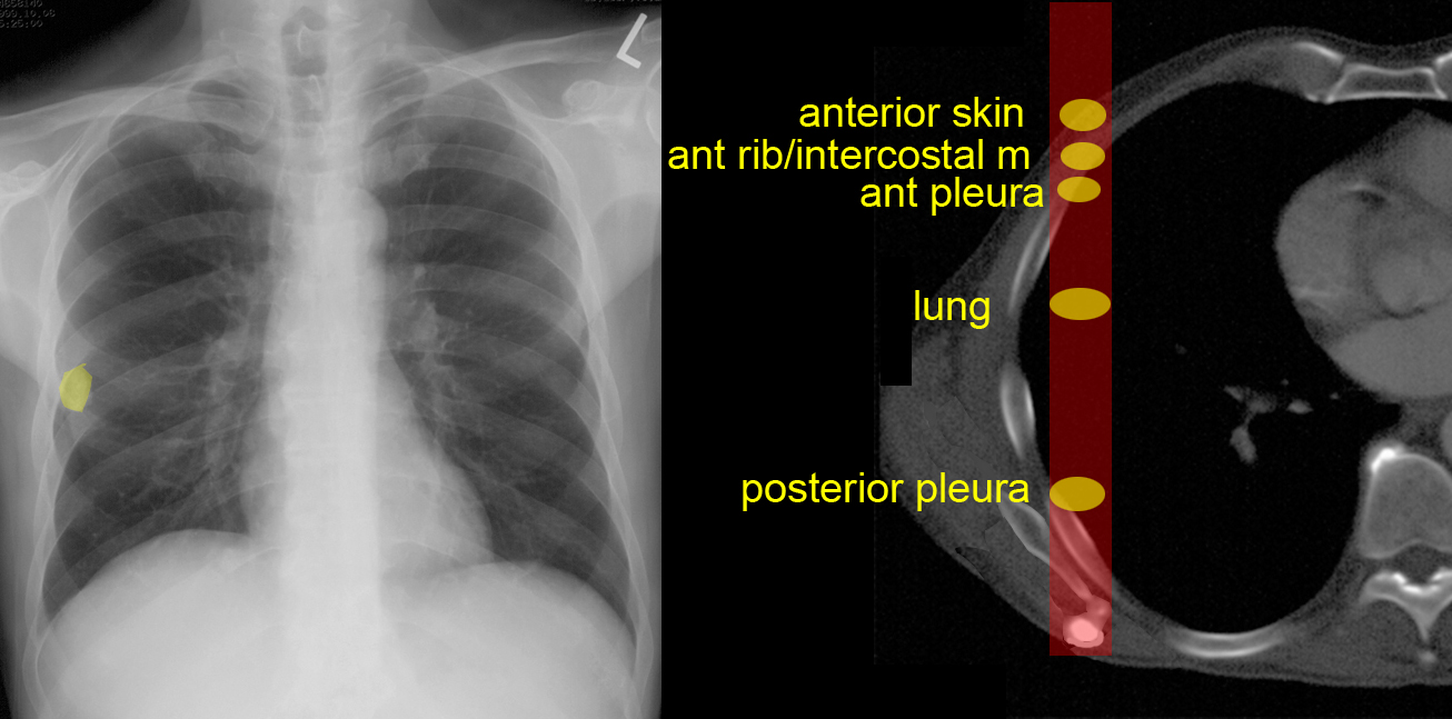
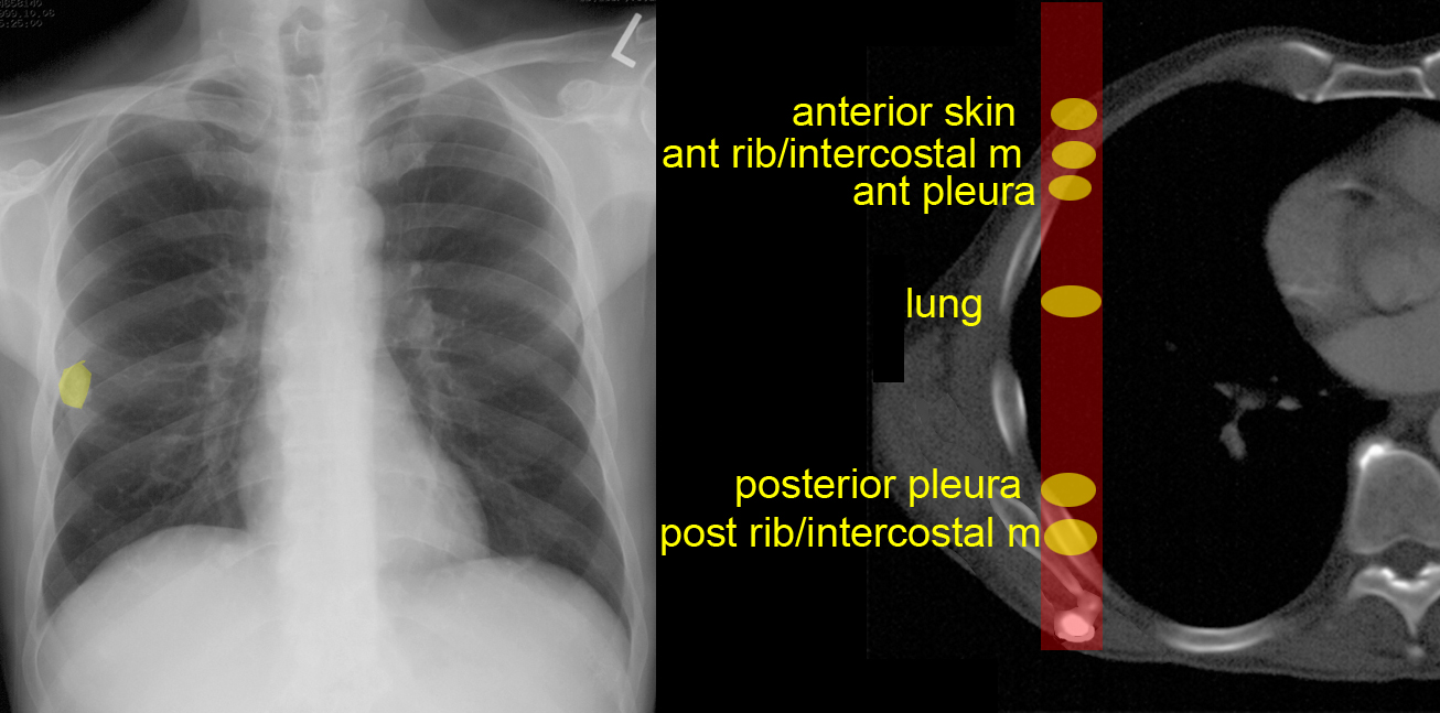
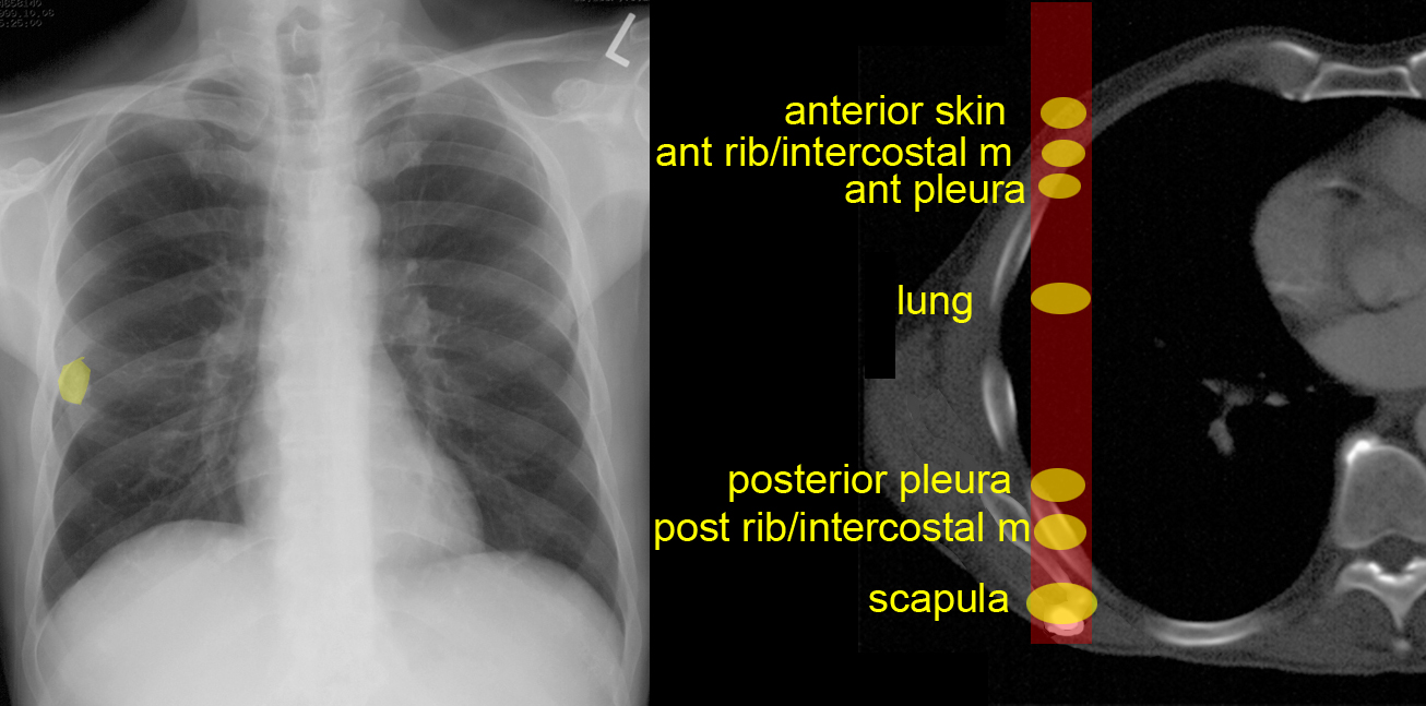
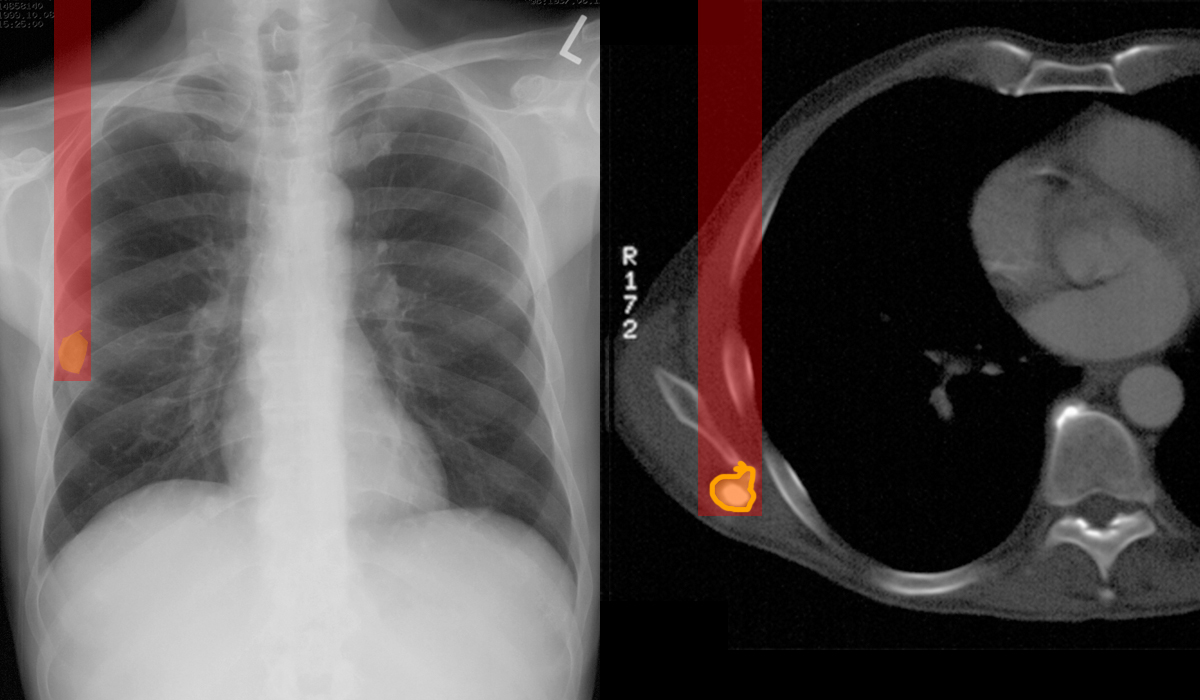
Now is your chance to think like a radiologist. The patient's right scapula and accompanying lesion has been moved on the CT image to approximately the position it was in for the chest radiograph.
Question 4:
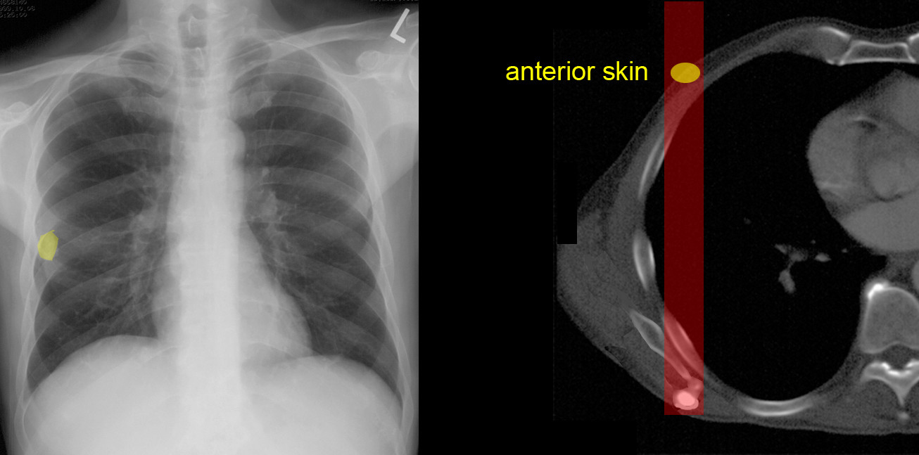
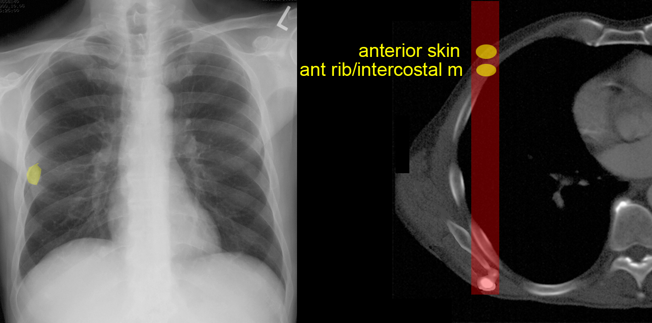
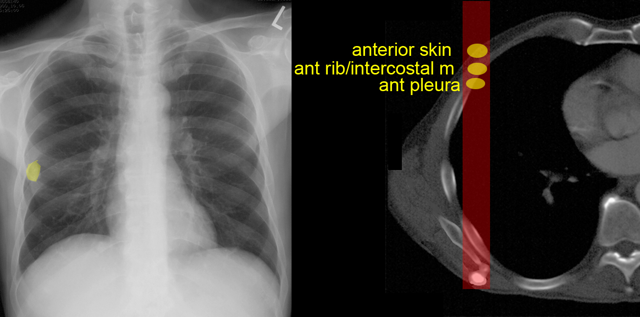
Given the location of the calcific density on the chest radiograph, how many different places can you think of where the lesion could be located? Think in terms of layers of body tissue.
The labels below list a number of possibilities. The lesion could also be in a pocket (in an article of clothing).
Each pixel on a radiograph represents a summation of all of the tissues that group of photons encountered on their way through the patient. So in order to interpret a radiograph, one must think geometrically. For the previous 'CT windows', image A shows bones best and is called a 'bone window'. Image B shows soft tissues (especially muscles) best and is called a 'soft tissue window'. Image C shows lungs best and is called a 'lung window.
