

Thorax Case 1
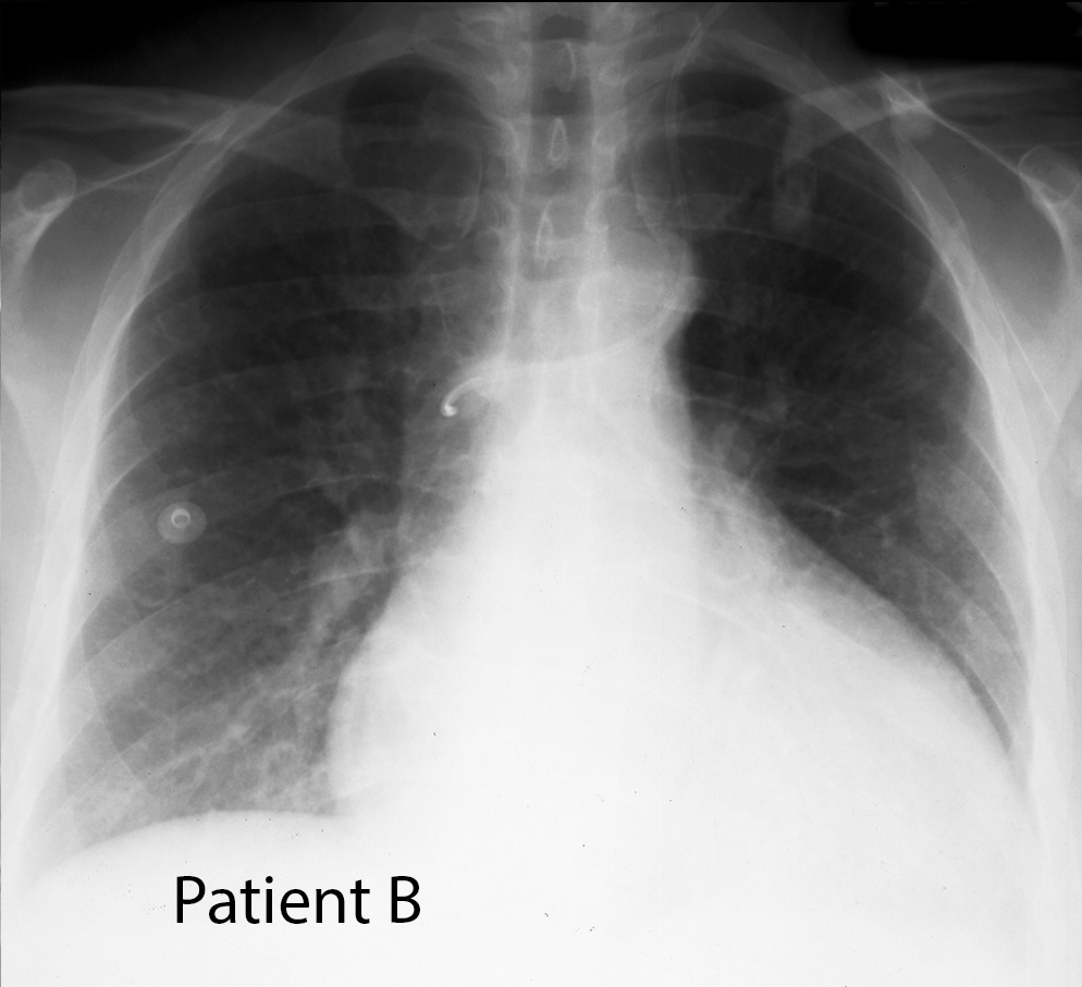
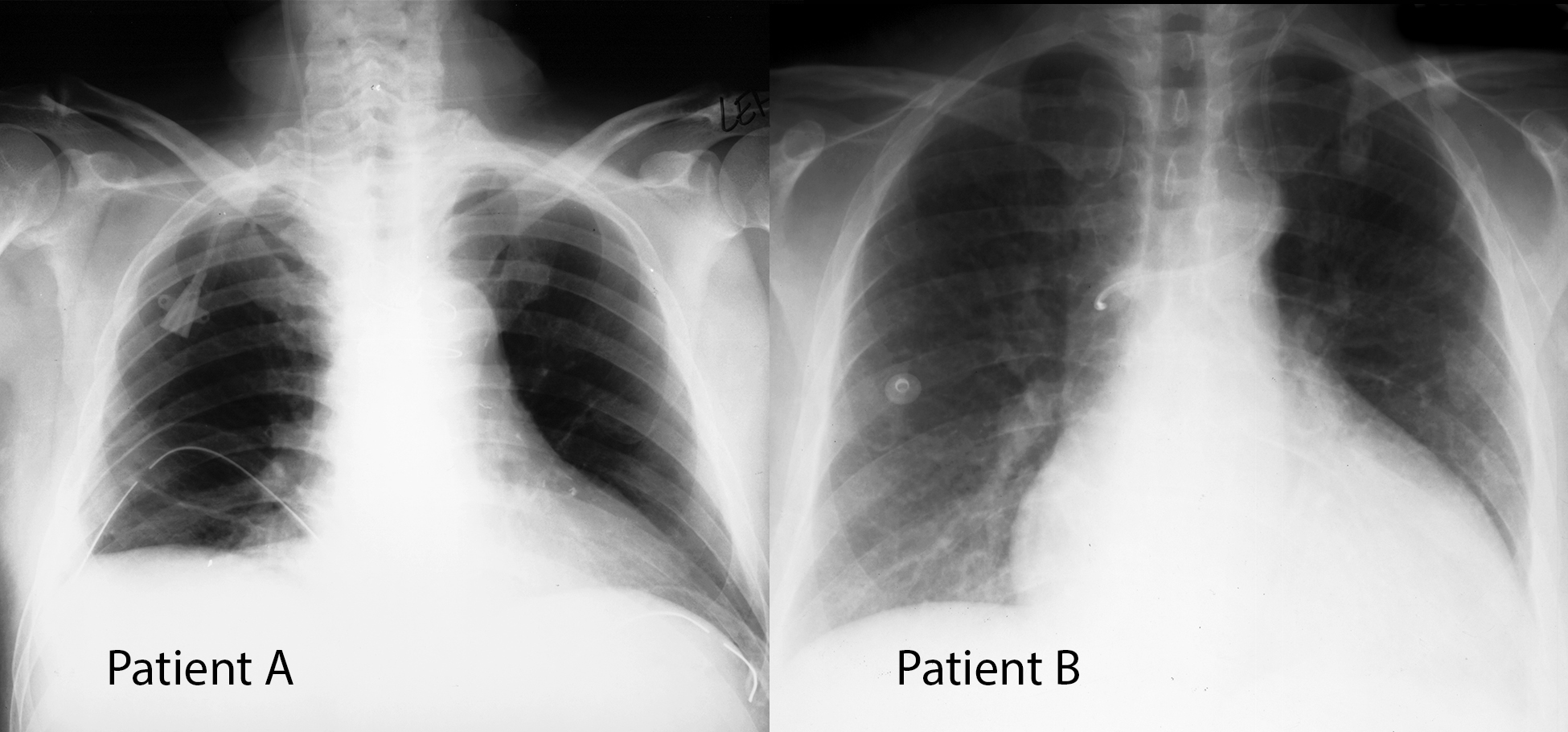
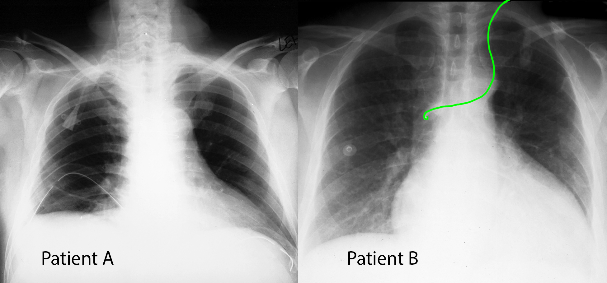
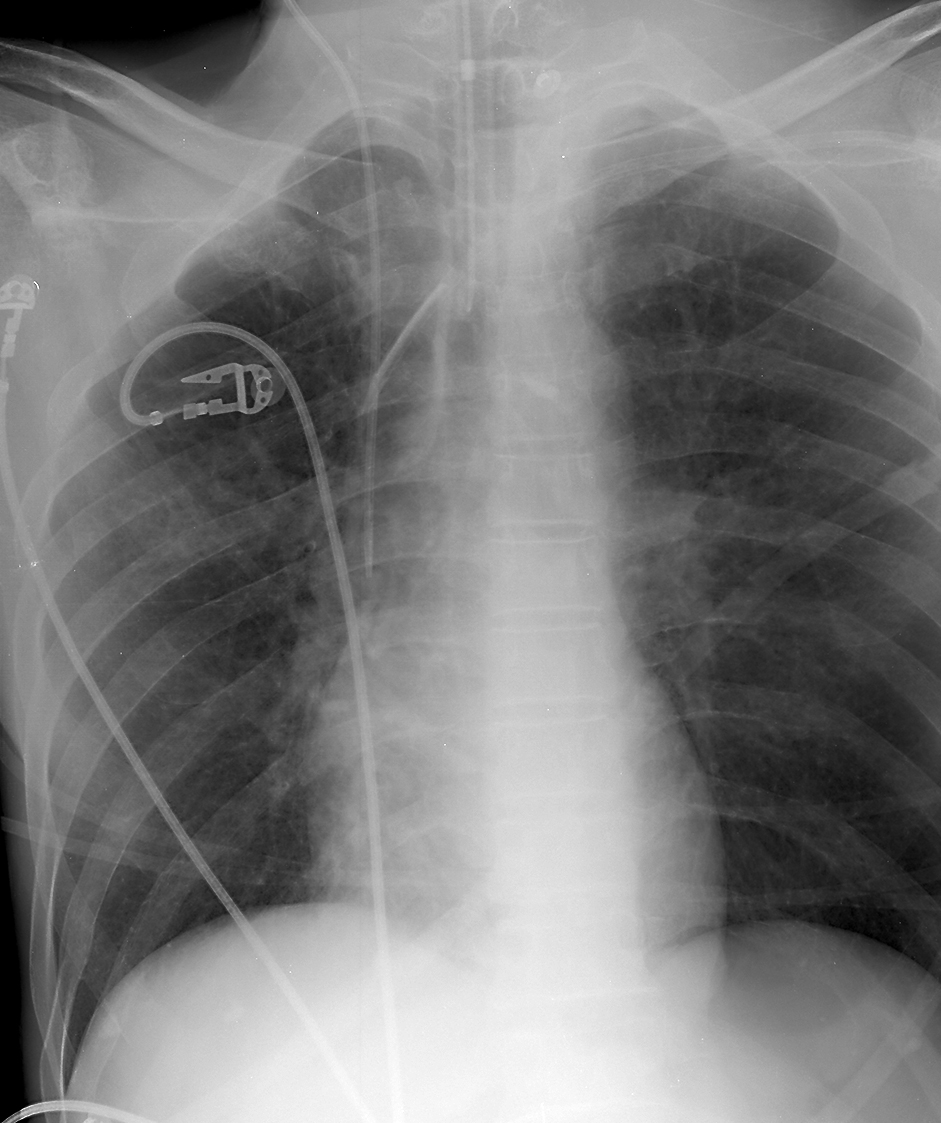
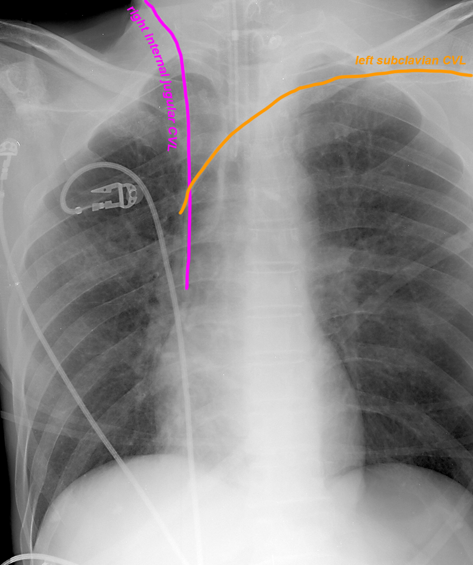
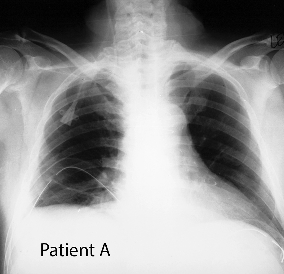
Two different patients are shown here, both of whom had central venous lines (CVLs) inserted. The frontal chest radiographs were performed to check the position of the CVLs.
Question 1:
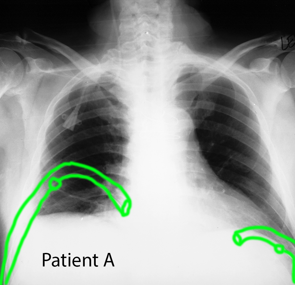
a) What do you think of the CVL positions for Patient A and Patient B? If you are having trouble seeing the CVLs, click on the labels below (but try to find them on your own first), and remember which is the RIGHT vs LEFT side of the patient.
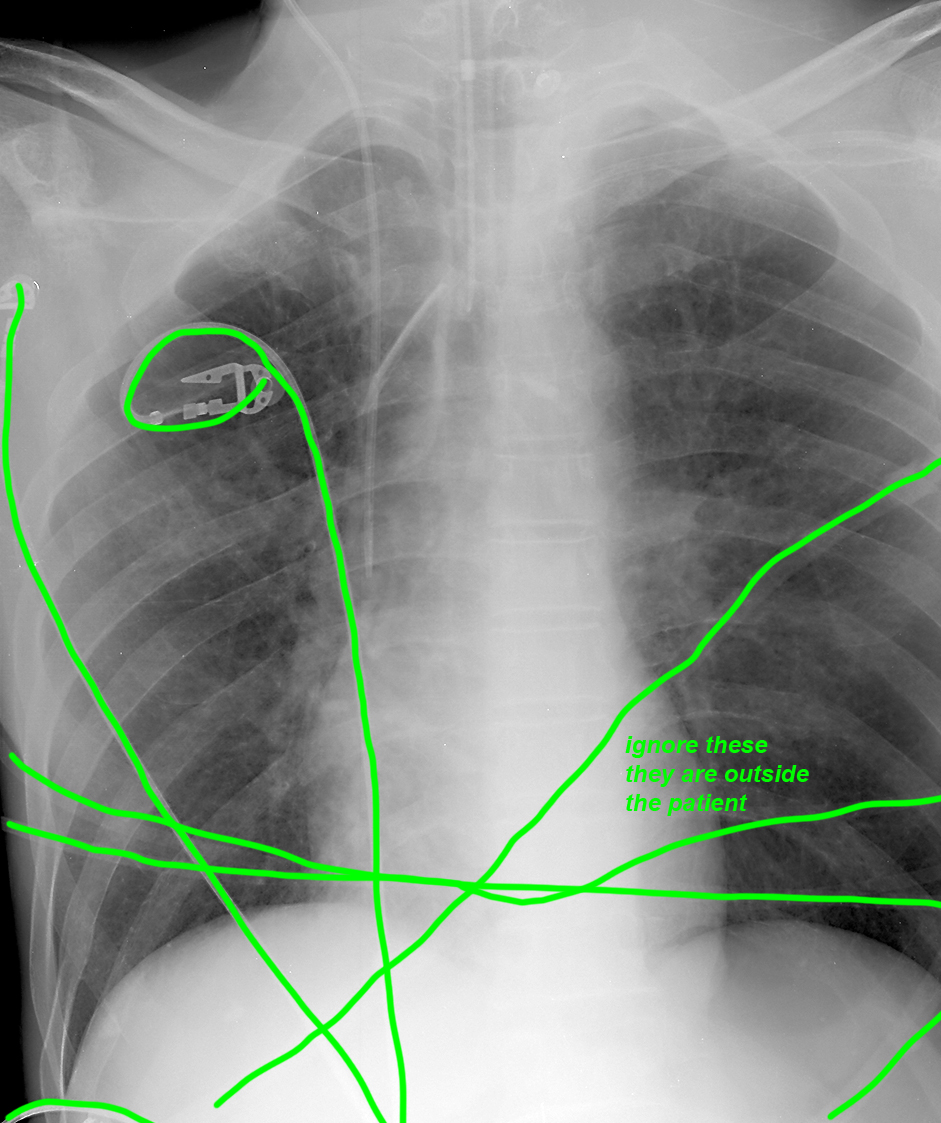
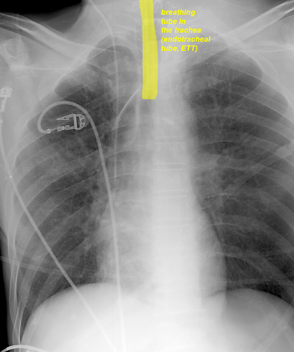
The CVL for Patient A is entering via the right subclavian vein and is then directed upward, which is NOT the position we expect for a properly placed CVL. The CVL for Patient B is entering via the left internal jugular vein and is directed toward the expected location of the superior vena cava, but has an unusual course near its tip. Chest radiographs are viewed as if you are facing the patient, so the RIGHT side of the patient is on the LEFT side of the image.
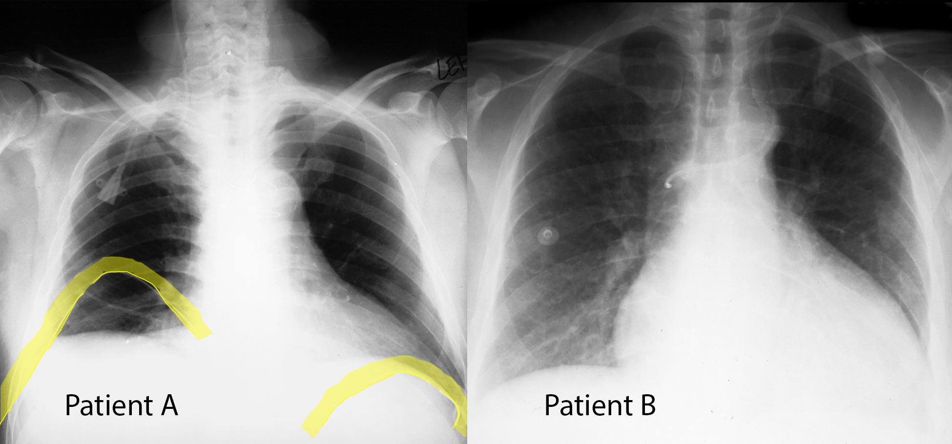
b) What are chest tubes inserted for?
Chest tubes are large caliber catheters that are inserted through the chest wall tissues into the pleural space. They may be used to drain fluid, air or both from the pleural space.
The commonest vessels used to place a CVL are the right and left subclavian veins and the right and left internal jugular veins.
Thorax Case 1
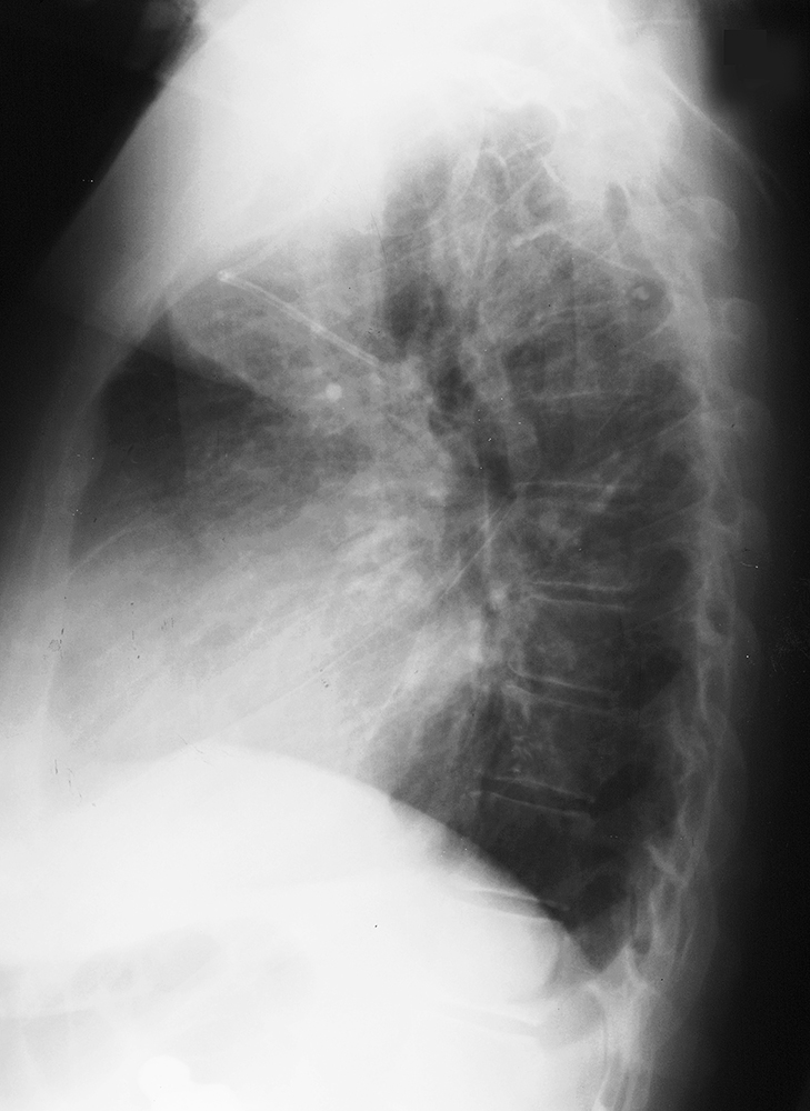
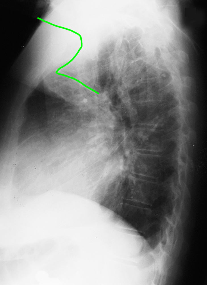
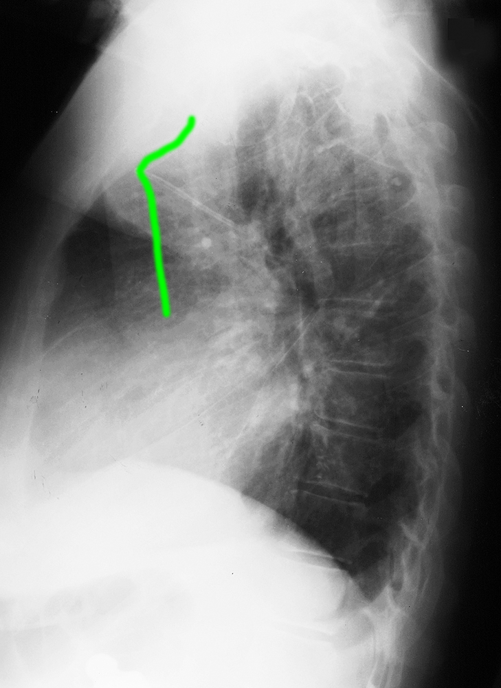
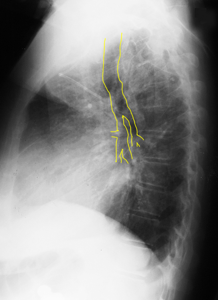
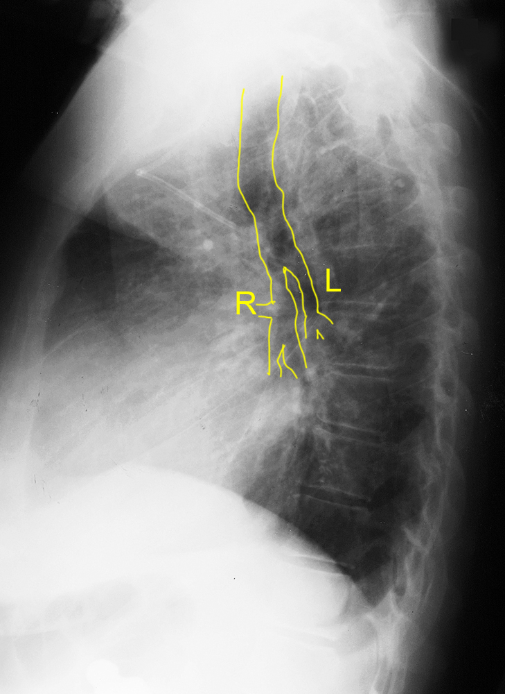
This is a lateral view for Patient B.
Question 2:
a) Which of the four common veins mentioned is the most straight approach to position a CVL in the desired vessel (superior vena cava)?
The right internal jugular (IJ) approach is the straightest path from insertion to superior vena cava (SVC). However, in some situations, other approaches are used, as in both of these cases. Other approaches require the catheter to make one or more turns to reach the desired position, and as can be seen in these two examples, the catheter may make wrong turns.
b) What vessel might have the course that this catheter follows?
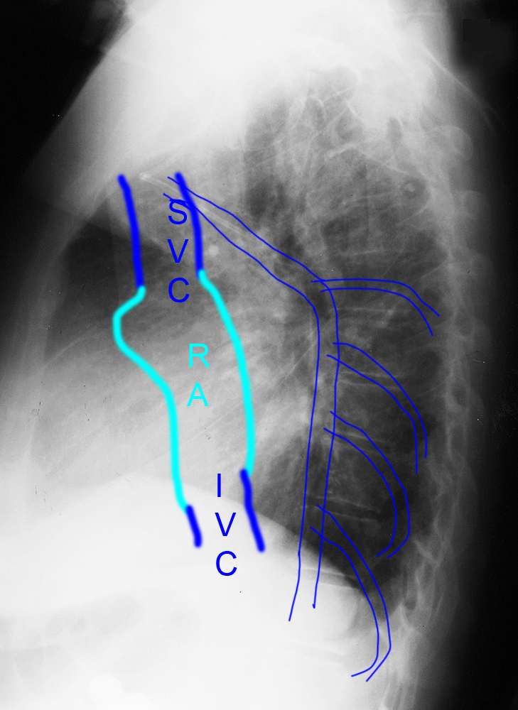
The course shown on this lateral view is consistent with a vein that is running from the posterior mediastinum forward to empty into the back of the SVC. This fits best with the azygos vein. The sharp curve that was seen on the frontal chest radiograph is because you are viewing a foreshortened view of the arching posterior course of the catheter within the azygos vein. Click on the label 'tributary' to see a drawing of the approximate location of this vessel.
Thorax Case 1
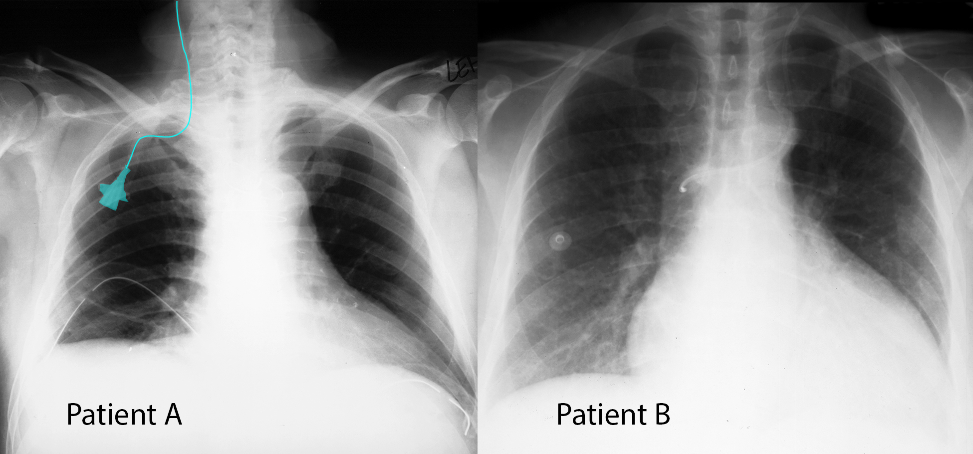
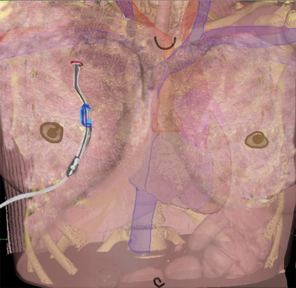
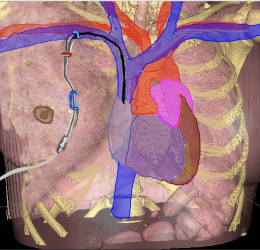
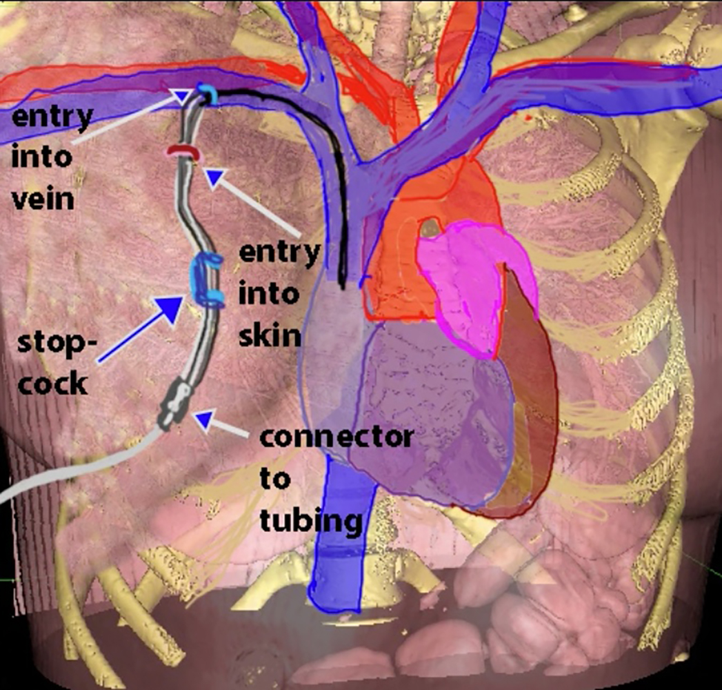
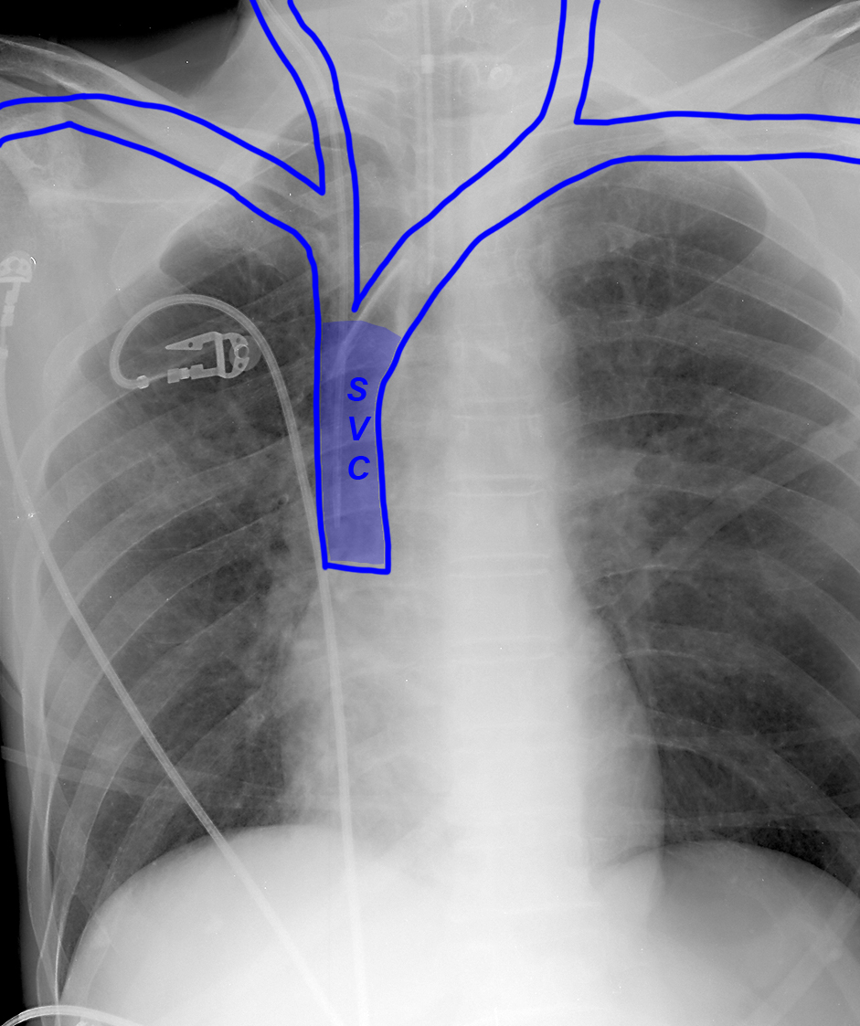
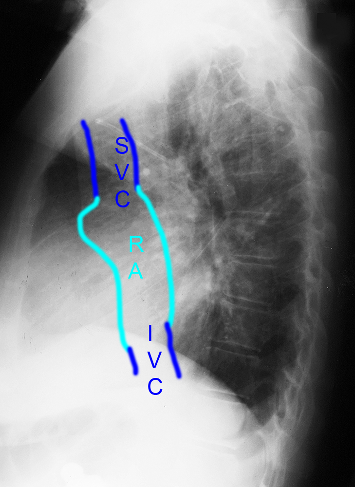
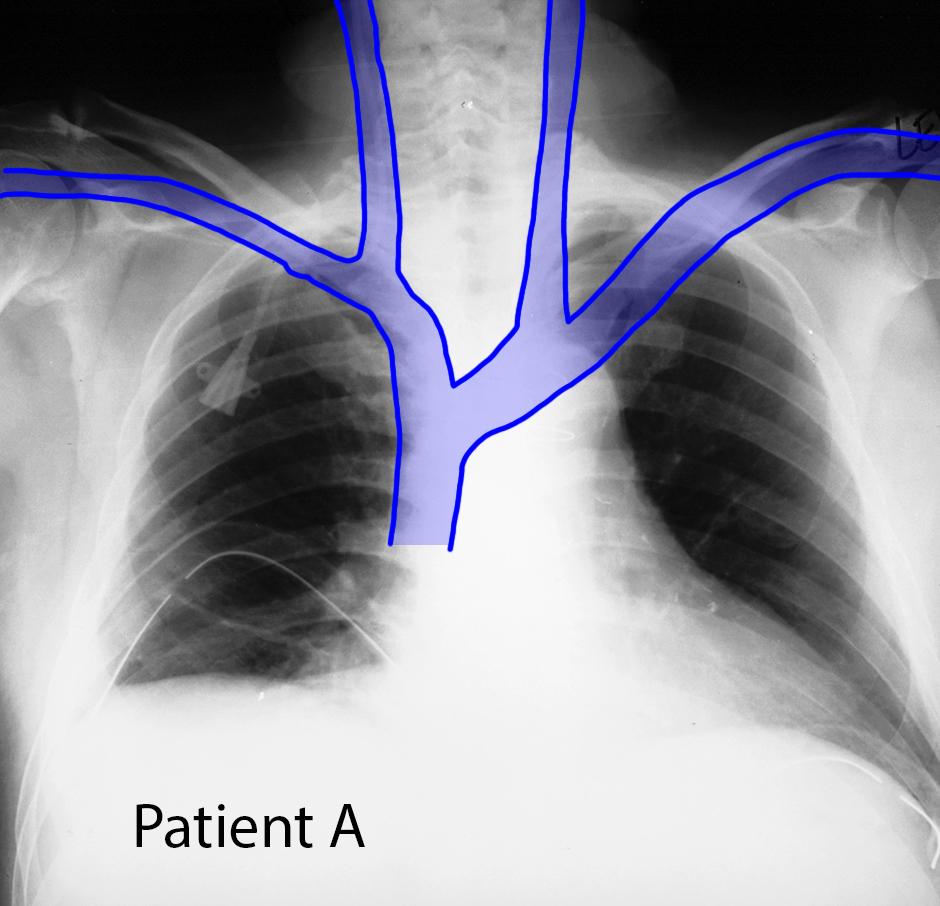
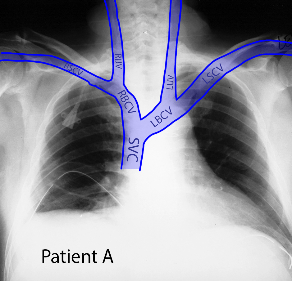
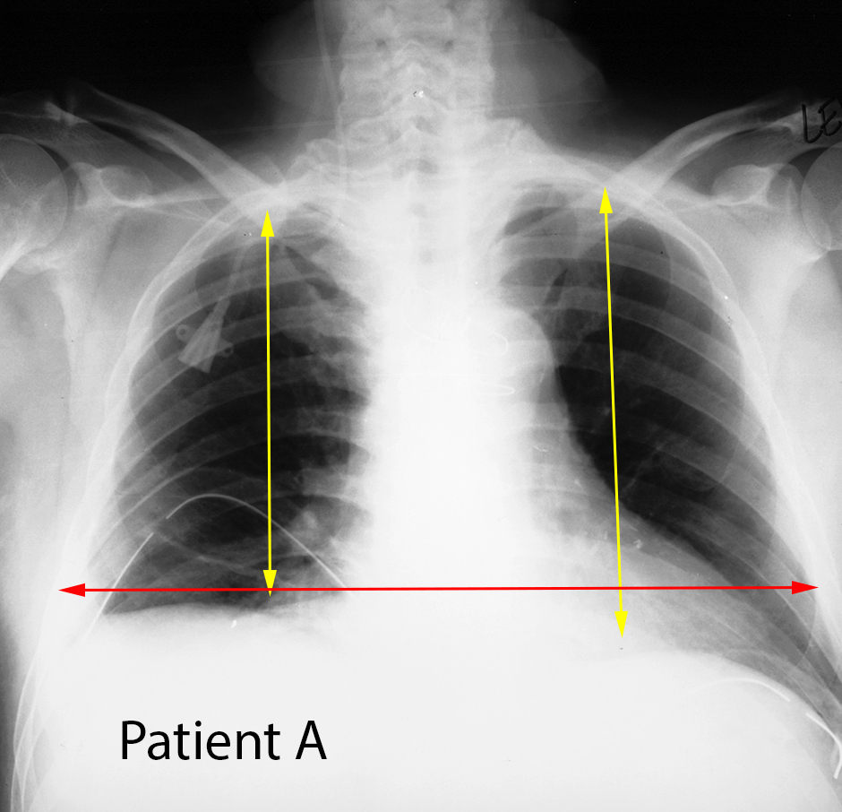
This is the frontal view of Patient A again. Before clicking on the veins label, try to recall what veins come together to form the SVC, and where you would expect them to be on a chest radiograph.
Question 3:
a) What vessel has this right subclavian CVL entered?
The sharply angled upward course along the right side of the neck is consistent with placement into the right internal jugular vein.
b) Does this catheter need to be repositioned?
Yes. Depending on what it is being used for, you would likely not want to infuse any strong compounds directly into the upper internal jugular vein, where they might reflux upward to the base of the brain. Positioning of CVL's in the SVC allow rapid dilution of infused liquids within the right atrium, mixing with blood entering from the IVC.
Thorax Case 1
This is the frontal view of Patient B again.
Question 4:
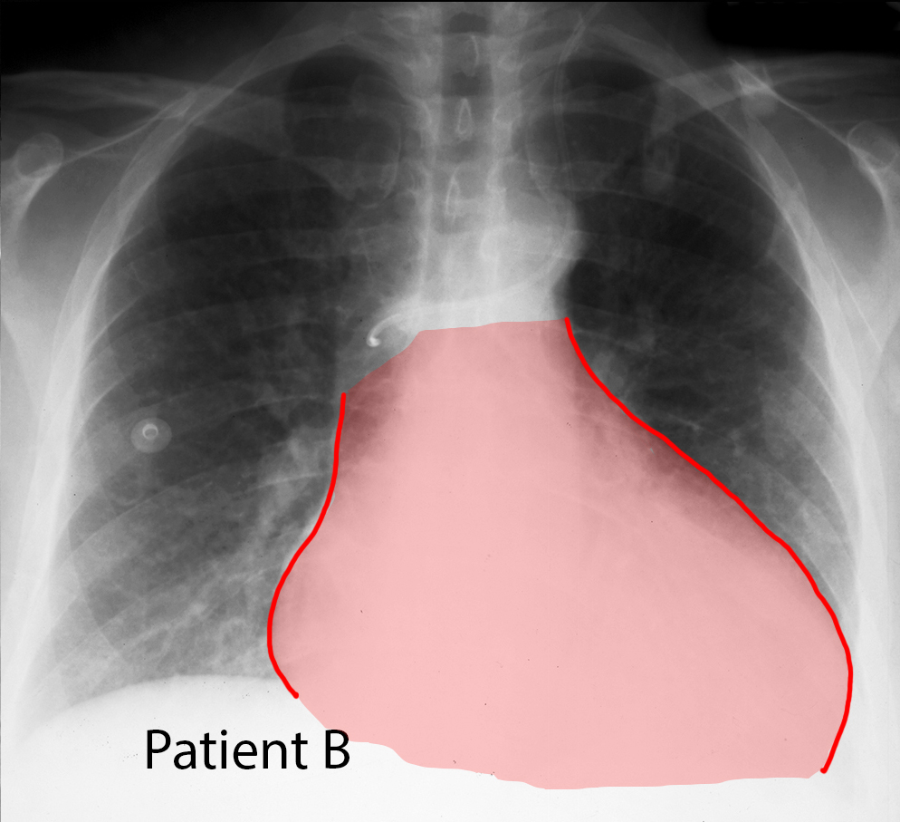
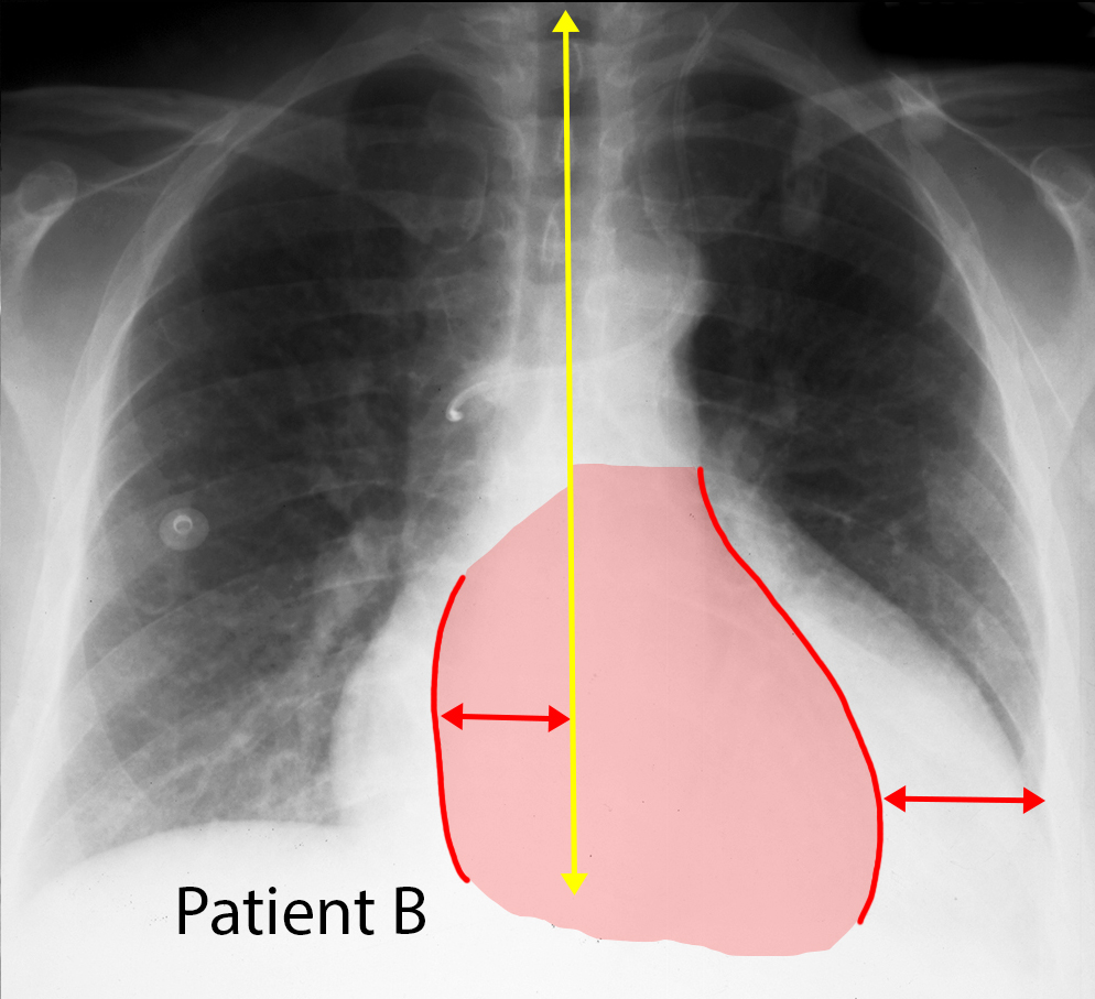
a) How do you assess heart size on a chest radiograph?
As is illustrated in the labels for 'heart' and 'normal', we use an internal control for heart size, rather than a specific number (which would vary a lot from one person to another based on their age and overall size). The normal heart transverse diameter should be half or less than half of the internal diameter of the chest itself. The measurements show in 'normal' in effect allow you to eyeball this relationship by drawing the two line segments and deciding which is longer.
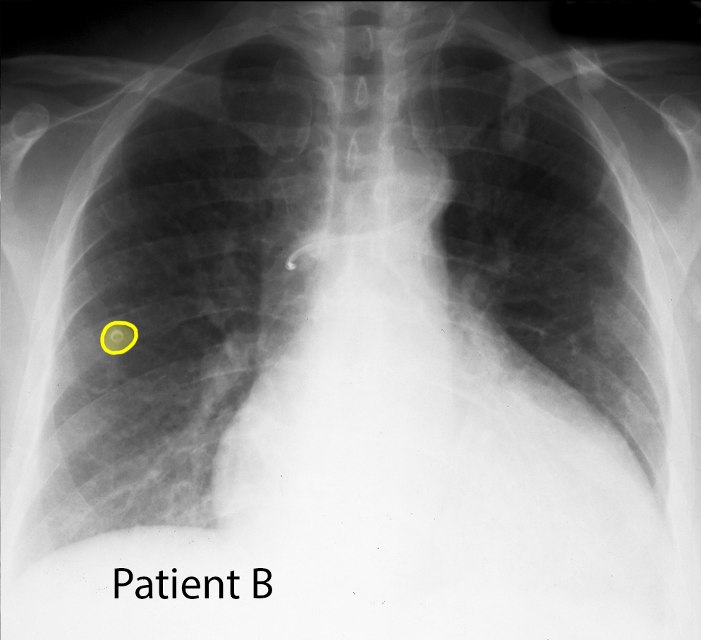
b) What is 'mystery 2'?
This has the appearance of a snap, probably on the patient's hospital gown.
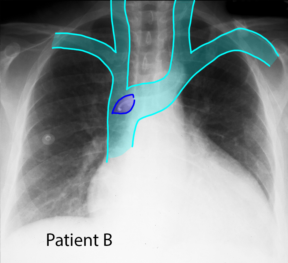
c) What vessel is shown on the veins diagram in dark blue?
The dark blue lens-shaped structure is the azygos vein, seen end-on on the frontal view. It is collecting blood from the posterior intercostal veins, running up in the posterior mediastinum, and then arching forward to enter the back side of the SVC. The curled appearance of the tip of the CVL indicates that we are seeing it end-on within the azygos vein. This matches the course shown on the lateral chest radiograph.
