


Case 3
57 year old female with positive family history of breast cancer in mother at age 51, vague firmness in outer left breast
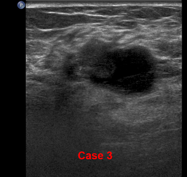
Question 1:
What terms would you use to describe the ultrasound findings
×
Answer:
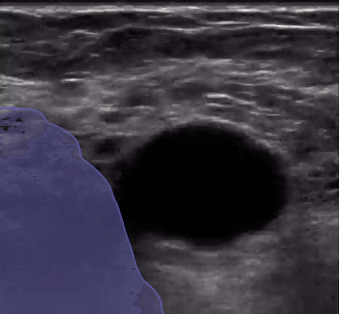
contour--irregular, shape--poorly defined, echodensity--hypoechoic, but with some anechoic areas suggesting fluid
contour--irregular, shape--poorly defined, echodensity--hypoechoic, but with some anechoic areas suggesting fluid
Case 3
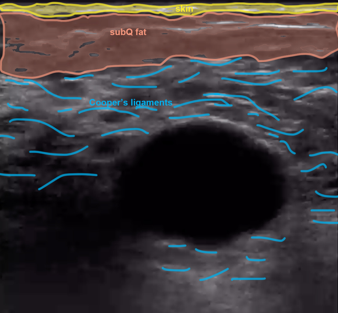
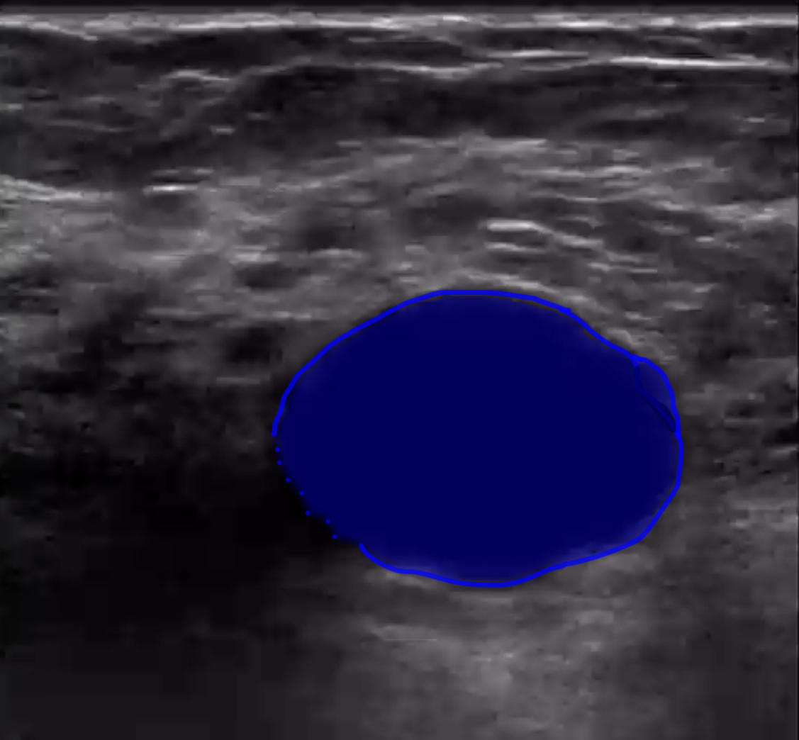
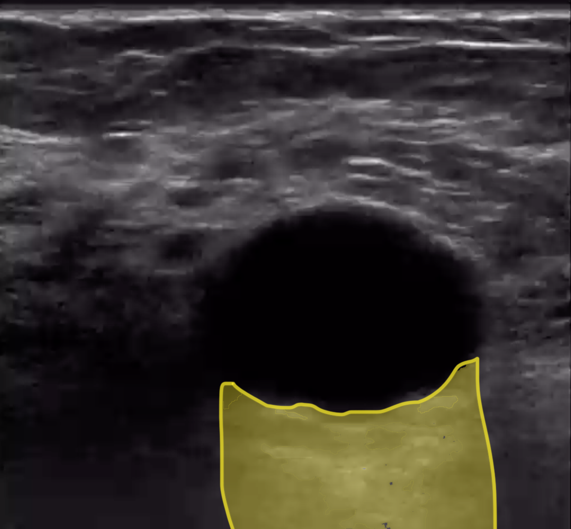
For normal structures, identify skin, subcutaneous fat, and Cooper's ligaments before clicking on the labeled image.
Further Explanation:
Case 3
Case 3
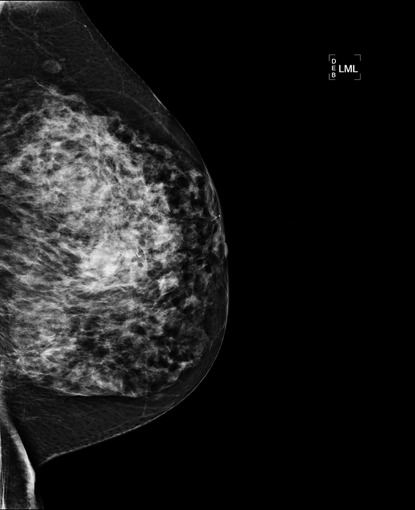
Question 2:
How easy is it to see these masses and cysts on this mammogram?
×
Answer:
Not easy at all, because the breast stroma is so dense. Dense stroma makes lesions much harder to detect, but is also a separate risk factor for development of breast cancer.
Not easy at all, because the breast stroma is so dense. Dense stroma makes lesions much harder to detect, but is also a separate risk factor for development of breast cancer.
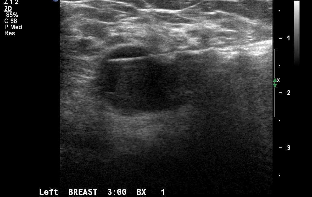
Question 3:
What is this study?
×
Answer:
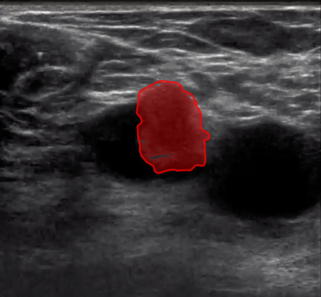
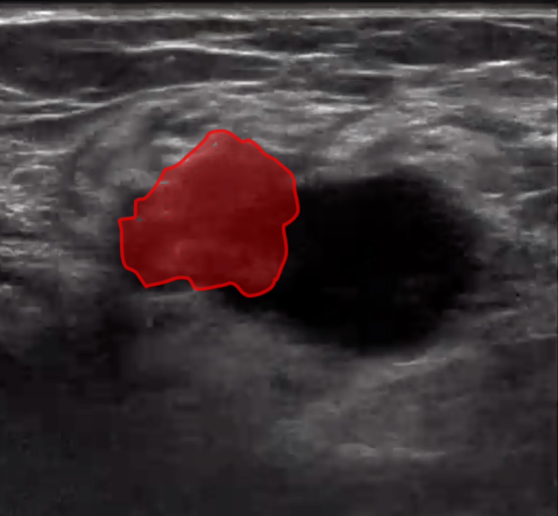
This is an image from an ultrasound-guided core biopsy of one of the bresast masses. It revealed invasive lobular breast cancer.
This is an image from an ultrasound-guided core biopsy of one of the bresast masses. It revealed invasive lobular breast cancer.
Click on  to return to Case List