


Case 9
51 year old female for routine screening mammography
Question 1:
Do any of the sites marked as moles look abnormal to you?
×
Answer:
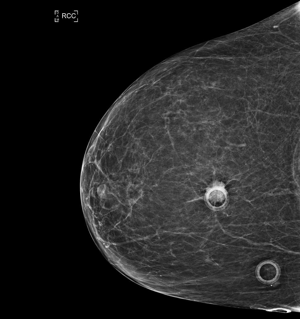
Most moles are only faintly visible on mammography, and often have a round or lobular contour. One of these mole markers seems to have a very irregular lesion underlying it, which should lead to further imaging.
Most moles are only faintly visible on mammography, and often have a round or lobular contour. One of these mole markers seems to have a very irregular lesion underlying it, which should lead to further imaging.
Case 9
Case 9
Question 2:
How was this mammographic view done?
×
Answer:
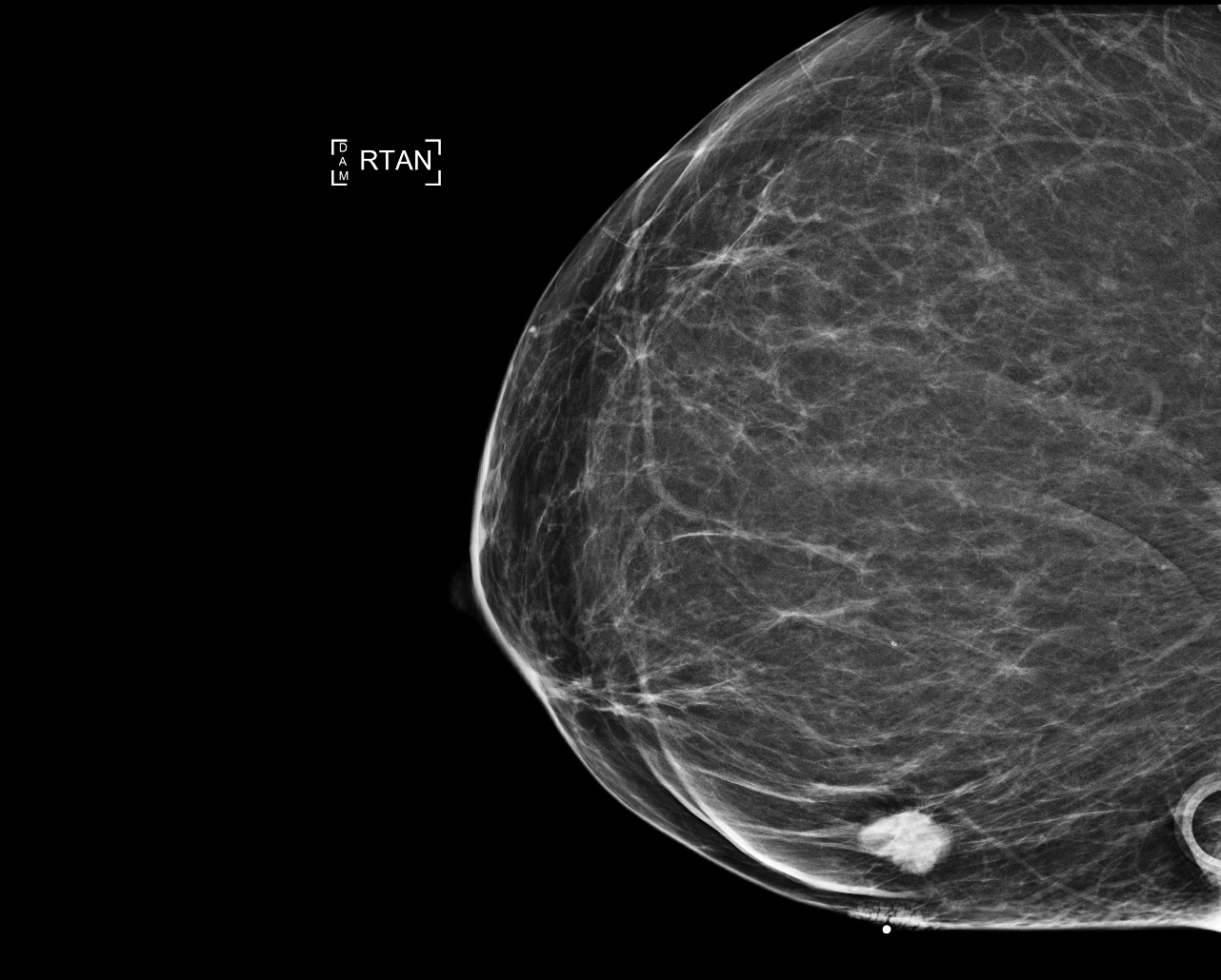
This is a tangential view. The breast was turned in such a way that the skin marker (the one overlying the irregular mass) is right at the edge of the breast. In this way, you can see the underlying tissue spread out. The mole that was being marked is faintly visible at the edge of the image, but there is a concerning mass in the underlying breast that has nothing to do with the mole.
This is a tangential view. The breast was turned in such a way that the skin marker (the one overlying the irregular mass) is right at the edge of the breast. In this way, you can see the underlying tissue spread out. The mole that was being marked is faintly visible at the edge of the image, but there is a concerning mass in the underlying breast that has nothing to do with the mole.
Case 9
(click on 'X' to return to Case List)
Question 3:
What is the most likely diagnosis in this case?
×
Answer:
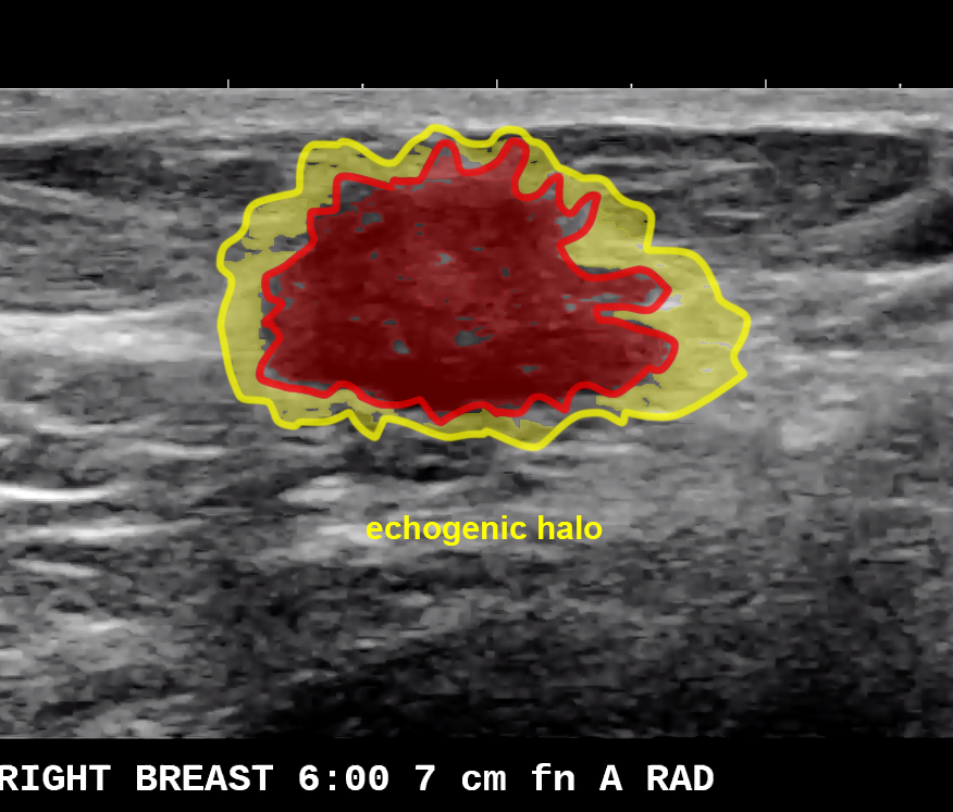
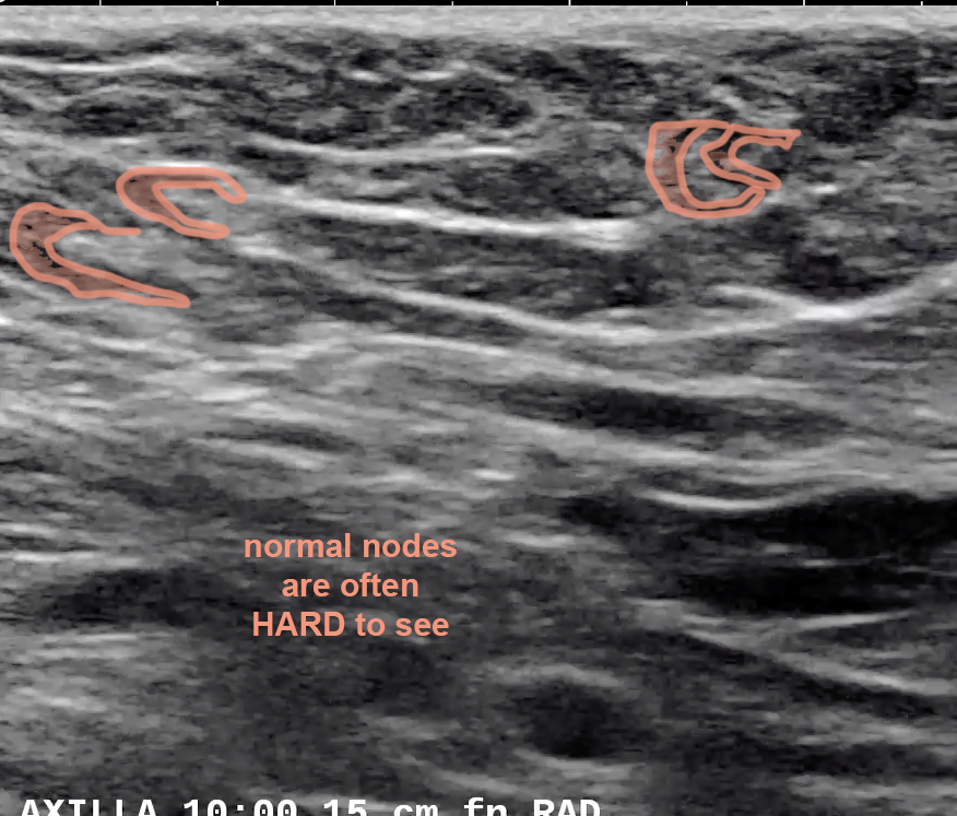
This was another invasive ductal carcinoma. The irregular border and the echogenic halo are both worrisome US features. Luckily the axillary nodes appeared normal, and no metastases were found to the axilla at surgery.
This was another invasive ductal carcinoma. The irregular border and the echogenic halo are both worrisome US features. Luckily the axillary nodes appeared normal, and no metastases were found to the axilla at surgery.