

Thorax Case 2
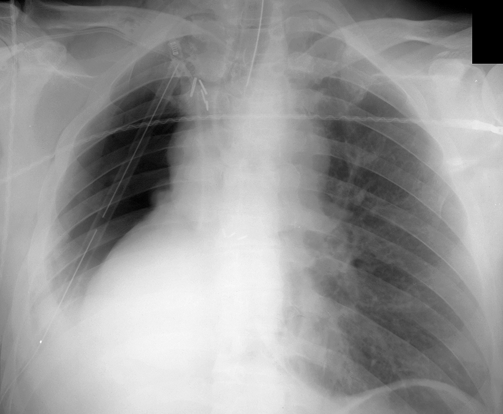
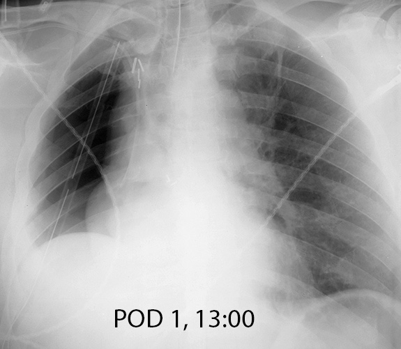
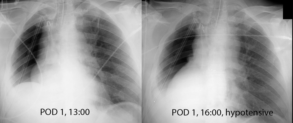
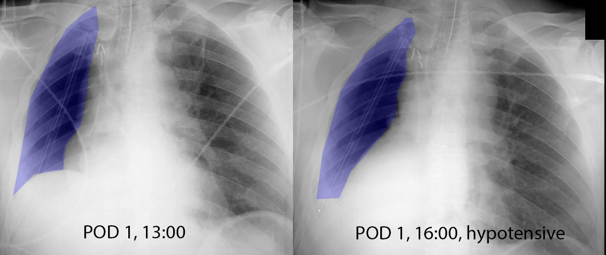
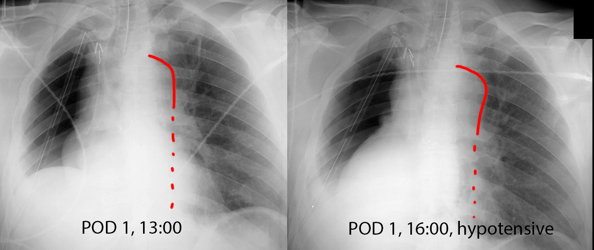
These two images are of the same patient, two hours apart. Try to determine what has changed between the two images. "POD 1" means 'post-operative day 1', or one day after surgery. "hypotensive" means that the patient's blood pressure has dropped and they are likely losing consciousness.
Question 1:
a) What surgery do you think the patient has had?
There are no lung markings on the right at all. The right ribs are closer together than the left and the right hemidiaphragm is elevated on the first image. This combination of findings suggests that they have had a pneumonectomy, or removal of an entire lung, usually done for cancer.
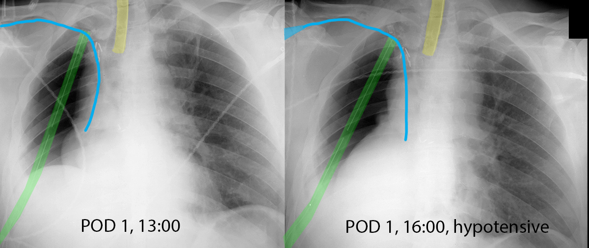
b) What support devices are present? Try to identify them on your own but if you need help, click on the label below to show their position and courses.
There is an endotracheal tube (labeled yellow), which is relatively large in caliber with a metallic marker along one side and projecting over the upper trachea. There is a right subclavian CVL (labeled blue), with the tip in the region of the lower SVC. And there is a right chest tube (labeled green), with tip near the apex of the right hemithorax.
Thorax Case 2
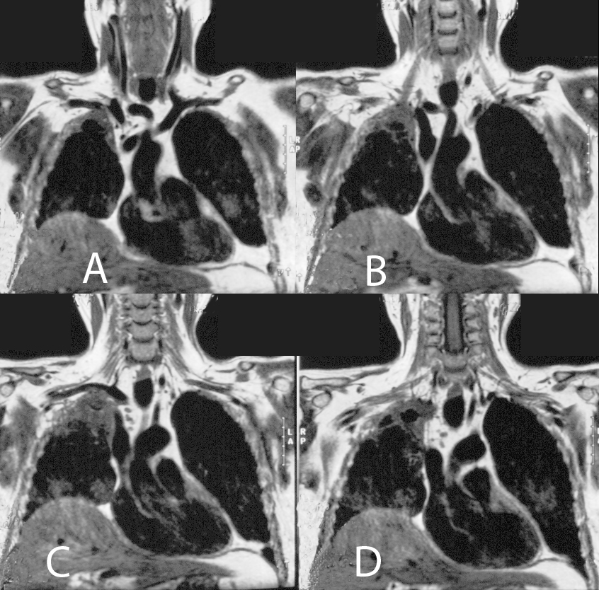
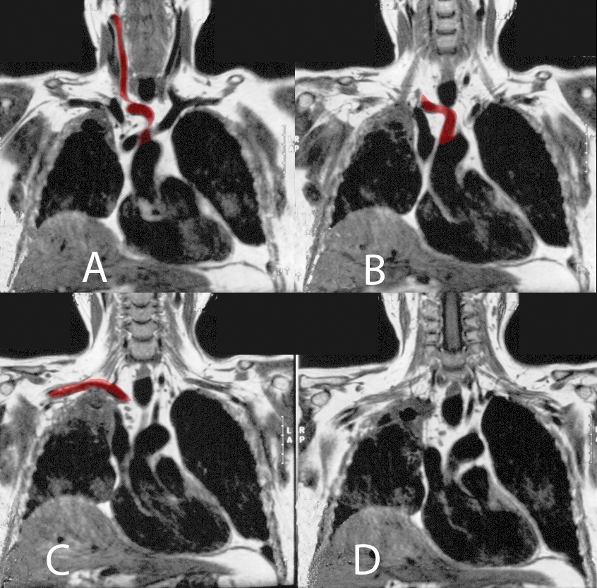
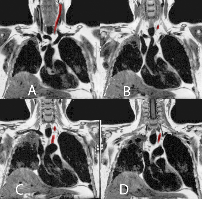
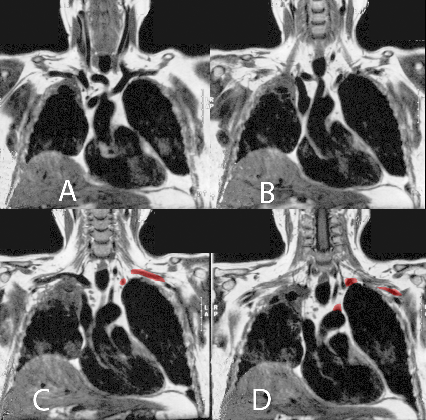
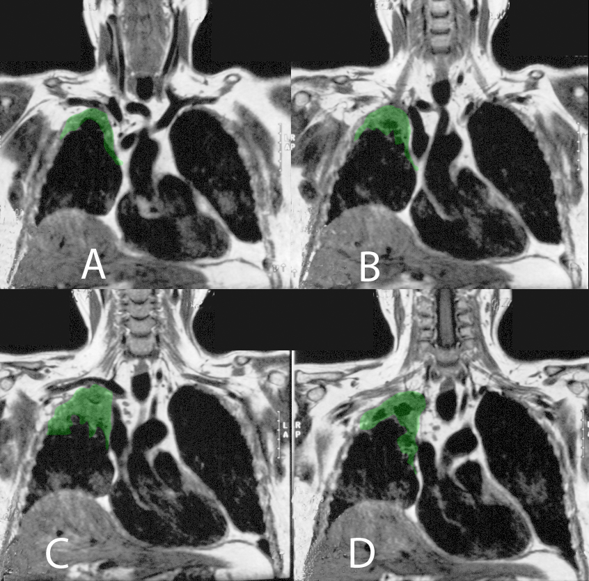
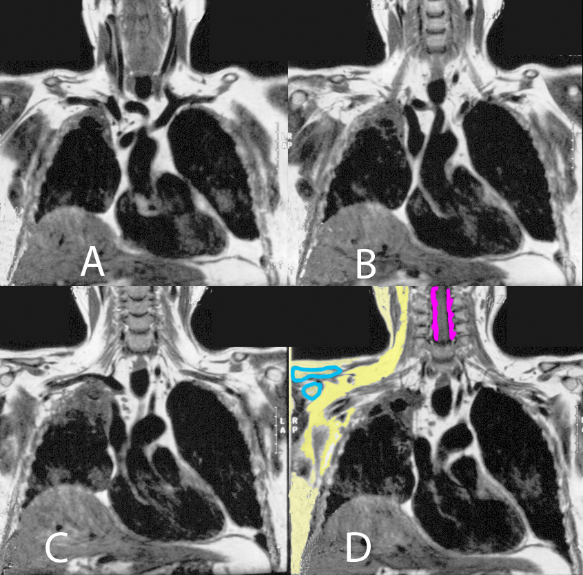
This is an MRI that was done prior to surgery on this patient
Question 2:
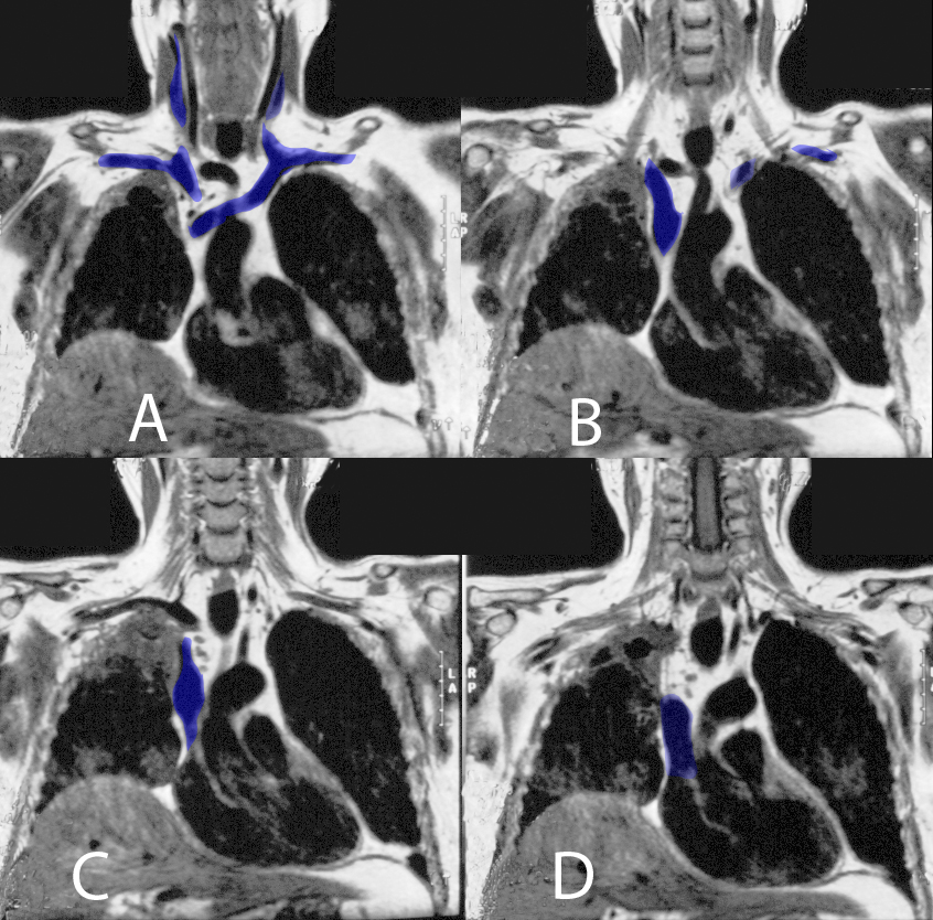
a) What are the three arteries indicated by the labels?
Artery 1 is the brachiocephalic artery. Artery 2 is the left common carotid artery. Artery 3 is the left subclavian artery.
b) What is the imaging plane for this MRI, how is it oriented (in terms of patient position), and how does the radiation dose compare to a CT?
This MRI is in the coronal plane. Each image is displayed just like a frontal chest radiograph (with the patient's RIGHT on the LEFT side of the image, as if you are facing the patient). There is no radiation in MRI imaging, so the dose is zero compared to a chest radiograph or a CT. The dose of a CT is roughly an order of magnitude higher than a radiograph.
c) What are the three tissues indicated by the label below, and how do they help you identify this study as an MRI?
Fat is shown in yellow and has high signal (white appearance). This is never the case for a CT. (However, some MRI sequences can have dark appearance of fat, depending on how they are set up). Cortical bone is shown in blue and has low signal (dark appearance). This is the best indicator that this is an MR, since cortical bone will always be white on CT and always dark on MR. Fluid (in this case, cerebrospinal fluid) is shown in purple, outlining the spinal cord in the cervical region. This tells you that this particular MR is a T1-weighted sequence, since it is dark. In a T2-weighted sequence it would be bright (white).
You can see that the tumor was quite extensive on the right side of the patient's chest, requiring removal of the entire lung rather than just a lobe for treatment.
Thorax Case 2
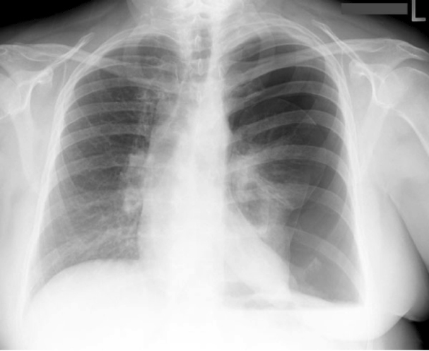
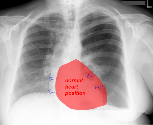
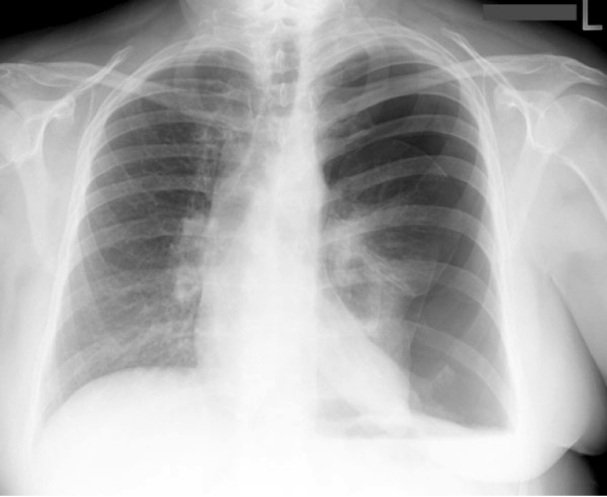
This is a different patient who also has absent lung markings, for a different reason. This 40 year old patient had pleuritic chest pain after several attempts to place a left CVL that were not successful.
Question 3:
a) What is pleuritic chest pain and what does it signify?
The term 'pleuritic' indicates chest pain that is worse on deep inspiration or coughing. This suggests that the pleura is irritated, since it moves most on deep inspiration or cough.
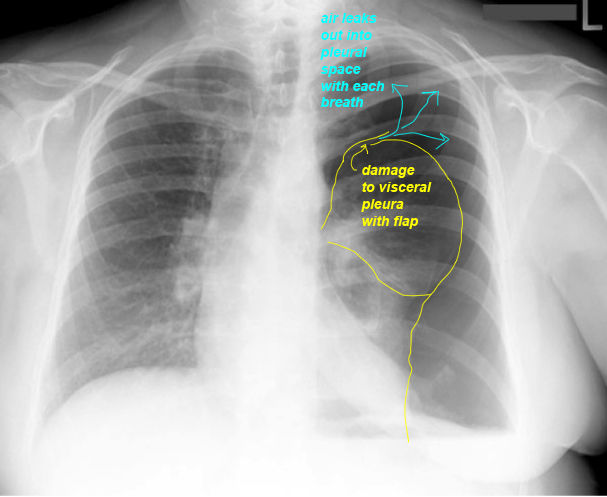
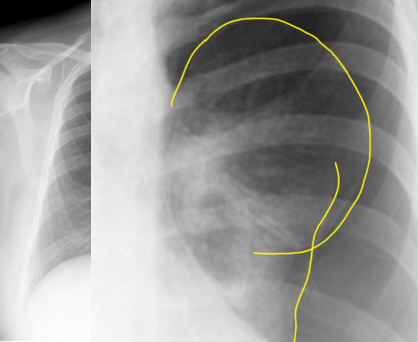
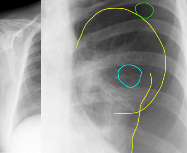
b) What do you think of the densities seen on the two areas indicated by circles (click on 'lung comparison' below)?
The area medial to the white pleural line (blue circle) appears denser (lighter) than the area outside the white pleural line (green circle). The area circled in blue is normal lung tissue, which is a mixture of air and other things like connective tissue, lymphatics and blood vessels. The area circled in green is pure air in the pleural space--a pneumothorax, or PTX for short.
c) What structure is represented by the white pleural line?
The white pleural line is the visceral pleura and underlying connective tissue. The parietal pleura hugs the inside of the ribs and blends with connective tissue there, so is not visible on imaging.
Thorax Case 2
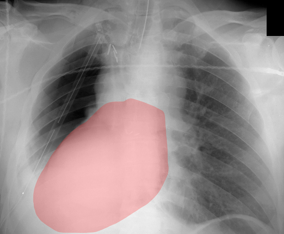
This is the later chest radiograph on our patient who had had a right pneumonectomy. The right side of the pericardium was also resected due to invasion by tumor. The heart position is very different compared to the earlier image.
Question 4:
a) What normally keeps the heart in its place within the mediastinum?
The coverings of the heart, the serous and fibrous pericardium, help to keep the heart in its usual position. The connections of the SVC and IVC to the right side of the heart also provide some stability from superior or inferior motion. In the normal heart position, the ventricles are rotated far to the left, relative to the axis of the SVC and IVC.
b) In both the pneumonectomy case and the pneumothorax case, the heart has been displaced from its normal position. What effect could that have on the inflow of blood to the heart?
In the pneumonectomy case, the heart has not only shifted but rotated from the left side of the chest around to the right, which can twist the SVC and IVC, and severely compromise return of blood from the body to the heart. In the pneumothorax case, the air in the left pleural space was under pressure (called a 'tension' pneumothorax), and was pushing the heart to the right without twisting it. This also can compromise venous return, but usually not as severely as when the heart is twisted.
This is the last page of the mini-consolidation Chest Imaging Anatomy cases. To return to the main page for Foundations 2 Imaging Anatomy, click HERE. To return to the first page of the Chest cases for mini-consolidation, click HERE.
