


Upper Extremity Cases
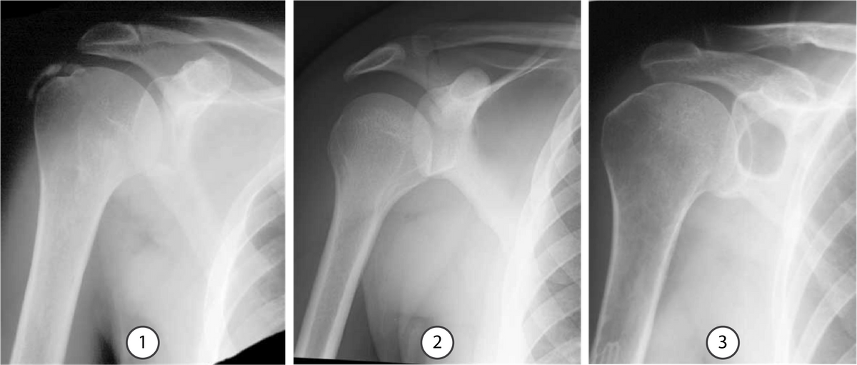
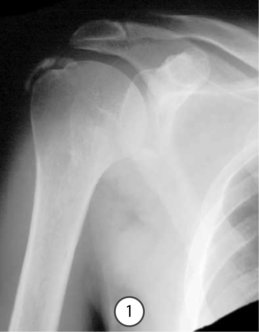
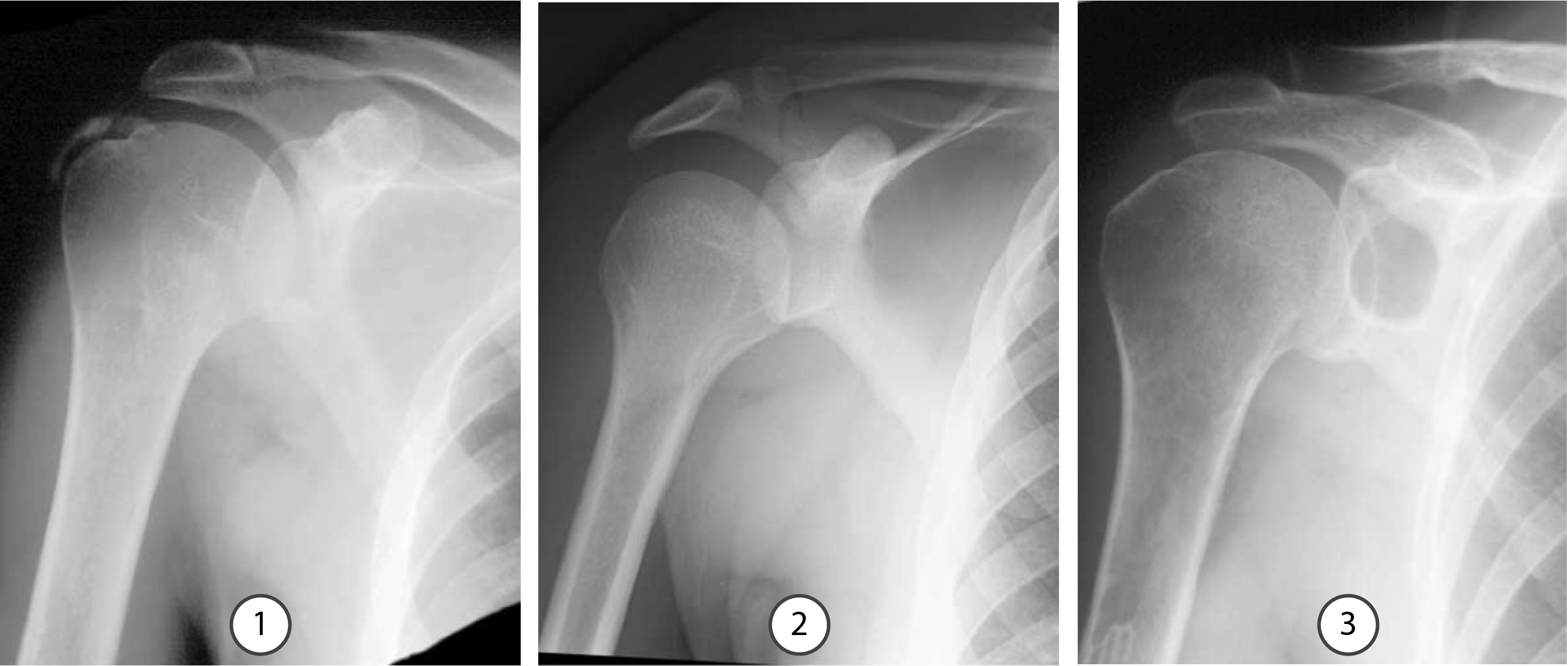
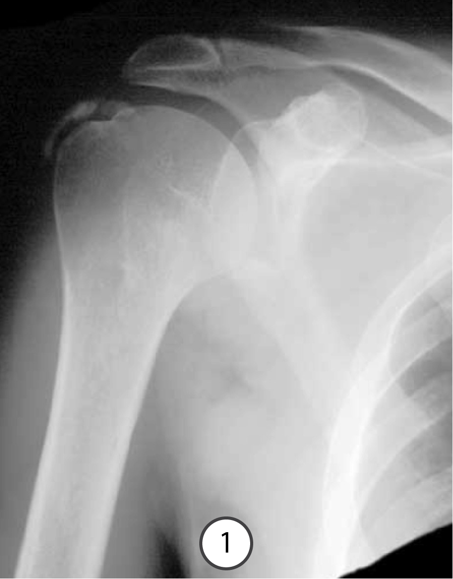
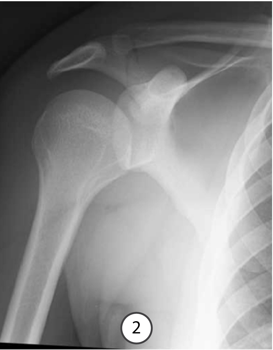
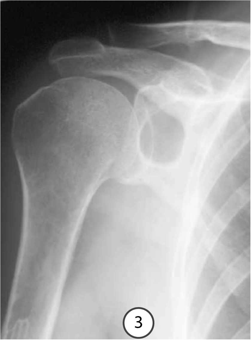
Three patients with shoulder pain, two chronic and one acute. For each image, state what the radiographic VIEW is, and what the ABNORMALITY is.
Question 1:
Which patient has ACUTE pain? Use the buttons below to look at each image individually.
Image 2 reveals an acute fracture. Thus, the patient in image 2 has acute pain. Image 1 shows chronic tendonitis and image 3 shows a chronic, benign lesion. Advance to the next slide for further explanation.

Further Explanation:
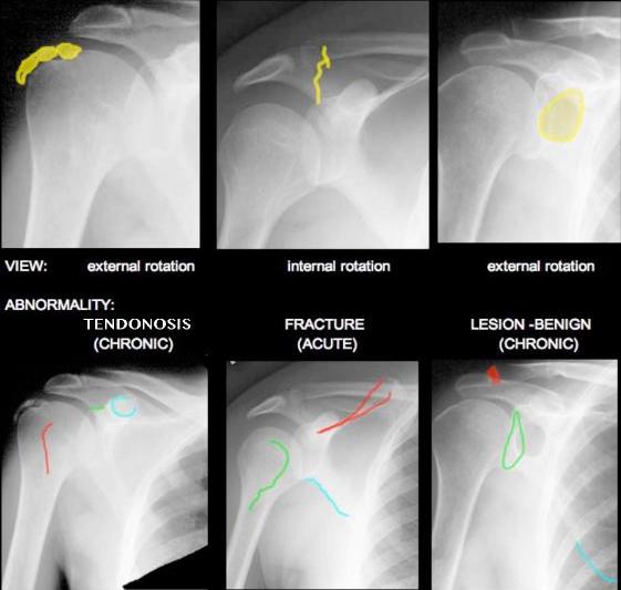
Have a look at the images shown to the right. For each image, be able to identify what attaches in the regions indicated in blue, red, and green.
You will be led through each image in the following slides.
Question 2:
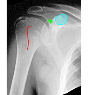
a) What attaches in the regions indicated in blue?
Blue (coracoid process)- pectoralis minor, coracobrachialis, short head of biceps
b) What attaches in the regions indicated in red?
Red (lesser tubercle)- subscapularis
c) What attaches in the regions indicated in green?
Green (supraglenoid tubercle)- long head of biceps
Question 3:
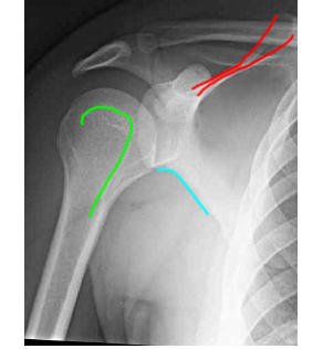
a) What attaches in the regions indicated in blue?
Blue (inferior margin of scapula)- teres minor
b) What attaches in the regions indicated in red?
Red (spine of scapula)-
superficial layer- trapezius from above and deltoid from below
deep layer- supraspinaus from above and infraspinatus from below
c) What attaches in the regions indicated in green?
Green (greater tubercle)- supraspinatus, infraspinatus, teres minor
Question 4:
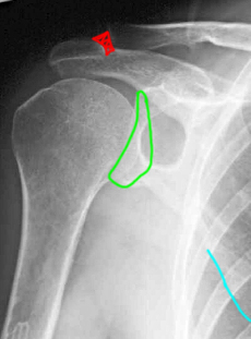
a) What attaches in the regions indicated in blue?
Blue (inferior angle of scapula)- teres major
b) What attaches in the regions indicated in red?
Red (AC joint)- acromioclavicular ligament
c) What attaches in the regions indicated in green?
Green (glenoid)- labrum
Question 5:
Which of these patients needs more imaging? What type of imaging?
Image 1: The calcifications are well seen on radiography so no further imaging is needed for the diagnosis, although ultrasound might be an option for treatment.
Image 2: The fracture is very faint and it is hard to be sure exactly how it is running through the scapula. When there is a need for better view of bony structures, CT is often best, although MR could also be considered.
Image 3: The lesion seems to be in the bone, but there might be a soft tissue component, so MR is probably best for further characterization, although CT could also be used, particularly if there is any contraindication to MR.
Click the blue buttons below to consult the ACR Appropriateness Criteria. Advance to the final slide in this section to view CT images.
Question 6:
a) How can you tell that this is a CT?
You can tell that both sets of images are CT scans because they are obviously SLICES (not projections, like an X-ray), and the cortical bone is very white. If you look at image sequence A, you can see a lot of internal detail in every bone, such as where the medullary cavity is.
b) What is the window that is being used to display the data?
This is a bone window, a particular brightness and contrast setting that shows bone best. The soft tissues look washed out. In image sequence B, you can still see the bones, but most of them look completely white and you can no longer see much internal detail. However you can see the muscles and fat of the soft tissues much better, so these images are being displayed with a soft tissue window.


