


Web Cases
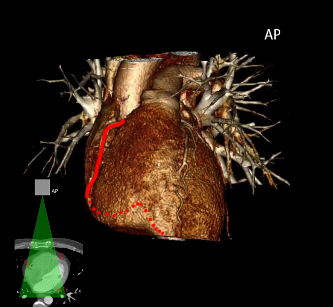
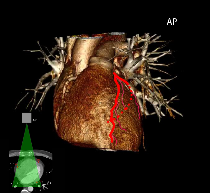
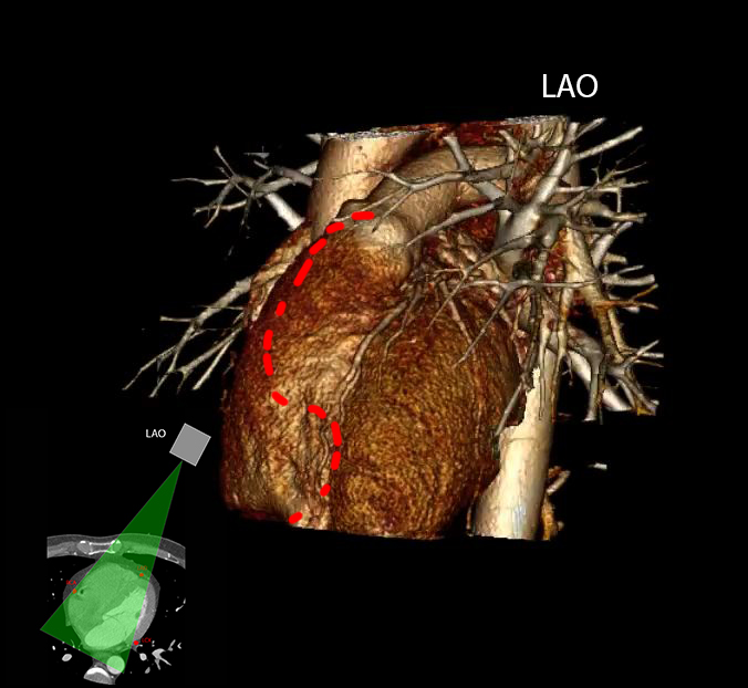
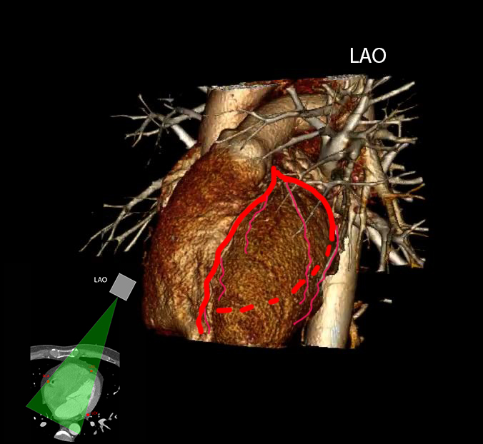
Here are images to demonstrate the orientation of the coronaries on various angiographic projections
Question 1:
Caudal and cranial angulation is also often used to better demonstrate the anatomy. What do these terms mean?
Caudal angulation means moving the X-ray source toward the patient's feet and angulating the beam so that it enters closer to the bottom of the heart. Cranial angulation means moving the X-ray source toward the patient's head, and angulating the beam so that it enters from the top of the heart. These refinements are not included in the diagrams. With cranial angulation, the diaphragm is often seen on the image, which can help to identify it.
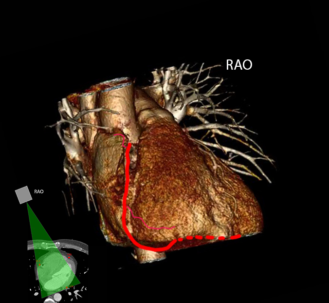
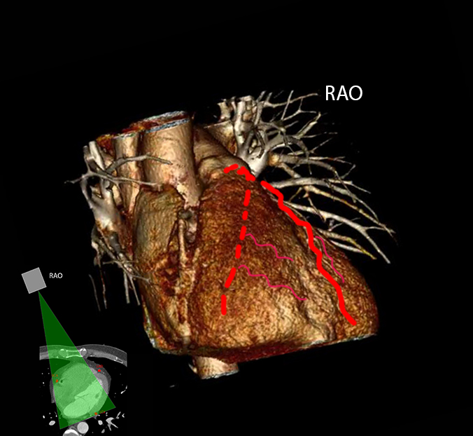
The initial image shows an AP view of the heart and shows a CT slice for reference. For other views, select the labels below. For LCA (left coronary artery) views, both LAD and LCX (circumflex) are shown. For the RAO view, if the spine is in the field of view it will be on the LEFT side of the image (patient's right side). For the LAO view, it will be on the RIGHT side of the image (patient's left side).


Web Cases
Case 1-61 year old male with intermittent exercise induce angina, getting worse.
Question 2:
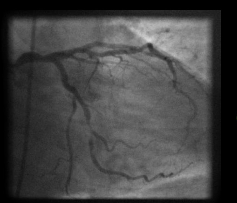
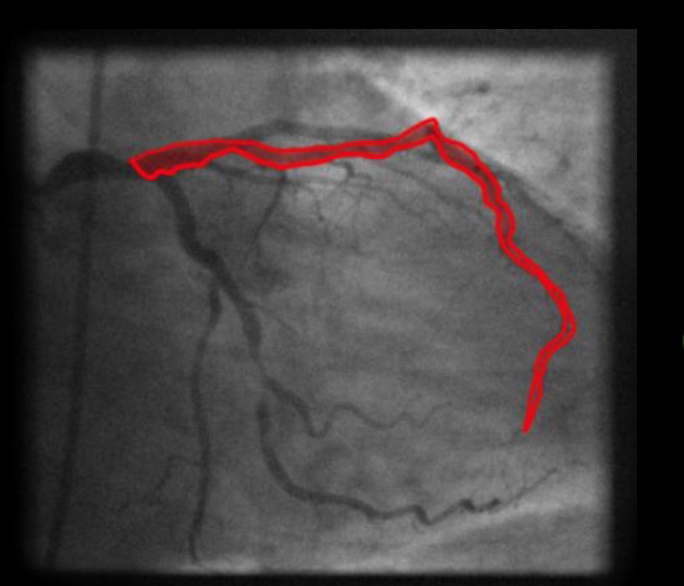
What is this study and what is the projection?
RAO view of left coronary injection--as on prior images, for an RAO view, the beam is coming in from the right, and this produces an image where the catheter (and spine, if visible) are on the LEFT side of the image (or the RIGHT side of the patient).
Web Cases
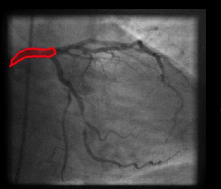
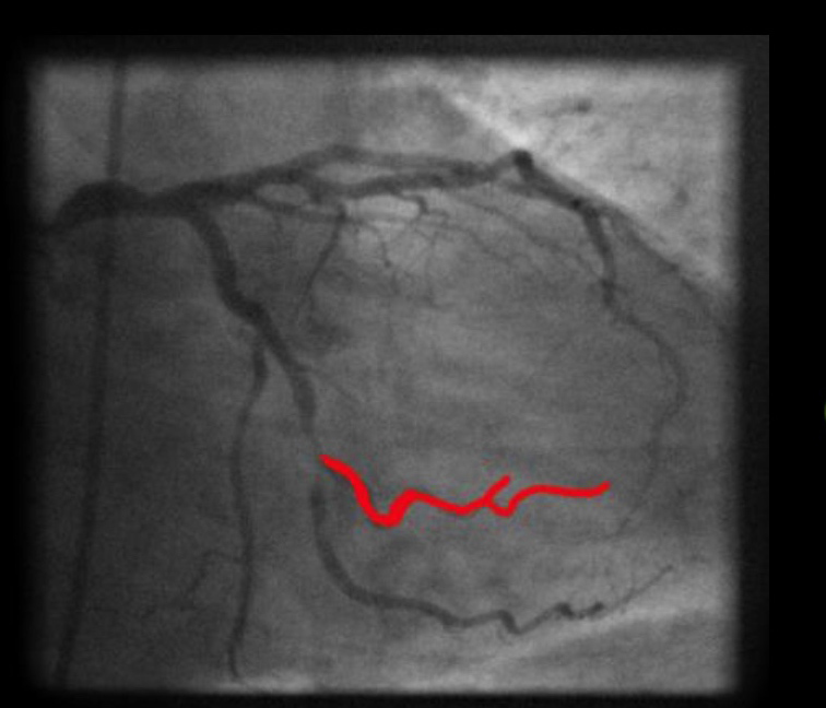
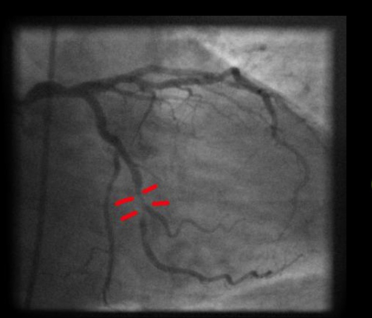
Try to identify the vascular structures listed below.
Question 3:
What is wrong?
There is narrowing of the circumflex, near one of the obtuse marginal branches.
Web Cases
This is the next image from this patient's series.
Question 4:
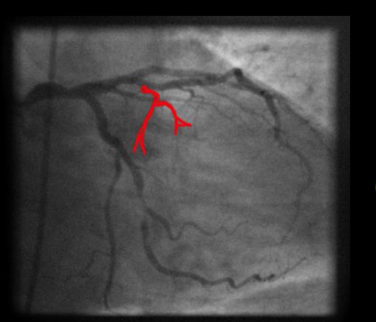
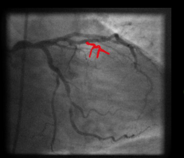
What has been done
A wire has been passed along the circumflex, past the stricture and a balloon is in place at the level of the narrowing.
Web Cases
This is the final set of this patient's imaging
Question 5:
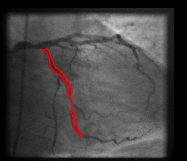
a) What is the projection
RAO view of the left coronary--cather is to the LEFT of the image, the patient's right. Spine is not included in this small field of view.
b) What has been done?
The area of narrowing is no longer seen. The balloon angioplasty has opened up the circumflex and it now appears normal.
Web Cases
Case 2-This is a 55 year old male with crushing chest pain and arrhythmias.
Question 6:
What is this study and what is the view
This one is tough because there is not much contrast getting past the origin of the vessel. There is just enough view of the ribs to tell that it is an LAO view, and probably the right coronary. There is a small branch off the proximal part of the vessel that may be the artery to the SA node.
Web Cases
Case 3-This patient was asymptomatic, having CT for other reasons. Pay close attention to the aortic root and coronaries on this exam.
Question 8:
a) What do you think of the esophagus
It appears to have a diffusely thickened wall
b) What is the timing of the contrast injection?
The scanning is later than for a PE study, with less contrast in the right atrium and pulmonary artery than in the left atrium, left ventricle and aorta.
c) What do you think of the left latissimus dorsi muscle?
There is a lipoma of the left latissimus dorsi muscle.
Web Cases
This is a labeled set of CT images of the same patient. Try to identify what is indicated in red, purple, green and blue.
Question 9:
a) What is abnormal about the coronary arteries?
The right coronary artery arises from the left coronary artery.
b) Is this a worrisome abnormality?
Yes. This aberrant right coronary has a 'malignant' course, running between the right ventricular outflow tract/main pulmonary artery and the aortic root. It may undergo sudden compression with vigorous exercise, leading to death. Sometimes the aberrant vessel can go in FRONT of the large vessels, which is generally asymptomatic, called a 'prevascular' course.