

Lower Extremity Cases
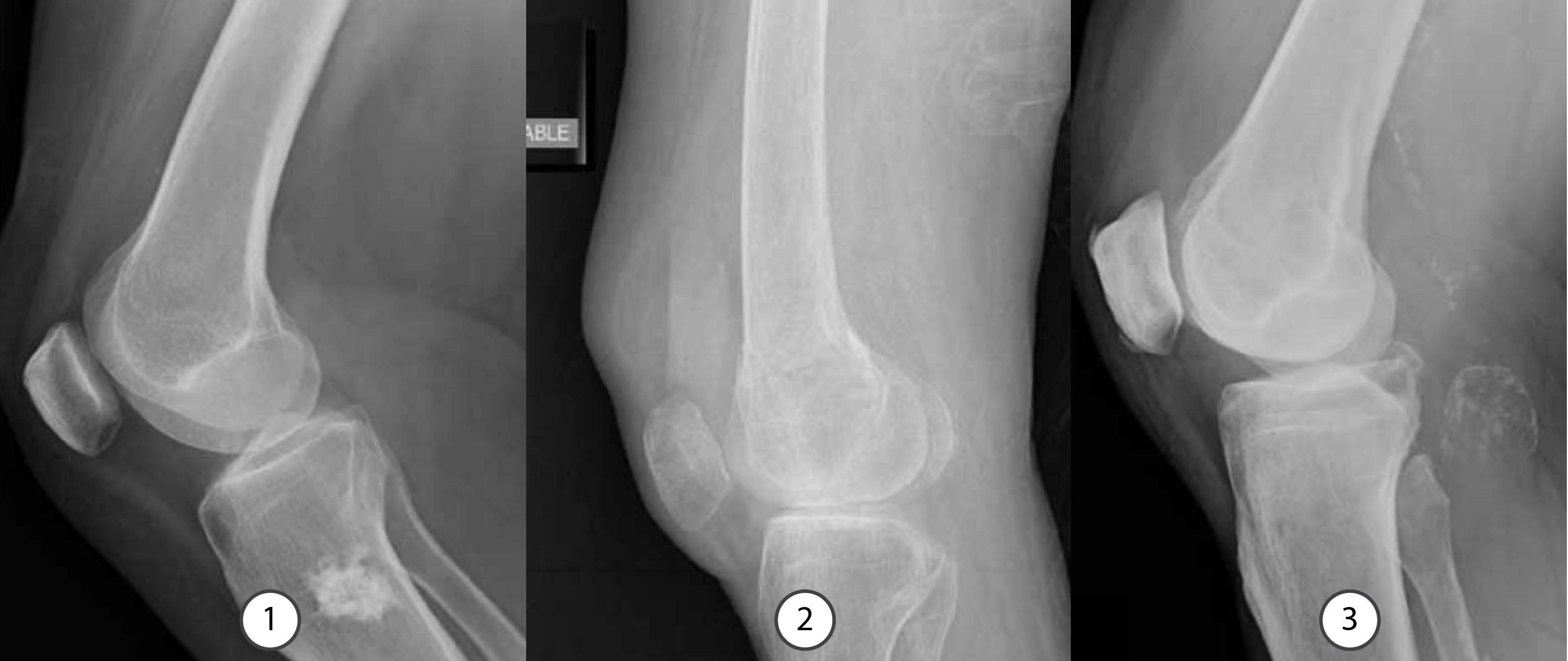
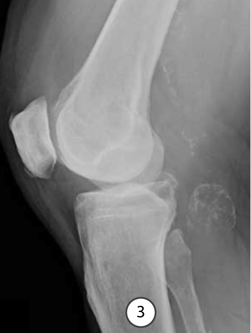
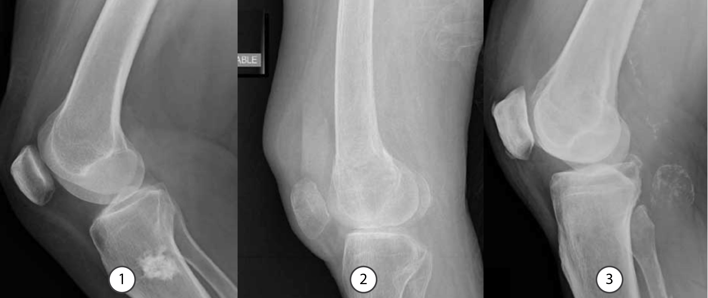
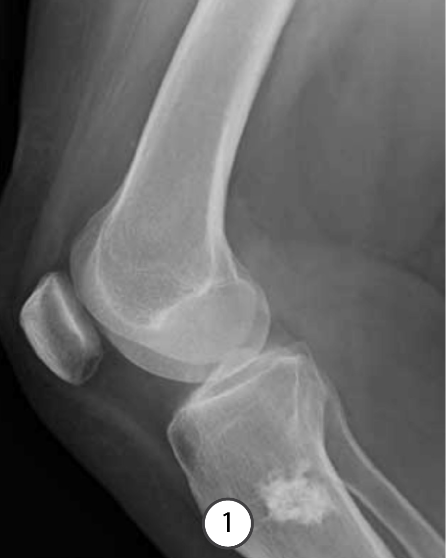
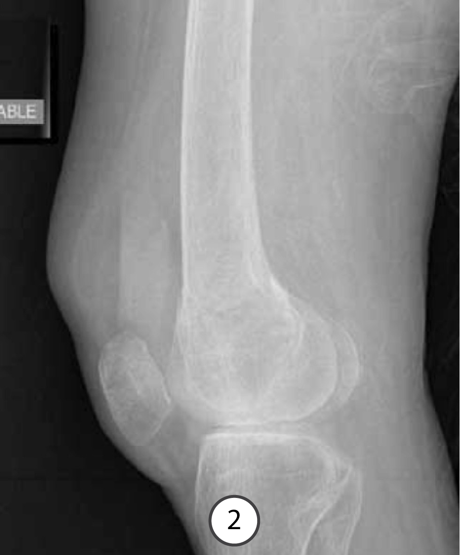
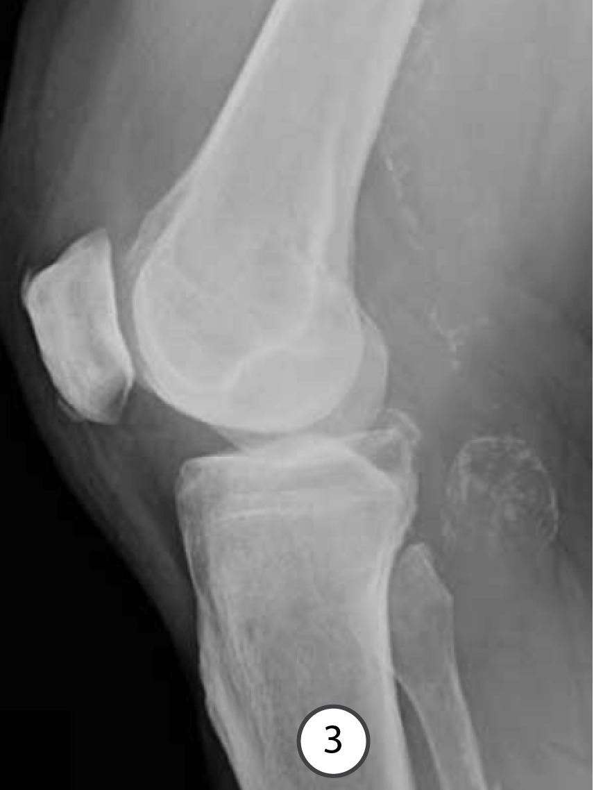
Three patients with knee pain, two chronic and one acute. For each image, state what the radiographic VIEW is, and what the ABNORMALITY is.
Further Explanation:
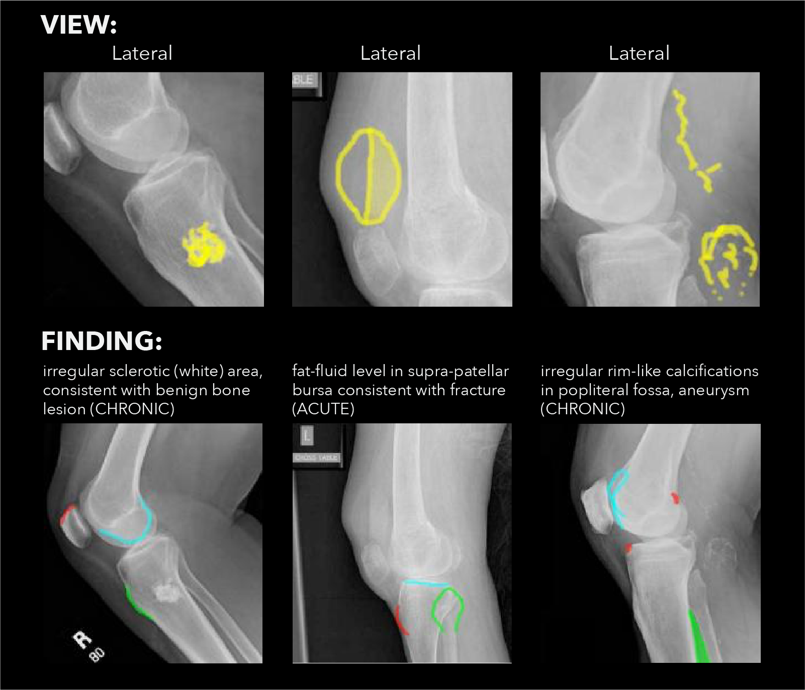
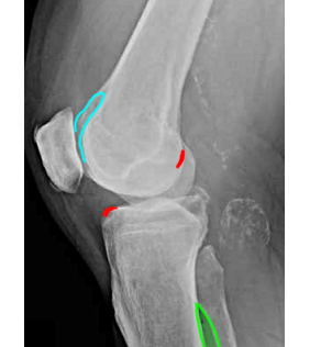
Have a look at the images shown to the right. For each image, be able to identify what attaches in the regions indicated in blue, red, and green.
You will be led through each image in the following slides.
Question 1:
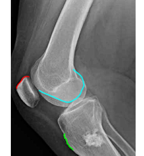
a) What attaches in the regions indicated in blue?
Blue- femoral condyle (articular surface of distal femur)
b) What attaches in the regions indicated in red?
Red (superior patella)- quadriceps tendon
c) What attaches in the regions indicated in green?
Green- tibial tuberosity
Question 2:
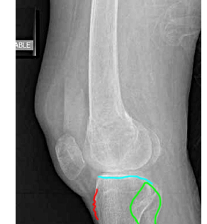
a) What attaches in the regions indicated in blue?
Blue-tibial plateau (articular surface of proximal tibia)
b) What attaches in the regions indicated in red?
Red (tibial tuberosity)- patellar tendon, continuation of quadriceps past patella, sesamoid bone
c) What attaches in the regions indicated in green?
Green- fibular head
Question 3:
a) What attaches in the regions indicated in blue?
Blue- patellar facet of femur (articular surface of patellofemoral joint)
b) What attaches in the regions indicated in red?
Red (anterior margin of tibia and posterior margin of femoral condyle)- anterior cruciate ligament
c) What attaches in the regions indicated in green?
Green- interosseous ligament (attachment point for many leg muscles)
Question 4:
Which of these patients needs more imaging? What type of imaging?
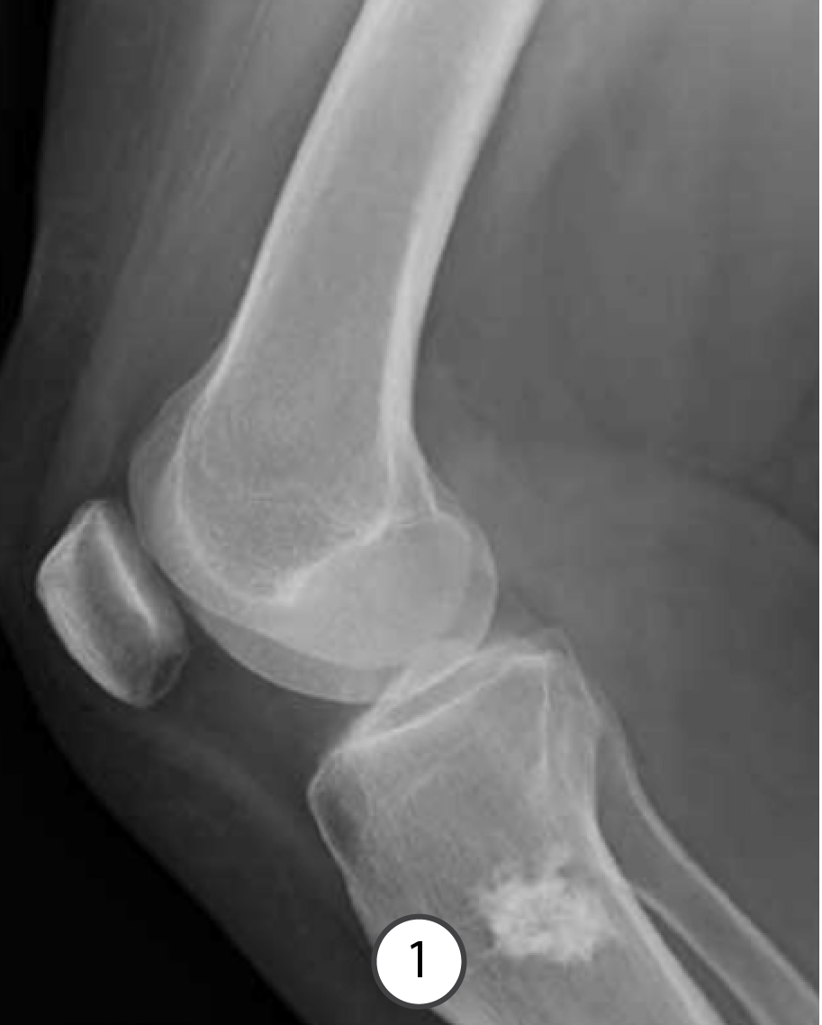
Image 1: This lesion looks relatively benign and no additional imaging is needed unless pain is localized to this area and increasing, then possibly a bone scan.
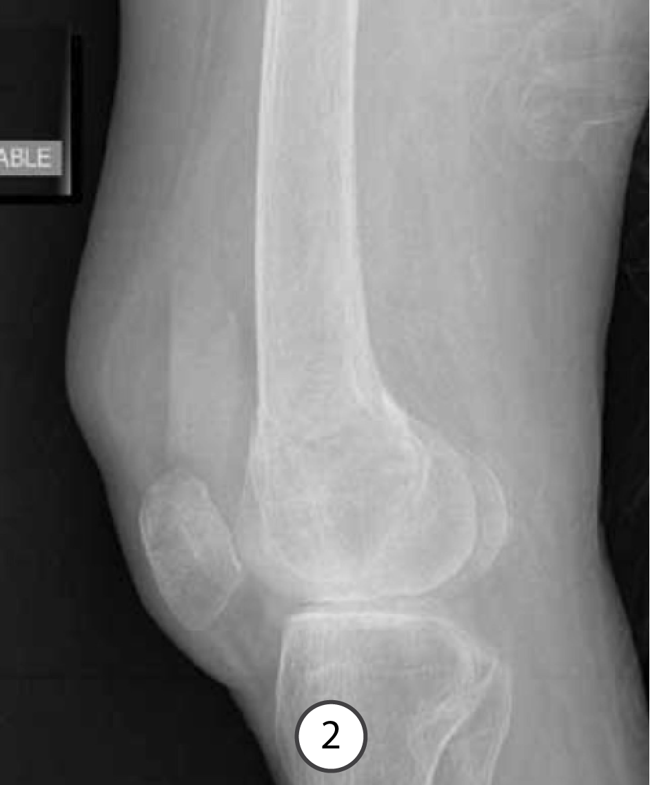
Image 2: The fat-fluid level tells you there is a fracture, but the fracture is not evident on radiographs so definitely needs additional imaging, and since it is mainly a bony problem, CT is the best choice.
Image 3: This appearance suggests a popliteal aneurysm, for which more imaging is definitely needed, either ultrasound or MR.
Click the blue buttons below to consult the ACR Appropriateness Criteria. Advance to the final slide in this section to view US images.
Question 5:
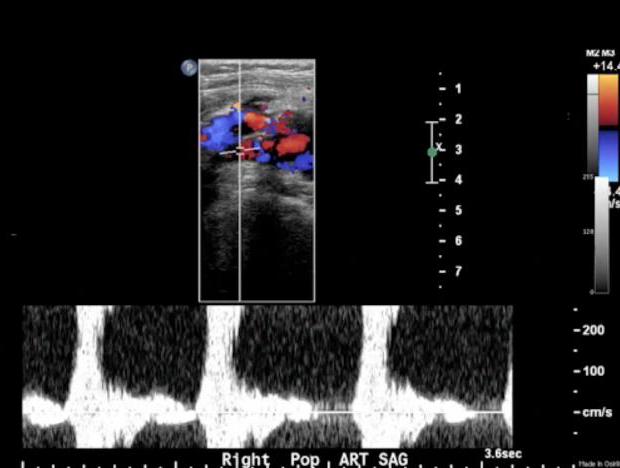
What are the colored parts of this image showing?
The top image is part of a popliteal ultrasound study, and the colored areas are Doppler flow analysis. The mixture of blue and red areas indicate tubulent flow. The scale on the right-hand side of the image shows that red colors indicate flow TOWARD the transducer and blue colors indicate flow AWAY from the transducer. The transducer is the mechanical apparatus that is used to send sound waves in and record reflected waves bouncing back. In this case, the transducer is touching the back of the patient's knee, in the popliteal fossa, so flow indicated in red is toward the back and blue is toward the front. Red and blue do NOT correlate to arterial and venous flow, just directions. The bottom tracing shows an arterial pulse in the area of the abnormality, indicating that it is a popliteal aneurysm.
You have finished the lower extremity section.
CLICK THIS LINK, to return to the topics list for PT cases.
