

Introduction Workshop - Case 1
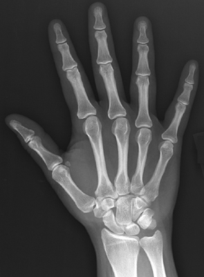
Here is an image of a 31 year old patient who has pain in the region of the base of her right thumb after playing tennis for the first time in years.
Question 1:
a) What is this image called and how is the body part oriented?
This is a radiograph (also sometimes called an 'x-ray') of the hand. The hand radiographic image is typically displayed with the fingers upward (which is NOT the anatomic position of the hand, which would have fingers downward), and includes a label to indicate which side is being imaged (which is not visible on this image). For this image, the beam passed from posterior to anterior (PA) because it is more comfortable for the patient to place their hand palm downward on the receptor. Because the hand is very thin from front to back, the PA vs AP view for the hand would not look noticeably different so we generally place the hand however is the most comfortable for the patient. We can also call this a 'frontal' view, without specifying PA or AP.
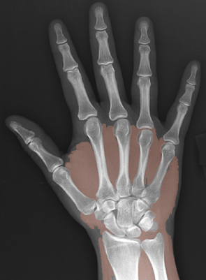
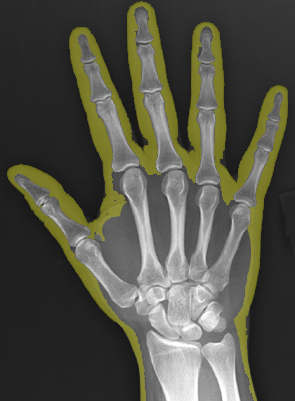
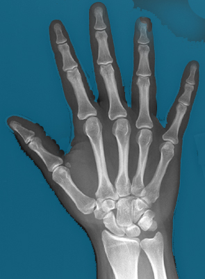
b) What tissues are indicated by the labels below?
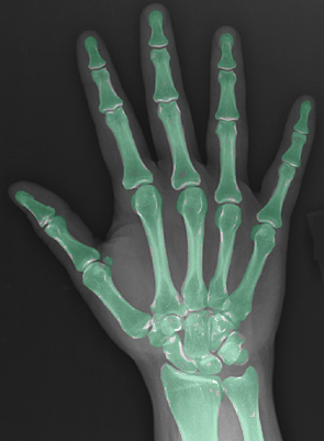
tissue 1 is bone (notice that the outer cortical bone is whiter than the inner medullary bone) tissue 2 is soft tissue or water density (which would include muscle, fascia, tendons, ligaments, vessels, cartilage, blood, and fluid) tissue 3 is fat tissue 4 is air
c) What do you think of this patient's thumb?
This is a normal hand radiograph. It does not show any bony abnormality, like a fracture or arthritis. However, this does not exclude soft tissue injuries, such as damage to cartilage, ligaments or tendons. To examine these structures in more detail, other types of imaging would be required, like ultrasound or MRI. But a radiograph is a quick, inexpensive and readily available way to exclude most serious injuries. The small rounded bone at the medial margin of the first metacarpophalangeal joint is a normal sesamoid bone--a bone lying within a tendon. The thumb and great toe usually have two sesamoid bones apiece. Extra sesamoids at other joints are fairly common.
Imaging Workshop cases provide optional additional examples and explanations on Imaging Anatomy. You may examine these cases on your own or in groups.
Introduction Workshop - Case 1
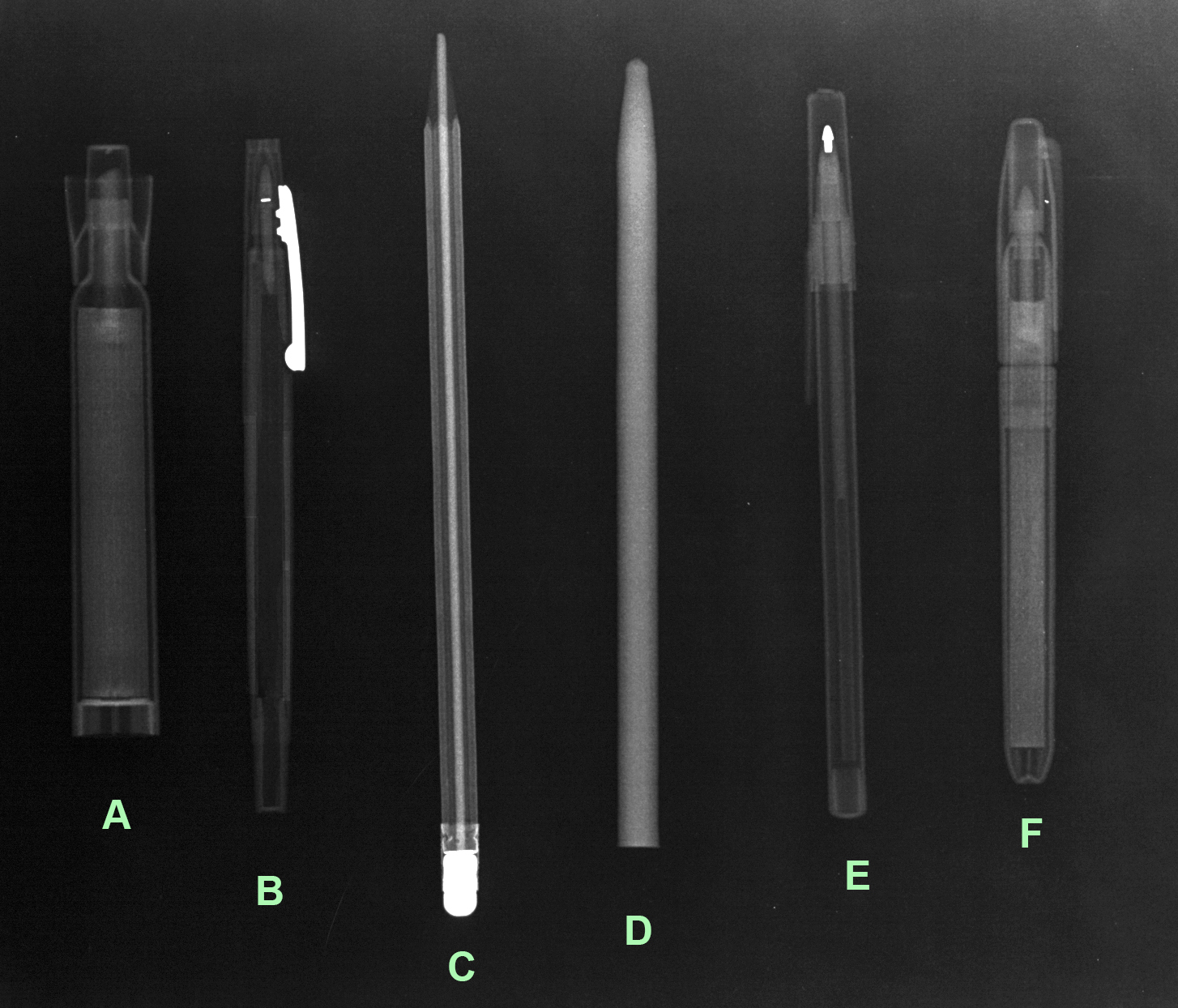
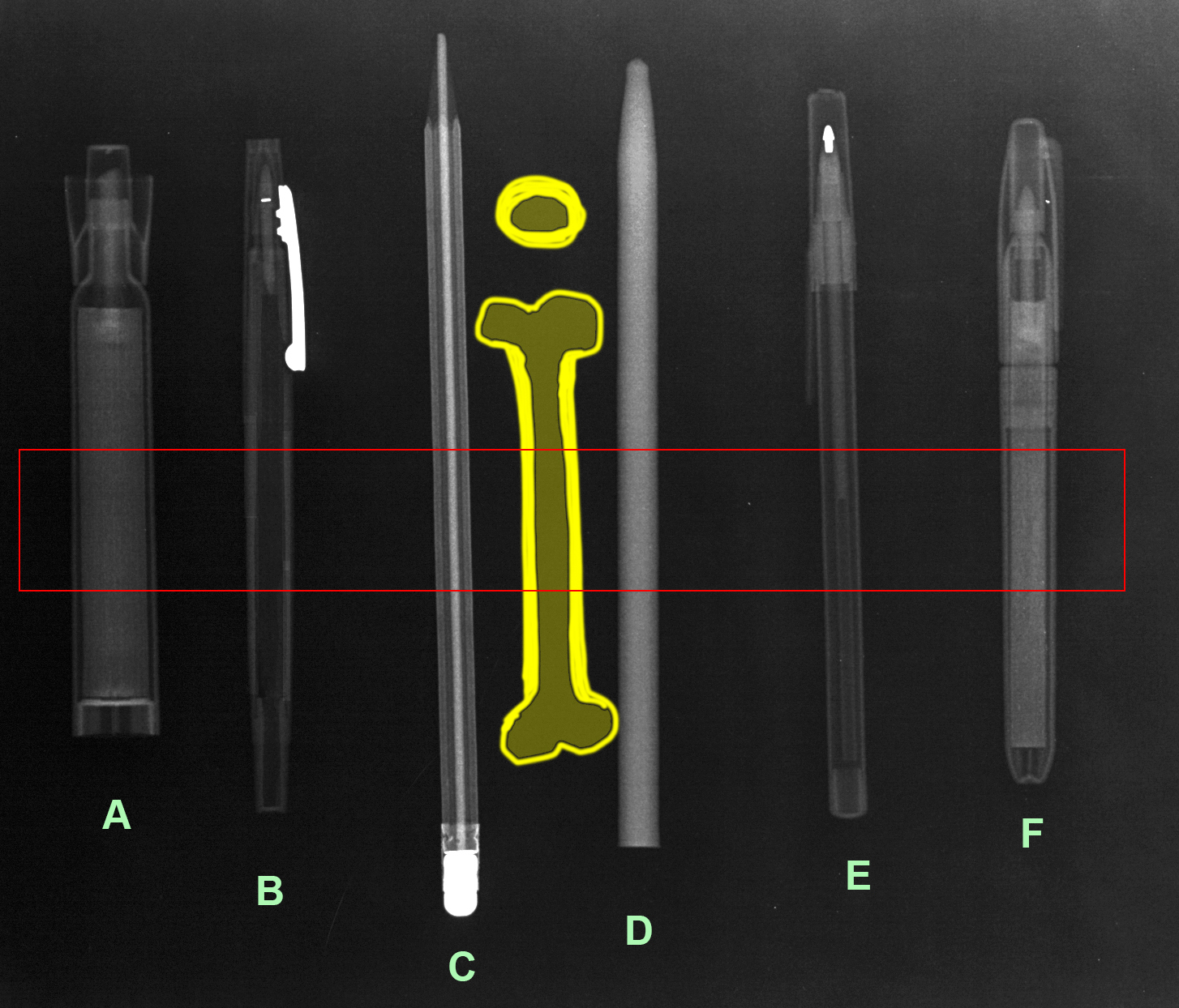
This is is a radiograph of some common familiar objects. Interpretation of two-dimensional radiographic images of three-dimensional objects requires some thought about how various shapes of internal structures will project onto the receptor.
Question 2:
a) A typical long bone consists of a shaft (with outer more dense cortex and inner less dense medulla), also called the 'diaphysis'. Thinking about how such an object would look on a radiograph, decide which of the everyday objects looks most like a bone in terms of geometric shape.
Object B (a Flair pen) seems to have a denser outer wall (made of plastic) and an inner portion that is less dense (filled with ink, which is less dense than the plastic walls). Object E looks a bit like this, but it is a ballpoint pen and if you look carefully, there is a second fainter structure inside which is the ink-holding plastic refill (with outer dense wall and interior filled with air or ink, depending on where you look along its length).
b) What is object C?
Object C is a pencil, with a more dense center (graphite) and less dense walls (wood).
c) What is object D?
Object D is a wax pencil, which is totally uniform throughout. As a result, it looks a bit less dense along the edges because it is round in cross-section, so at the edges, the beam is not going through as much wax as in the center.
Introduction Workshop - Case 1
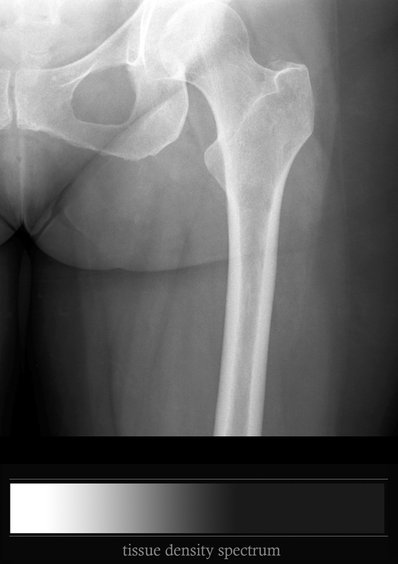
Our patient also had left hip pain, so this imaging was also done.
Question 3:
a) What is this study?
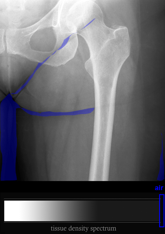
This is a radiograph of the upper femur and hip. It is typically done in an AP projection, again because it is easier than asking the patient to lie face-down. The femur is in anatomic position.
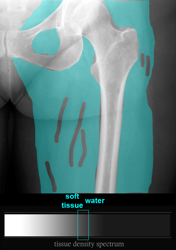
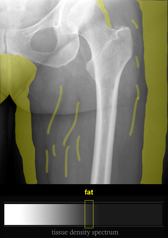
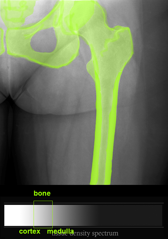
b) Which of the tissues listed below is LEAST dense and which is MOST dense?
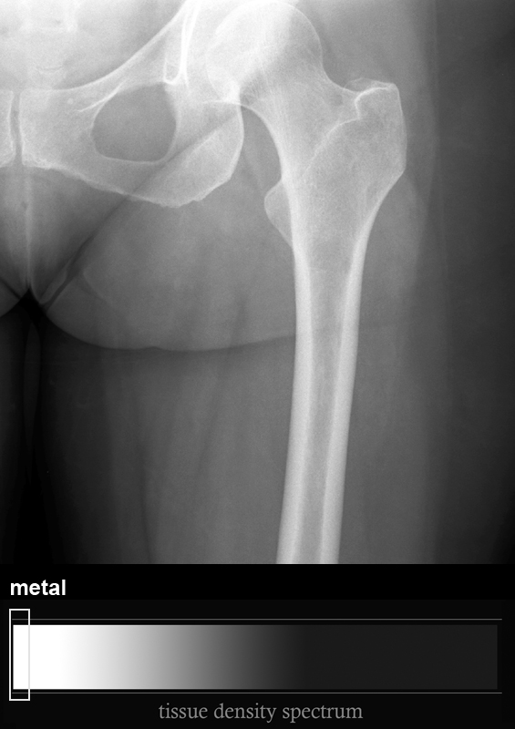
Air is least dense. There is no air inside the thigh, but you can see that there are small streaks of air caught along the groin crease and at the inferior margin of the buttock. The most dense material we sometimes see on a radiograph is metal, but this is not present in the normal patient and is not seen on this image. The next most dense tissue is anything that contains calcium, in this case bones. You can see that as on the prior example with the flair pen, the outer (cortex) of the bone is whiter (more dense) than the inner (medulla) of the bone became there is more tightly packed calcified material in the cortex. The medulla contains a mix of calcified spicules or network of bone, as well as the much less dense elements of the bone marrow (which contains blood-forming cells and fat). So the medulla is an average of a lot of different density tissues, overall much less dense than the cortical bone.
c) How different is fat from water/soft tissue?
These two tissues are very close together in the center of the density spectrum, so fat is easiest to distinguish from water/soft tissue where there is a lot of fat (along the inner and outer thigh) right next to lots of muscle. There are very faint streaks of fat seen more centrally, but they are not very obvious.
d) Why does the top part of the femur (called the 'head') appear particularly dense?
The actual degree of whiteness on any part of a radiograph depends not only on the type of tissue present, but the geometry of the structure. When two bones overlap on the image (like the head of the femur and the pelvis), the density will summate and appear whiter than either bone would appear alone. In fact, every part of the image is summing up a variety of material, since the beam passes through air, skin, fat, muscle, bone (sometimes), and then back through muscle, fat, skin and air. The density is a sum of all of those tissues for each pixel on the image.
