


Spine Cases
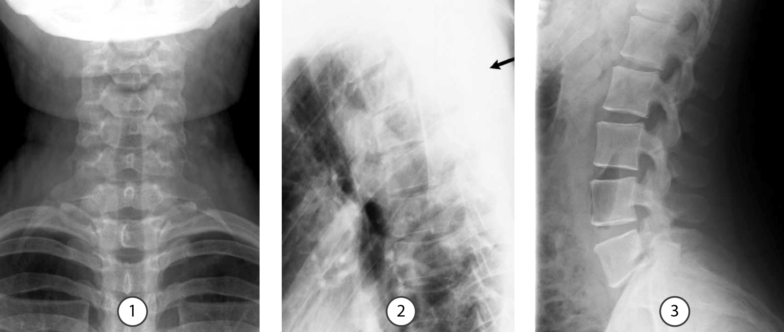
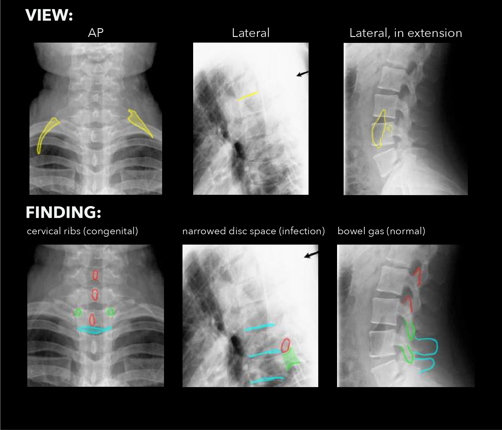
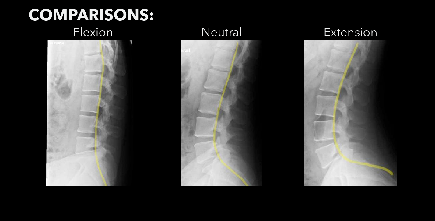
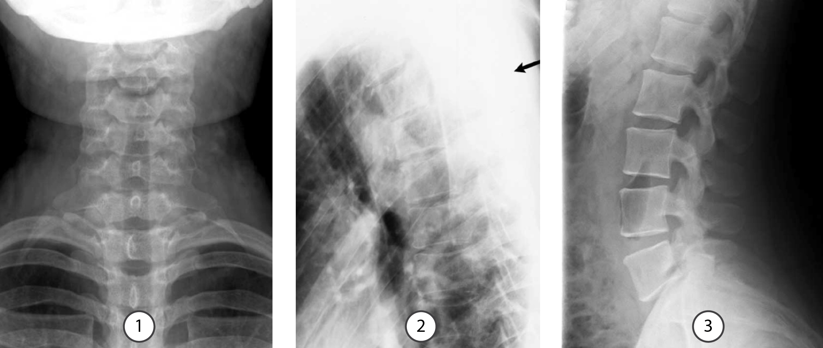
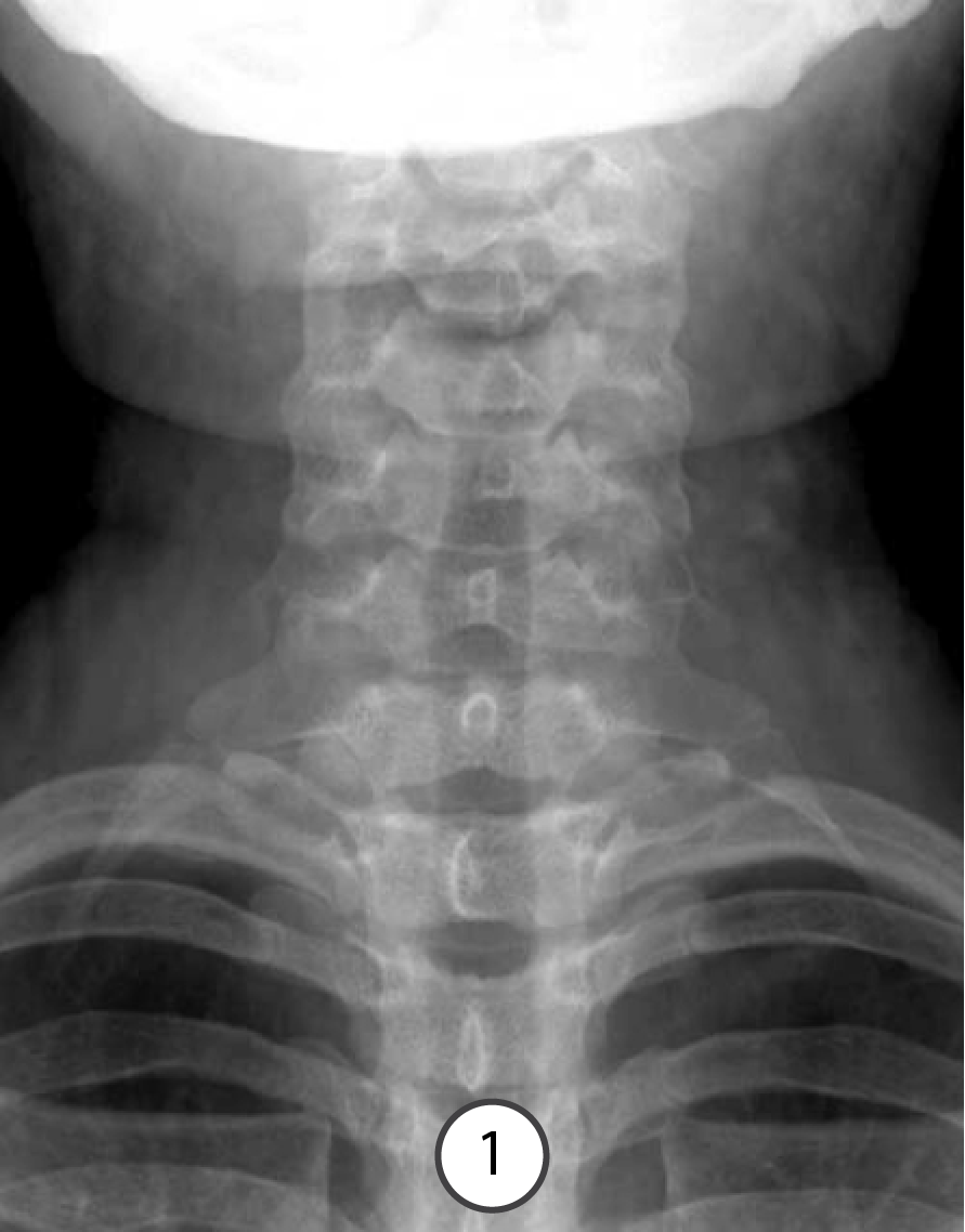
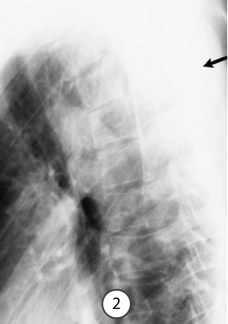
Three patients with back pain, all chronic. For each image, state what the radiographic VIEW is, and what the ABNORMALITY is.
Further Explanation:
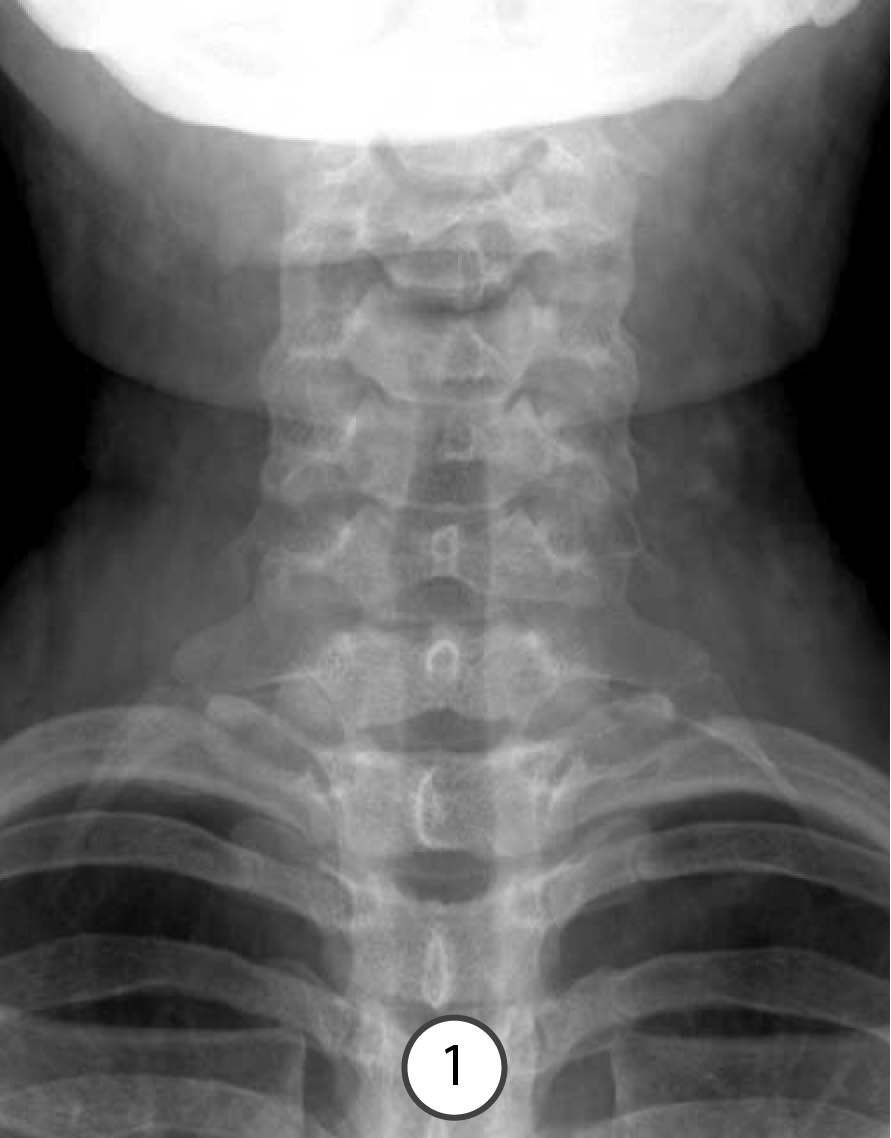
Image 1: Lower neck pain for many years, sharp, worsened with neck flexion or rotation, no other symptoms
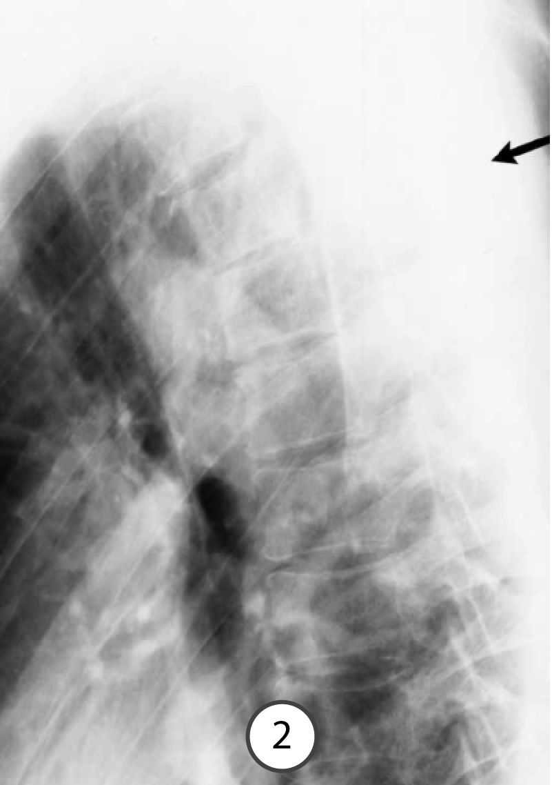
Image 2: Upper thoracic pain for several months, worst at the site of the black arrow, also fever and elevated white count
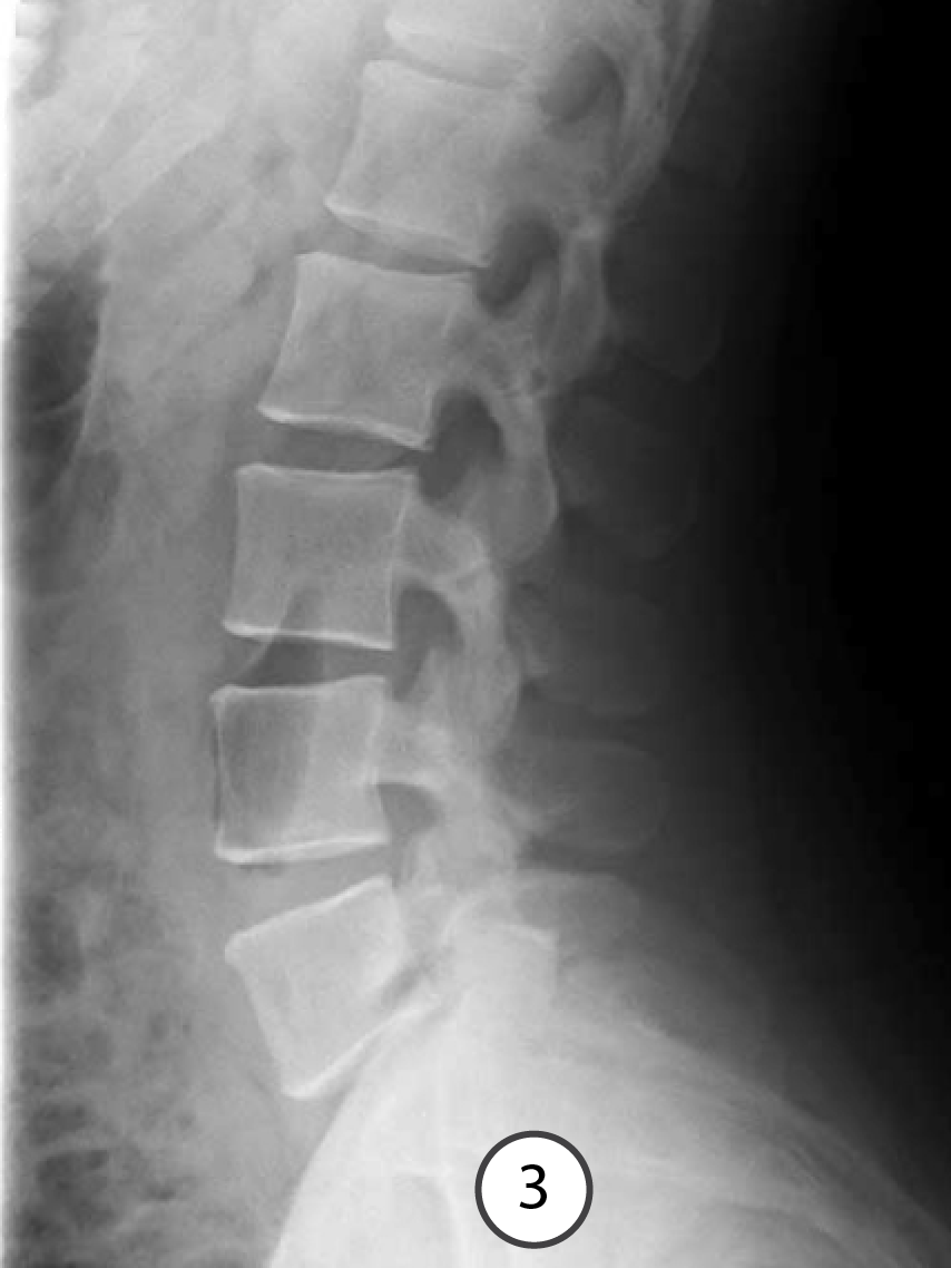
Image 3: Low back pain for several years, worse with flexion
Further Explanation:
Have a look at the images shown to the right. For each image, be able to identify what attaches in the regions indicated in blue, red, and green.
You will be led through each image in the following slides.
Question 1:
a) What attaches in the regions indicated in blue?
Blue- intervertebral disc space at T1-T2 level
b) What attaches in the regions indicated in red?
Red- spinous processes of C6, C7 and T1
c) What attaches in the regions indicated in green?
Green- pedicles of T1

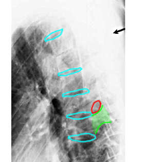
Question 2:
a) What attaches in the regions indicated in blue?
Blue- inferior end-plates of several adjacent thoracic vertebral bodies
b) What attaches in the regions indicated in red?
Red- neural foramen of the upper thoracic spine
c) What attaches in the regions indicated in green?
Green- pedicle of an upper thoracic verebral body
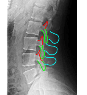
Question 3:
a) What attaches in the regions indicated in blue?
Blue- spinous processes of L4 and L5
b) What attaches in the regions indicated in red?
Red- superior articular processes of L3 and L4
c) What attaches in the regions indicated in green?
Green- inferior articular processes of L3 and L4
Question 4:
Which of these patients needs more imaging? What type of imaging?
Image 1: Probably no need for additional imaging, but if symptoms are severe enough to consider treatment, CT or MR could be performed to assess degree of compression of neural and vascular structures by the cervical ribs.
Image 2: Definitely needs more imaging, either CT, MR or both, to determine the extent of infection and how best to treat it.
Image 3: Probably no need for more imaging, but because the symptoms were so severe, the patient did go on to MR.
Click the blue button below to consult the ACR Appropriateness Criteria. Advance to the final slide in this section to view MR images.
Image 1: Lower neck pain for many years, sharp, worsened with neck flexion or rotation, no other symptoms
Image 2: Upper thoracic pain for several months, worst at the site of the black arrow, also fever and elevated white count
Image 3: Low back pain for several years, worse with flexion
Question 5:
a) How can you tell that this is an MRI?
You can tell that this study is an MR right away because FAT is bright (white) and the edges of BONES (cortical bone) are dark (the cortical bone of S1 is outlined above in dark blue). This is the opposite of how fat and cortical bone look on CT.
b) Is it T1 or T2-weighted?
This is a T1-weighted sequence because water is high in signal (white), such as the CSF, which is outlined above in light blue.
c) Is it abnormal?
There is an abnormal disc at L4-5 (outlined above in red) that is narrowed and lacks the normal central watery high signal of a healthy disc (outlined at other levels in yellow). This is a dessicated disc that has lost height and has herniated slightly posteriorly.
Advance to the final slide for a comparison and further explanation.
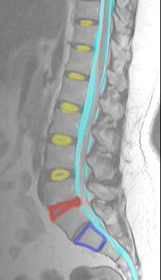
Further Explanation:
For comparison, to the right is the T1-weighted sequence on the same patient. Look at the CSF and you will see that it is now DARK. This is how you recognize a T1-weighted sequence--areas of water or watery fluid (like urine, bile or CSF) are dark on T1-weighted images, and bright on T2-weighted images.
The abnormal disc is only bulging posteriorly a small amount. Is this the cause of the patient's pain? It is almost impossible to tell. MR of the lumbar spine can be problematic for just this reason-it may detect abnormalities that may not have anything to do with the patient's symptoms. This patient improved with conservative treatment and never had any symptoms that specifically localized to the L4-5 level.


