

Case 5
This case concerns the imaging appearance and differential diagnosis of pleural disease.
Question 1:
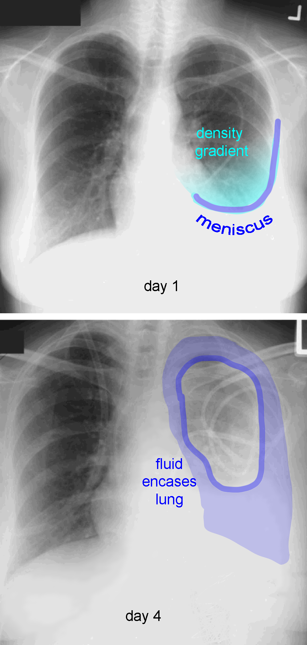
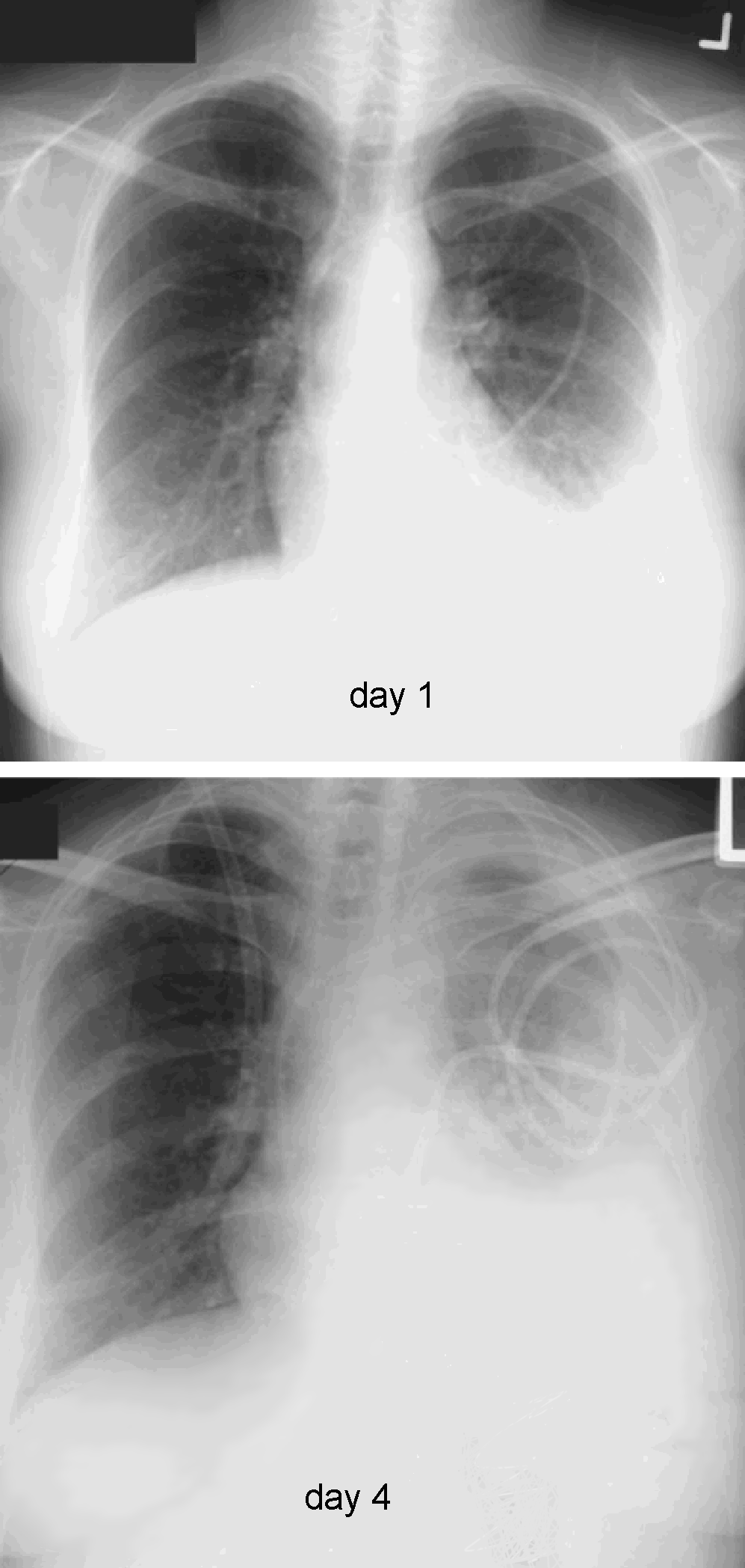
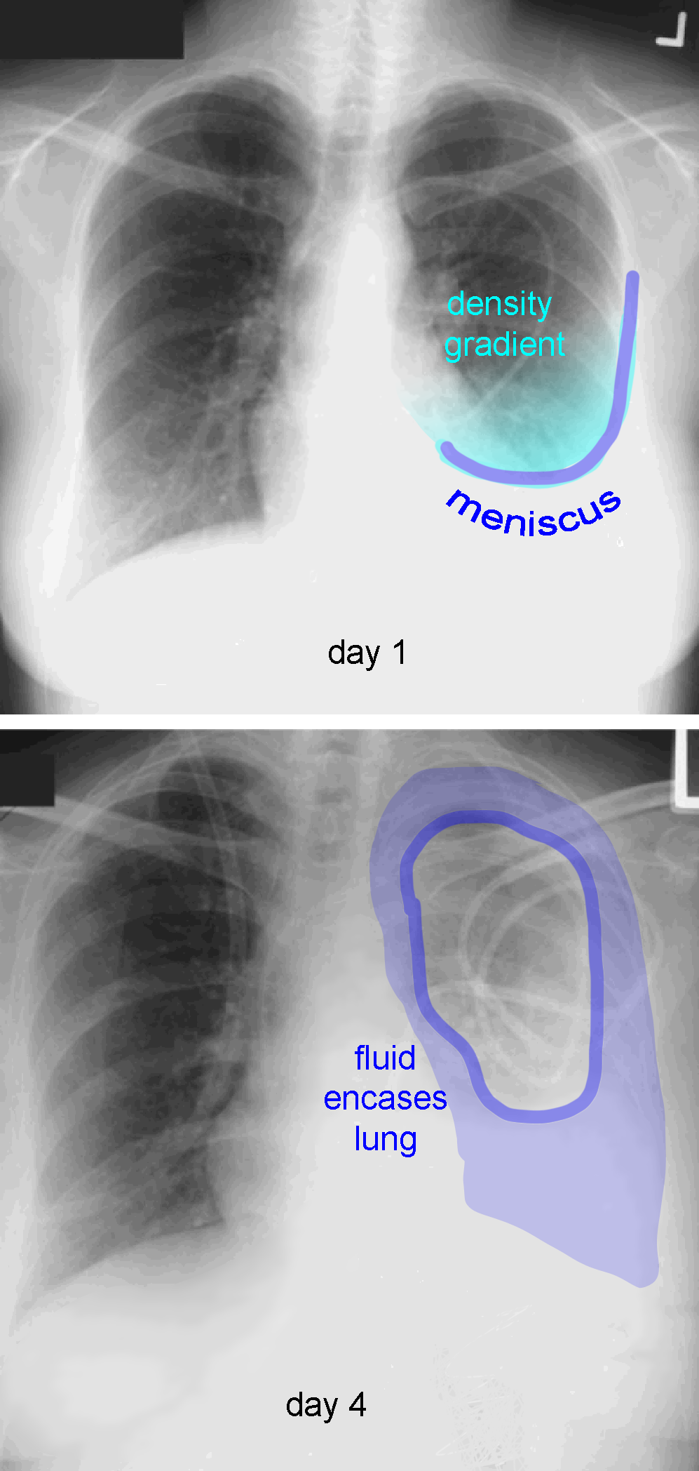
These are two chest radiographs of the same patient several days apart. How can you identify the opacity on the left as pleural rather than parenchymal? What has changed between the two images?
×
Answer:
Pleural fluid is often more homogeneous in density than lung disease, and may have a characteristic shape, particularly on an upright image. On day 1, there is a curved bottom to the opacity that creeps up the lateral chest wall, representing a meniscus. There is also a gradual gradient of increased density above the meniscus, due to fluid wrapping around the chest at that level. On day 4, the fluid collection is larger and now wraps around the top of the lung as well as inferiorly and laterally. This pattern of change would be very unlikely for disease involving the lung parenchyma. It is also important to remember that although we are assuming that the opaque material in the pleural space is FLUID, it could be other things and have the same appearance: blood, pus, fibrotic tissue, or tumor. Some of these would be unlikely to change this quickly, such as tumor or fibrosis.
Pleural fluid is often more homogeneous in density than lung disease, and may have a characteristic shape, particularly on an upright image. On day 1, there is a curved bottom to the opacity that creeps up the lateral chest wall, representing a meniscus. There is also a gradual gradient of increased density above the meniscus, due to fluid wrapping around the chest at that level. On day 4, the fluid collection is larger and now wraps around the top of the lung as well as inferiorly and laterally. This pattern of change would be very unlikely for disease involving the lung parenchyma. It is also important to remember that although we are assuming that the opaque material in the pleural space is FLUID, it could be other things and have the same appearance: blood, pus, fibrotic tissue, or tumor. Some of these would be unlikely to change this quickly, such as tumor or fibrosis.
Case 5
Other things besides fluid can be in the pleural space, as shown by the image below.
Question 2:
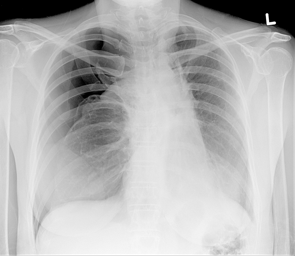
What density material is in this patient's pleural space? What other water density material could have been in the previous patient's pleural space besides fluid?
×
Answer:
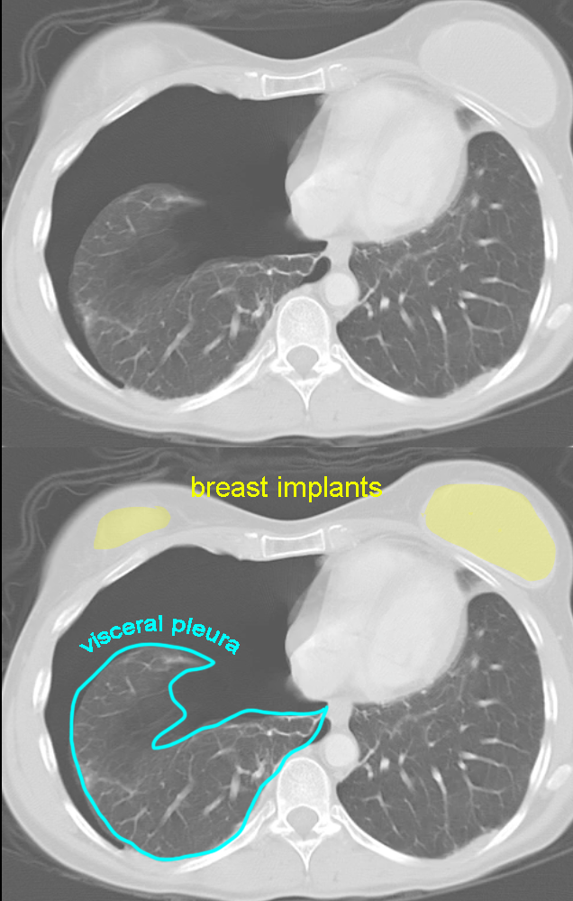
There is AIR in this patient's pleural space, consistent with a large pneumothorax. Their CT is shown below, with was done for another reason. The diagnosis is clear from the chest radiograph alone. For a patient with dense material in the pleural space, it is also important to realize on a CT that the air-filled lung will float above the dense material. In the case of a pneumothorax, the air in the pleural space will rise and the lung will fall into a more dependent position. This is another difference between dense material and air in the pleural space that can affect how these processes look on imaging in different positions.
There is AIR in this patient's pleural space, consistent with a large pneumothorax. Their CT is shown below, with was done for another reason. The diagnosis is clear from the chest radiograph alone. For a patient with dense material in the pleural space, it is also important to realize on a CT that the air-filled lung will float above the dense material. In the case of a pneumothorax, the air in the pleural space will rise and the lung will fall into a more dependent position. This is another difference between dense material and air in the pleural space that can affect how these processes look on imaging in different positions.
