

Orientation
It is important to remember orientation when looking at ultrasound images, which is the same for high resolution probes and curvilinear probes.
Question 1:
a) How are transverse, coronal and sagittal images displayed?
For both transverse and coronal images, the image is displayed as if you are facing the patient, so the patient's RIGHT side is on the LEFT side of the image. For sagittal images, the patient's HEAD is to the LEFT side of the image.
b) For the image below, showing a renal stone in the sagittal plane, is it in the upper or lower pole of the kidney?
It is closest to the LEFT side of the image, so it is in the upper pole.
Orientation
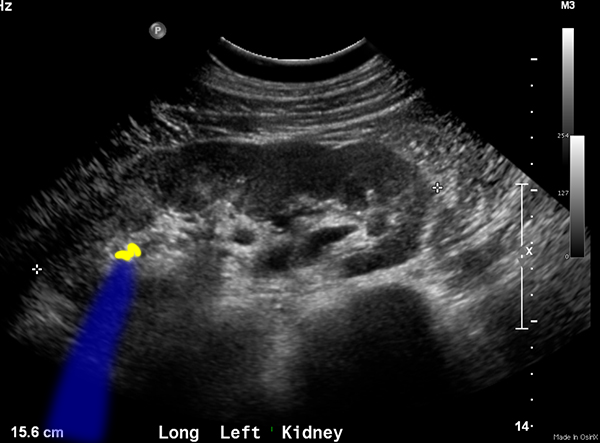
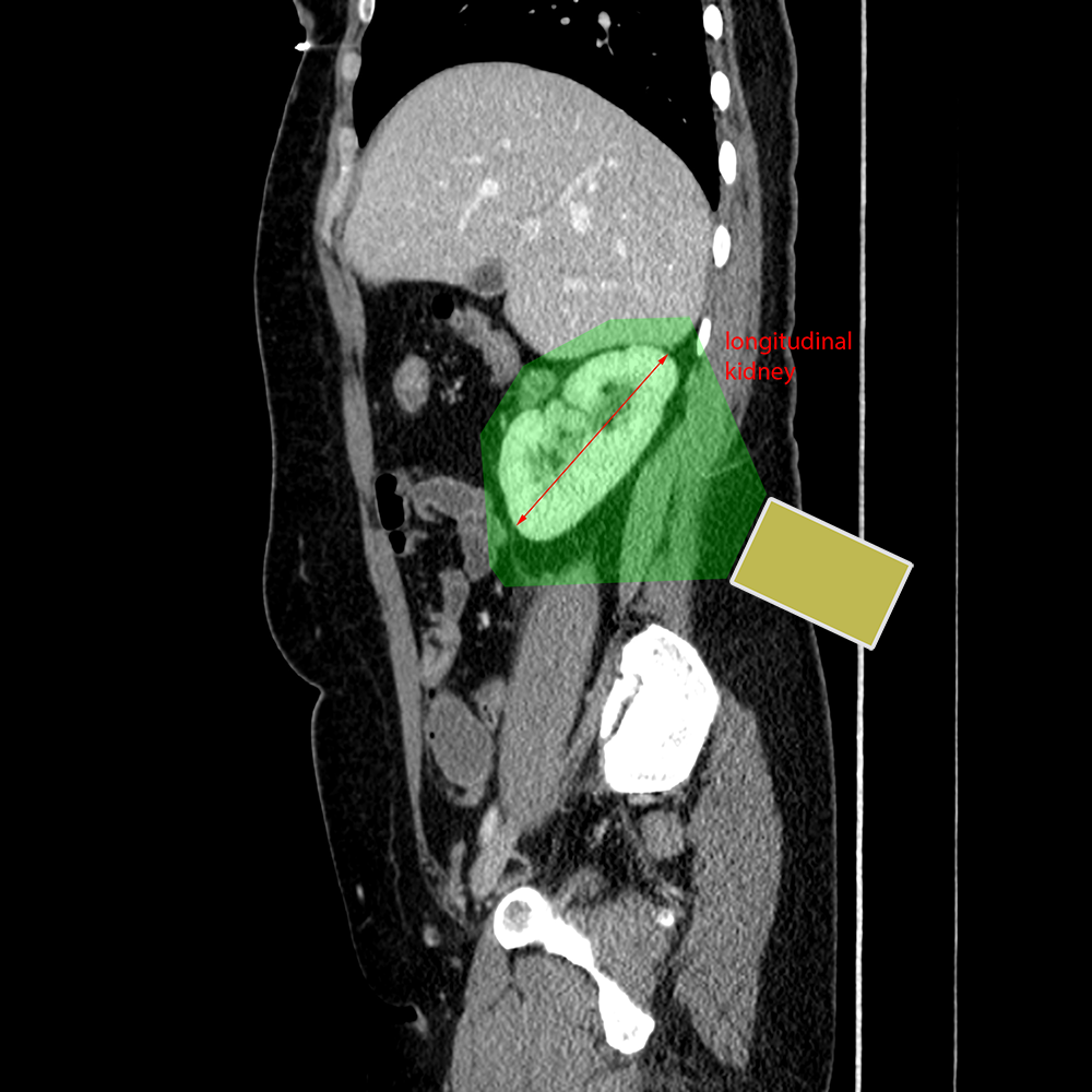
It is also important to remember that labels like coronal, sagittal, and axial are often angled to orient toward the actual organ being imaged, which are often not positioned in the exact anatomic plane. So sometimes images are labeled 'long' for longitudinal, and 'trv' for transverse instead of using the anatomic planes.
Further Explanation:
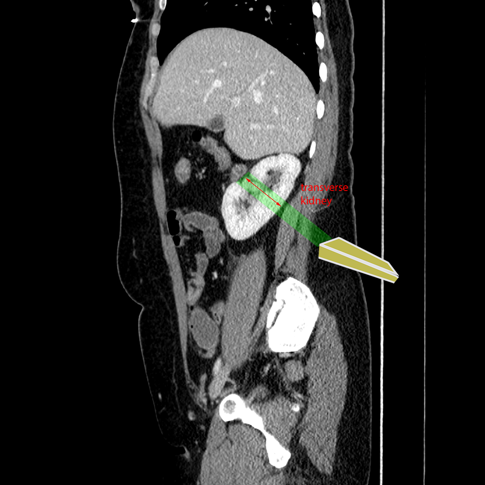
Orientation
For Doppler imaging, it is important to also be oriented toward the expected direction of flow.
Question 2:
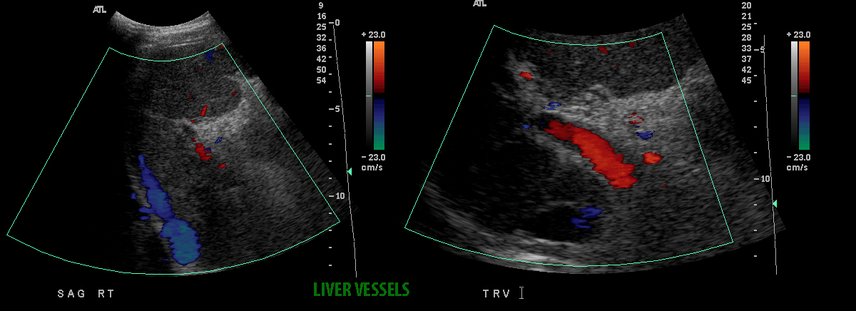
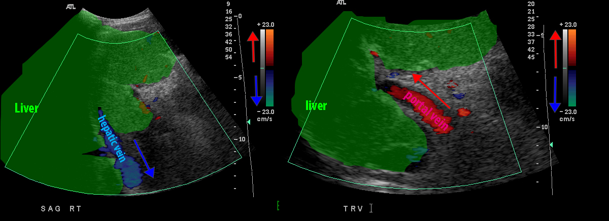
On the sagittal and transverse images shown, what do you think of the direction of flow in the imaged vessels?
The sagittal image shows a hepatic vein flowing into the IVC, so the flow is AWAY from the transducer (which is at the top of the image), and is colored blue. The transverse image at a lower point in the liver shows the portal vein, with flow TOWARD the transducer, into the liver, which is colored red.
