

Case 1-lung cancer
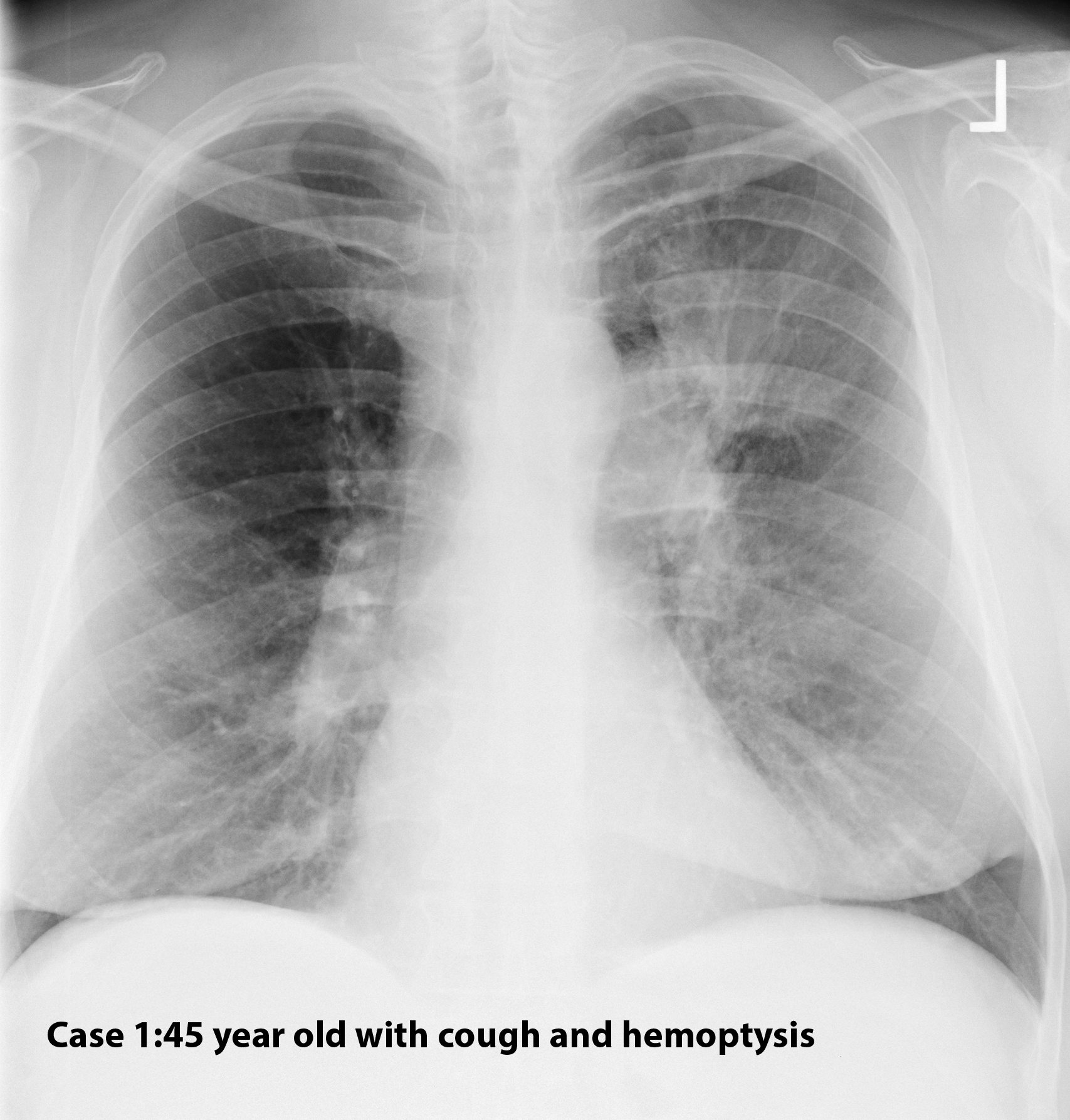
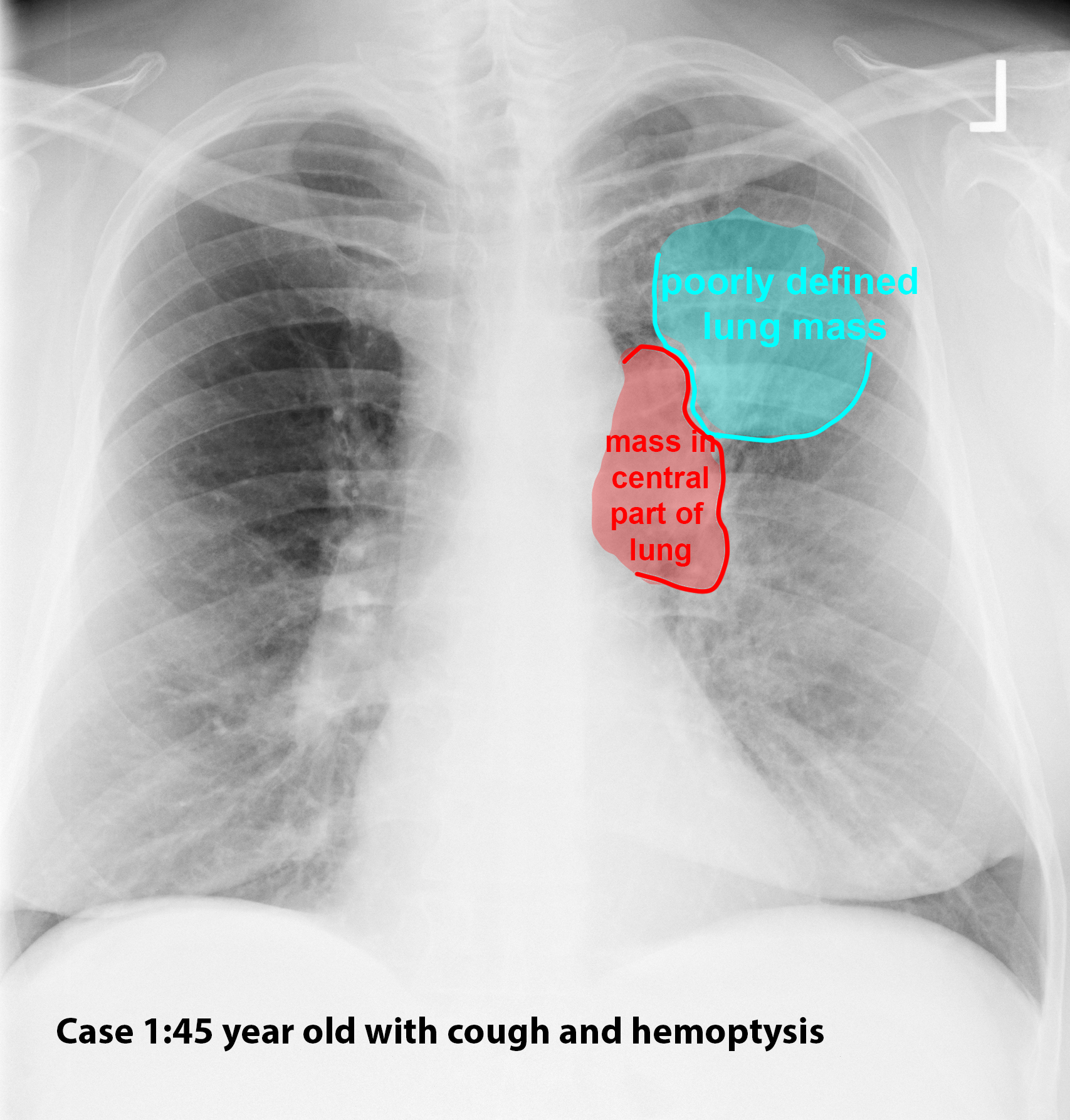
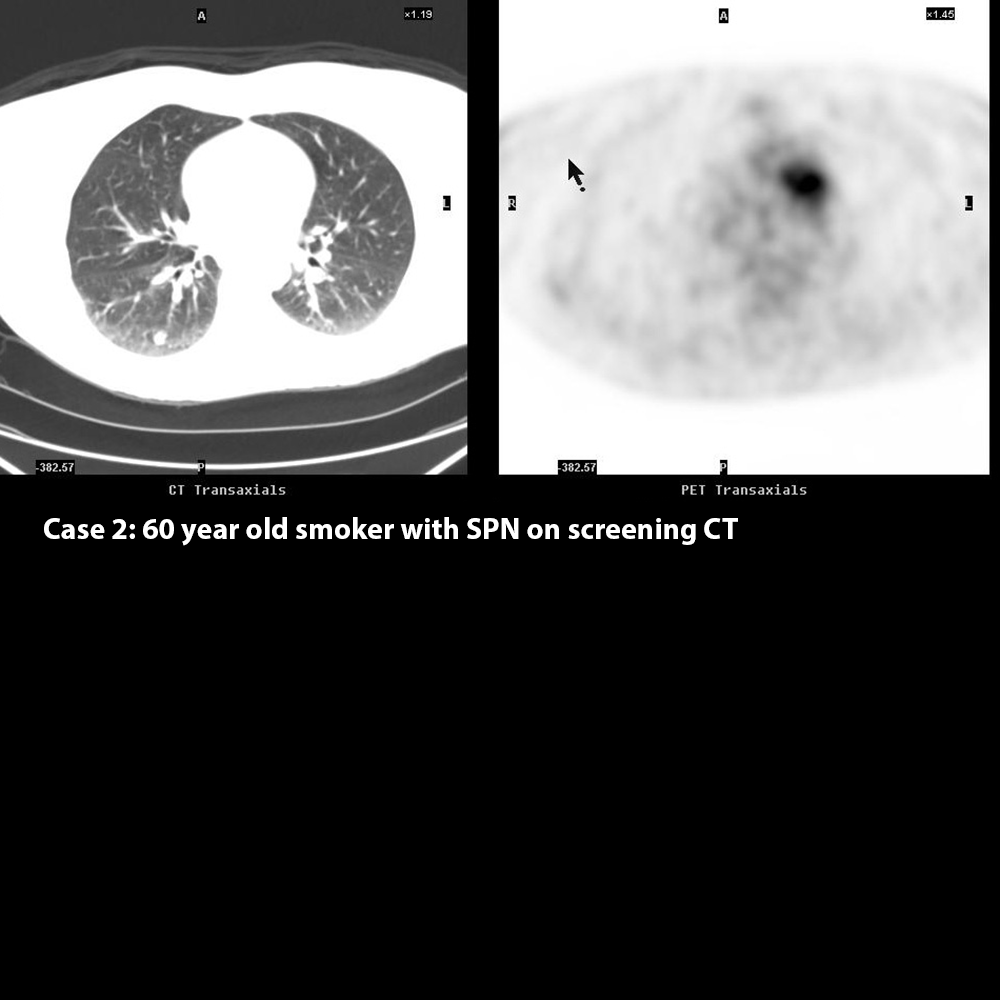
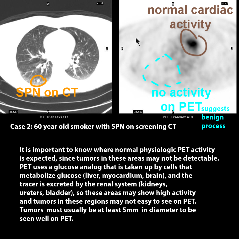
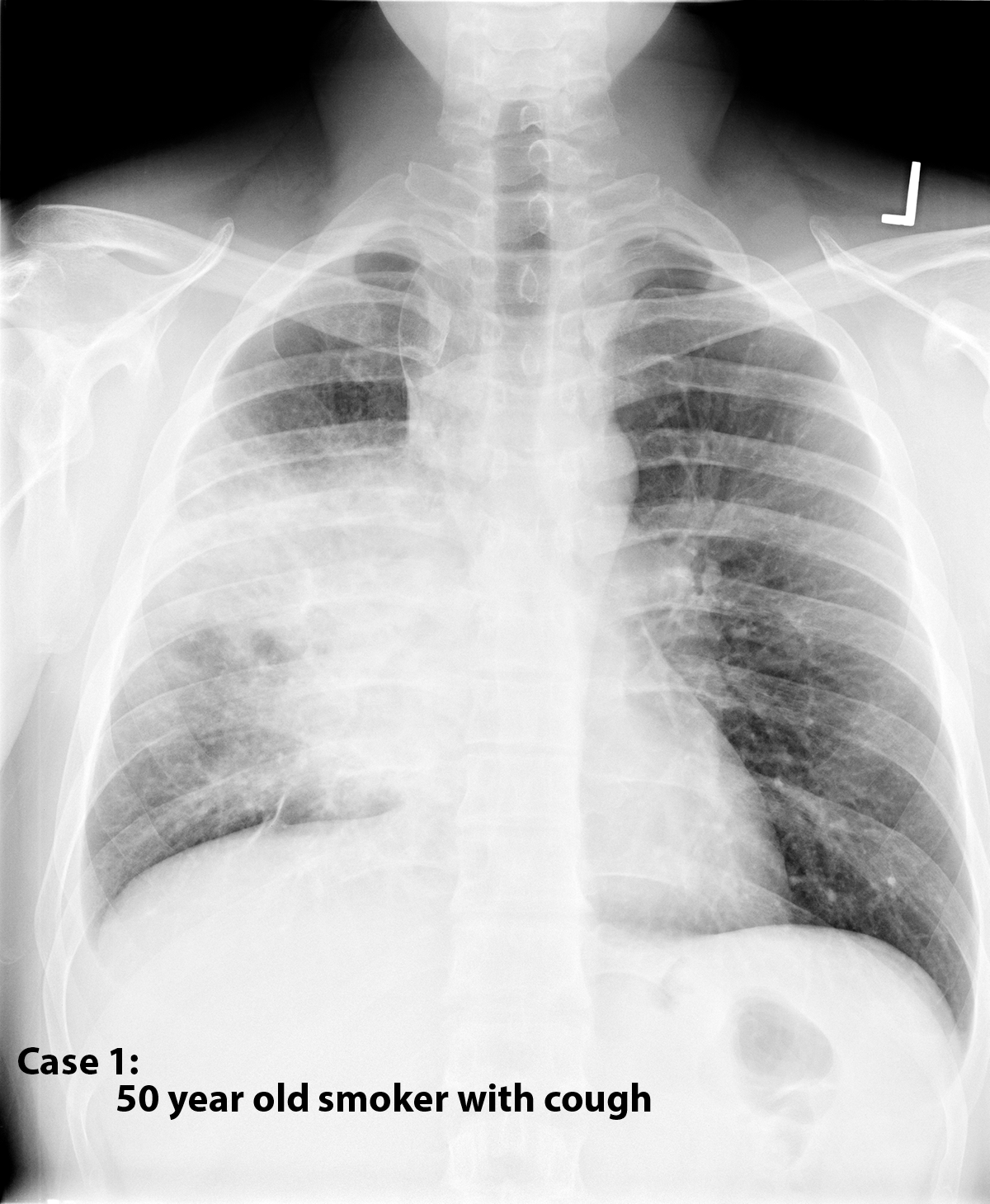
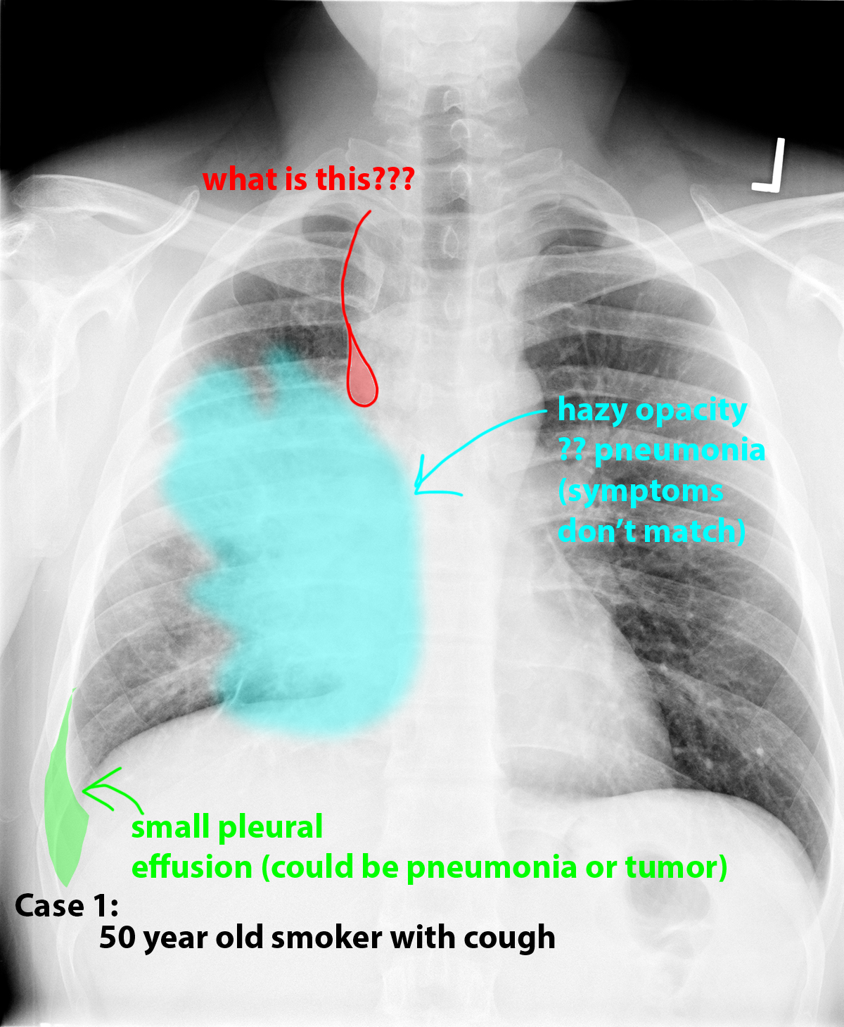
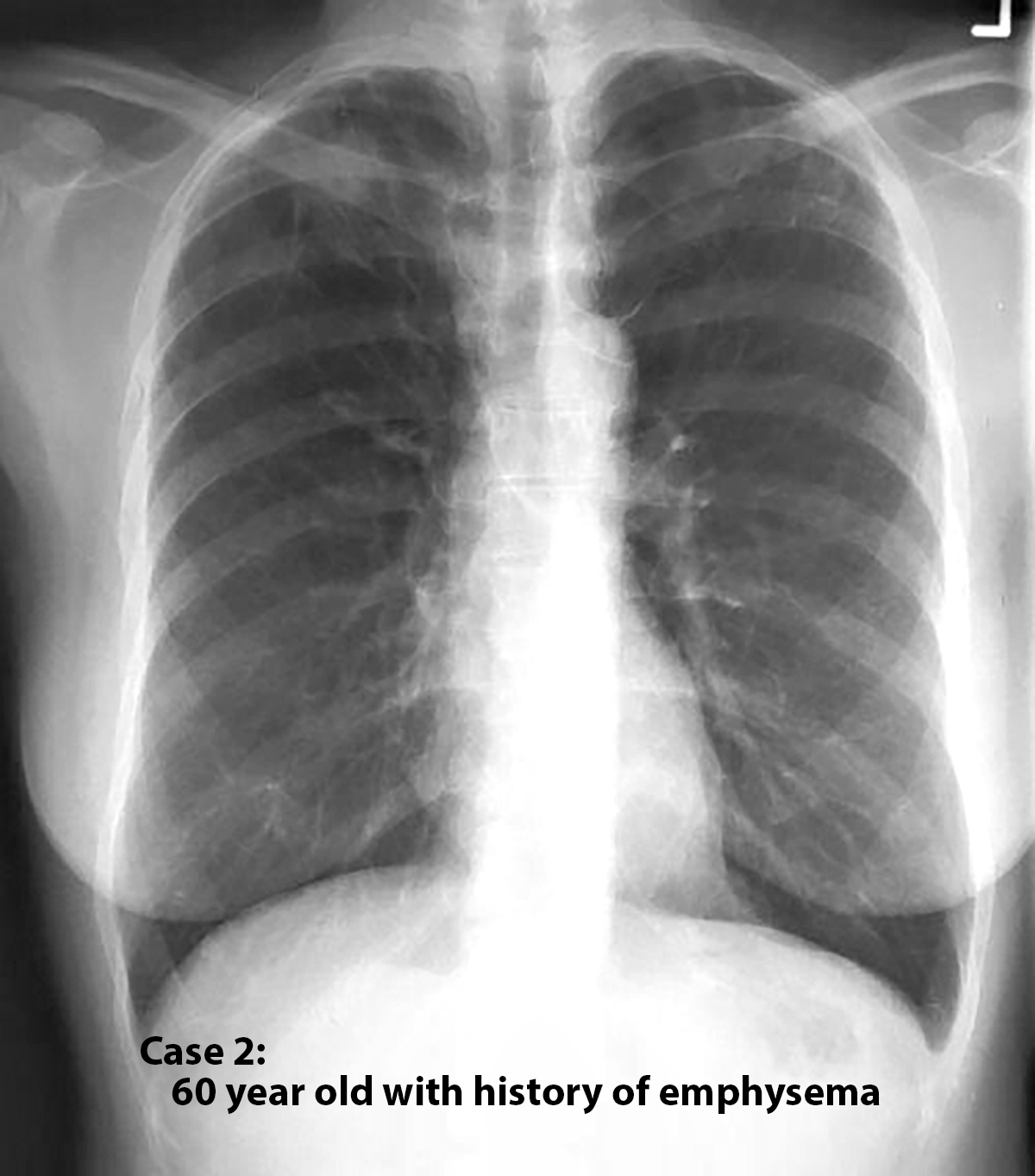
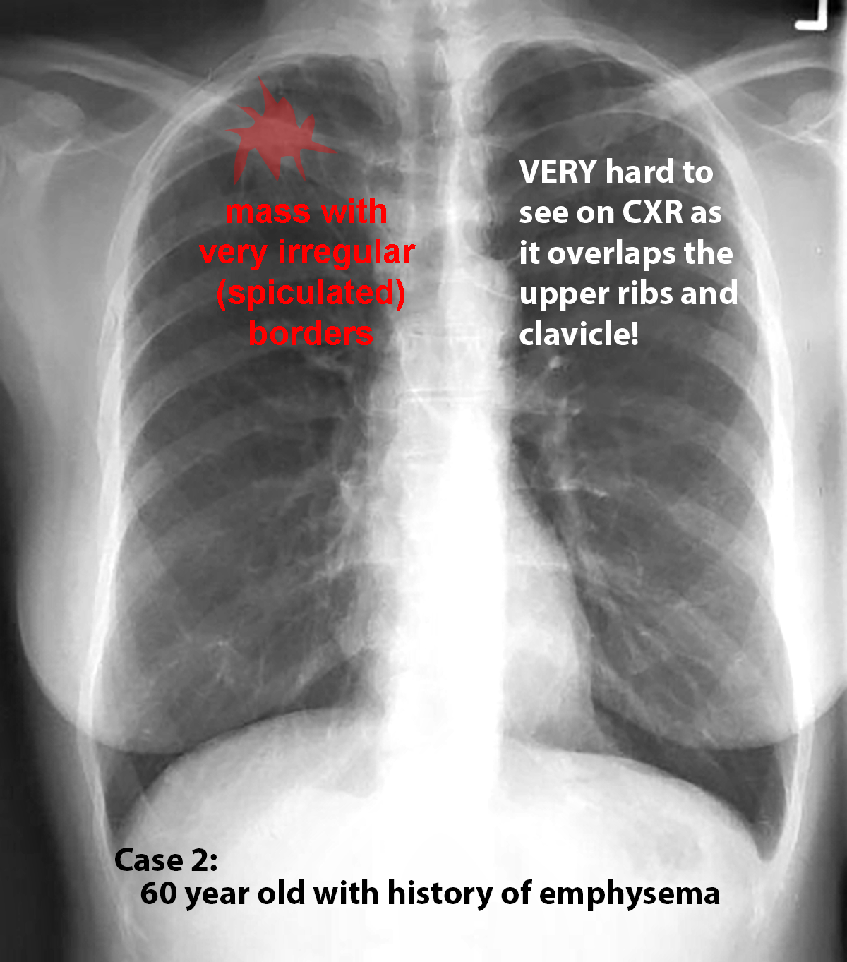
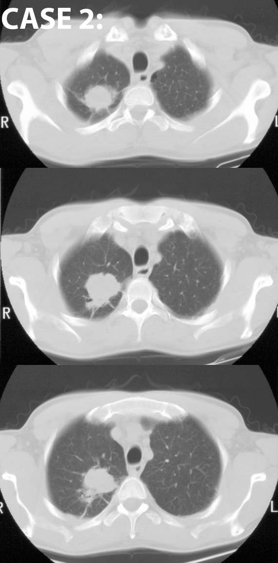
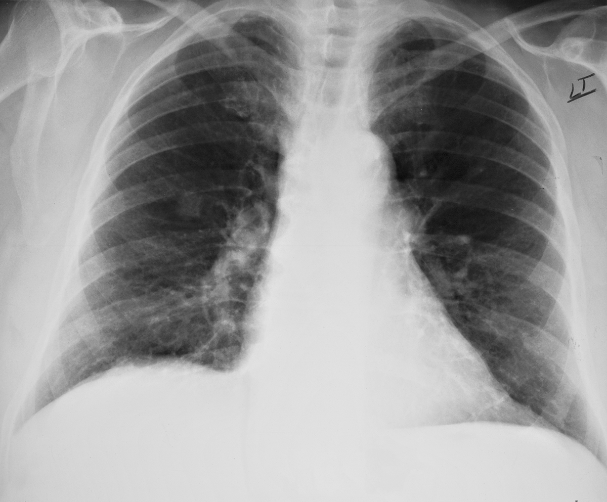
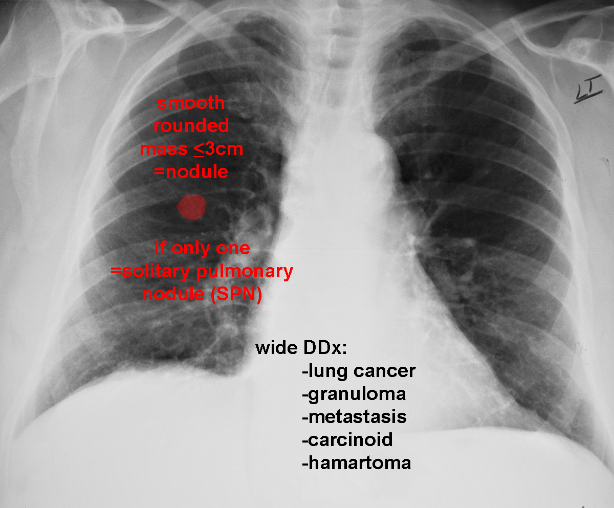
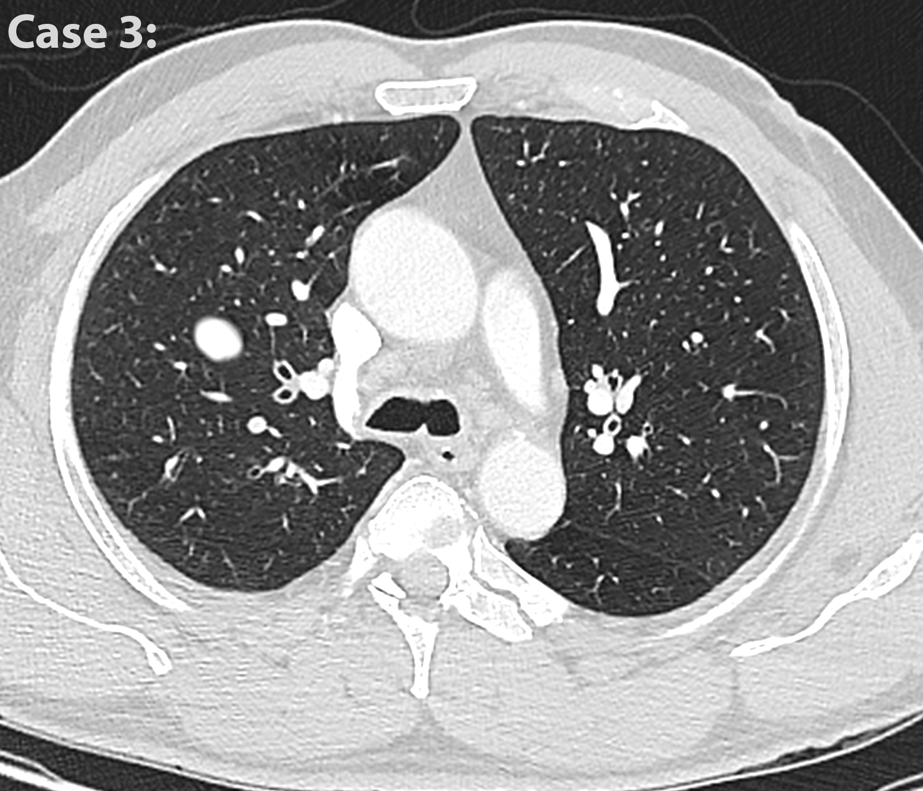
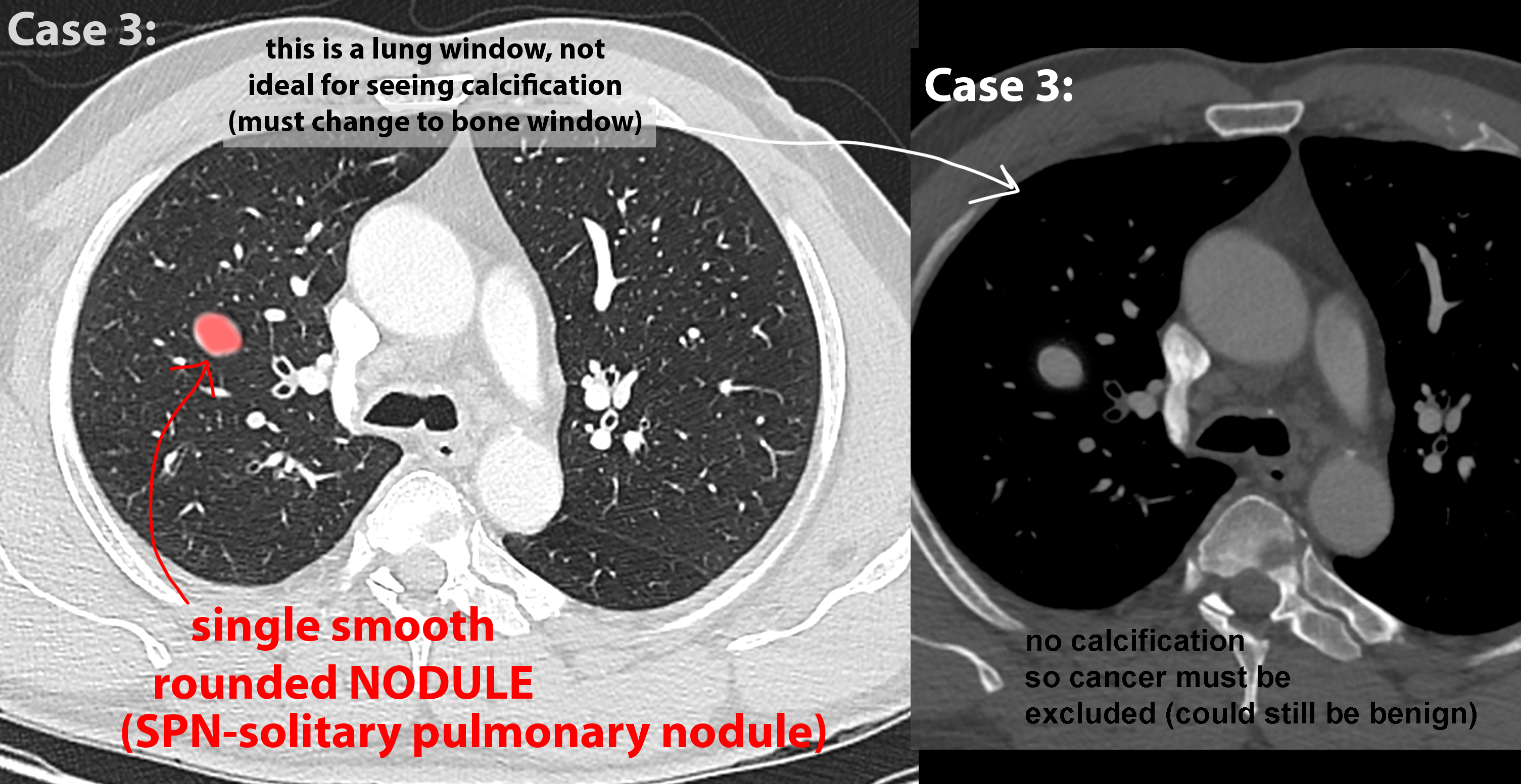
These three cases review imaging of lung cancer, and use of specific terminology to describe findings on chest radiography. By definition, 'nodule' refers to a rounded opacity that is under 3 cm in diameter. Anything else is called a 'mass'. Each case has a CT as well. Details of CT findings will be presented on the next page.
Further Explanation:
Case 1-lung cancer
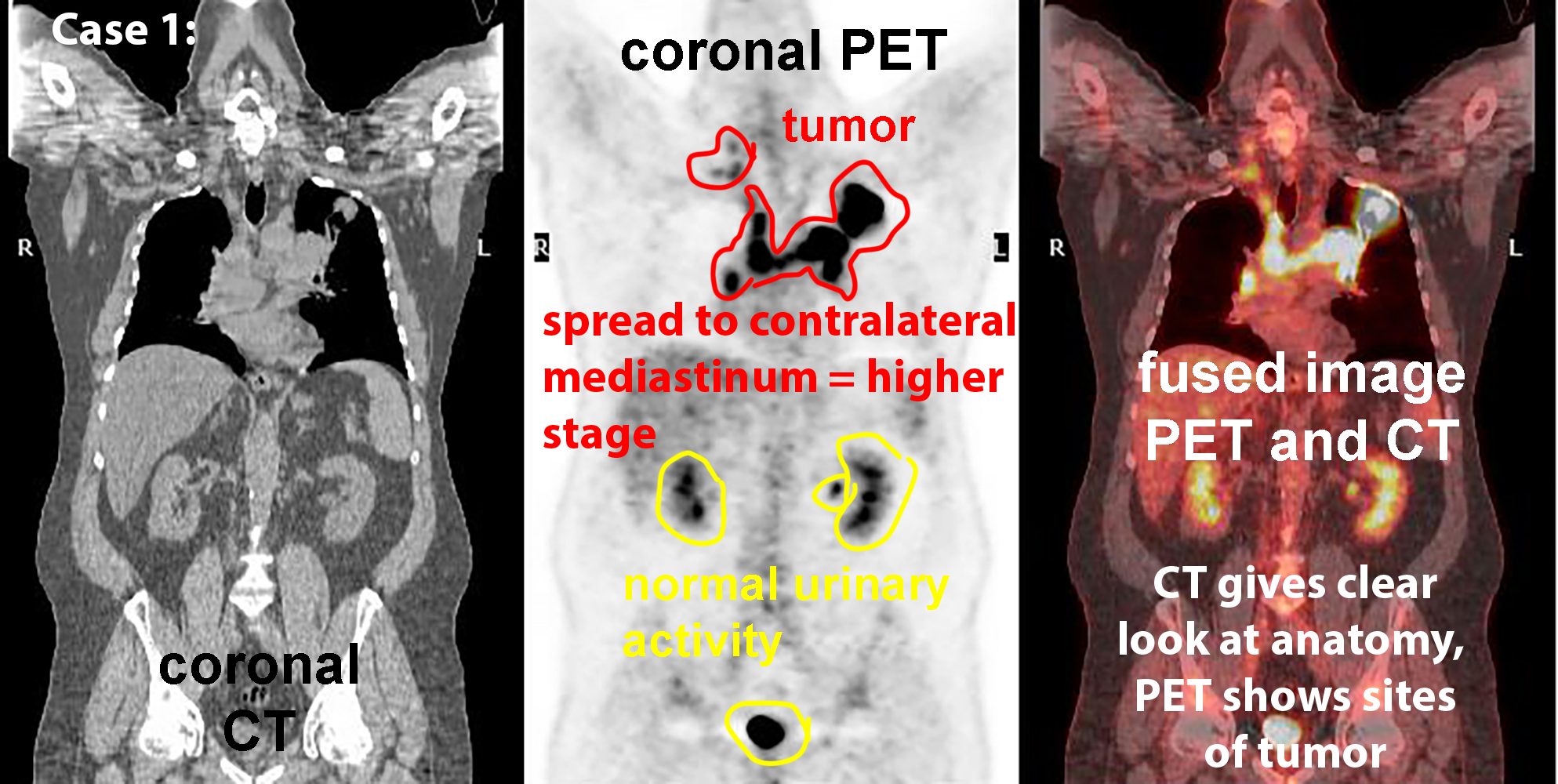
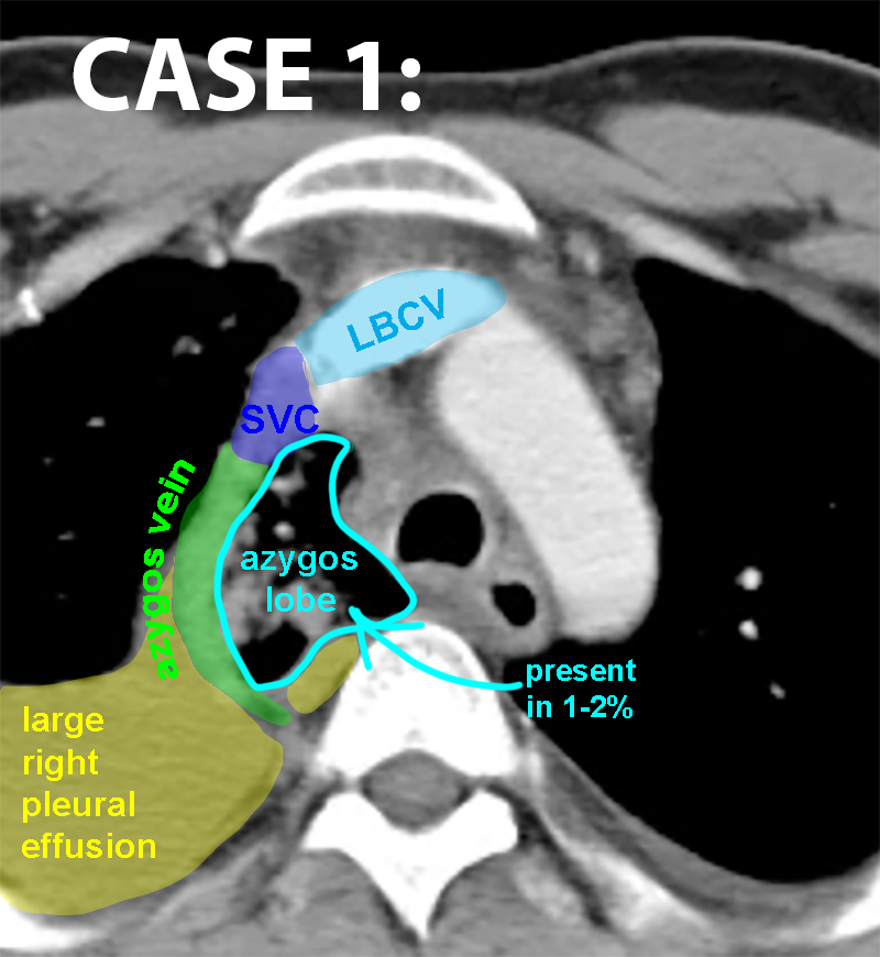
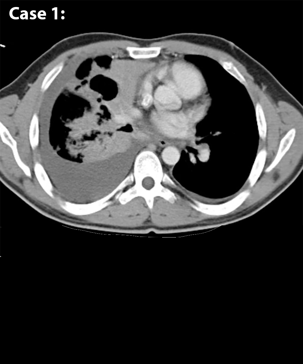
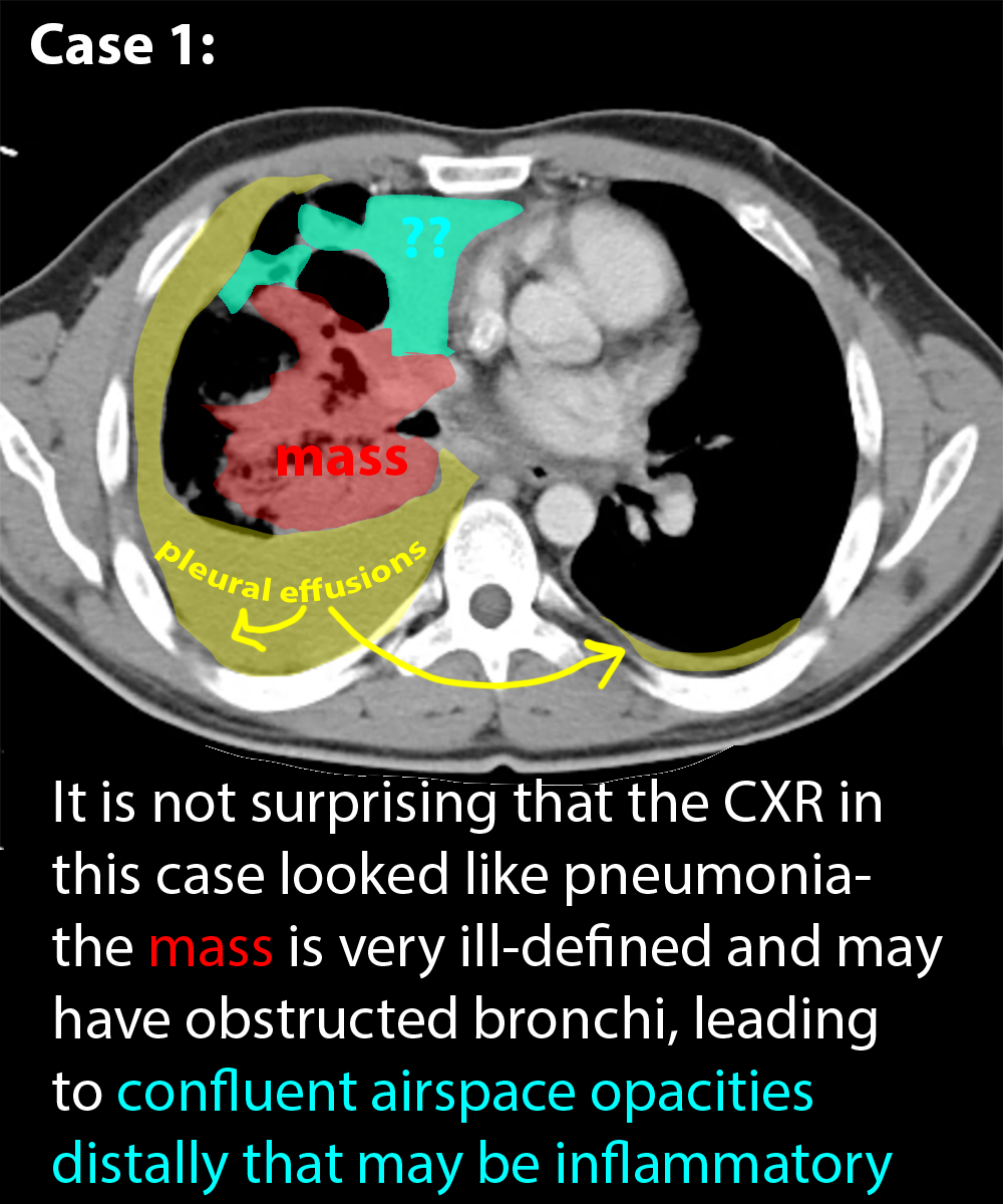
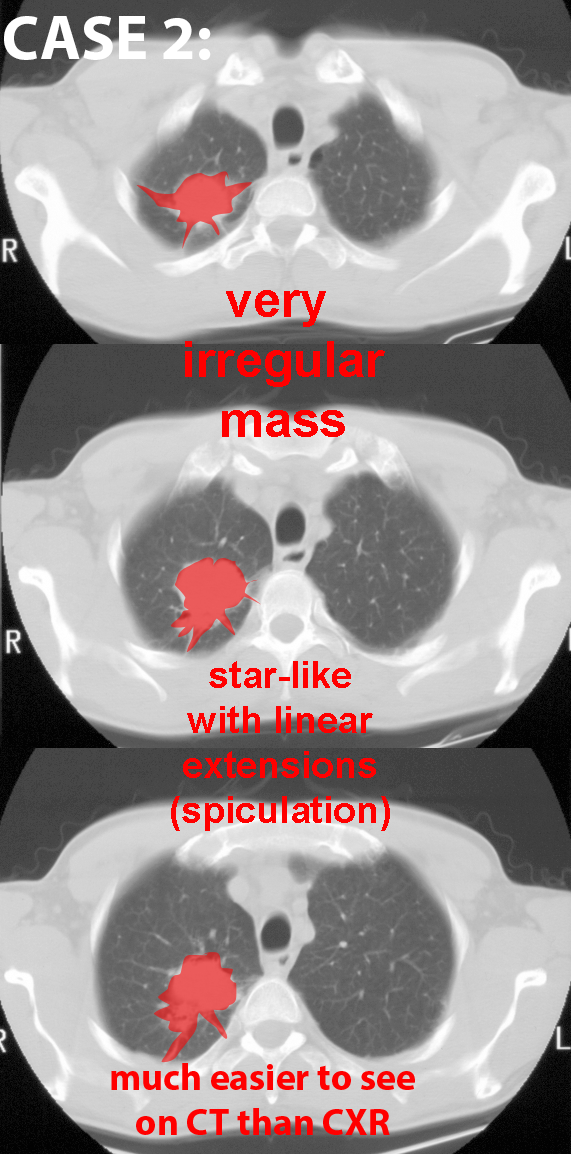
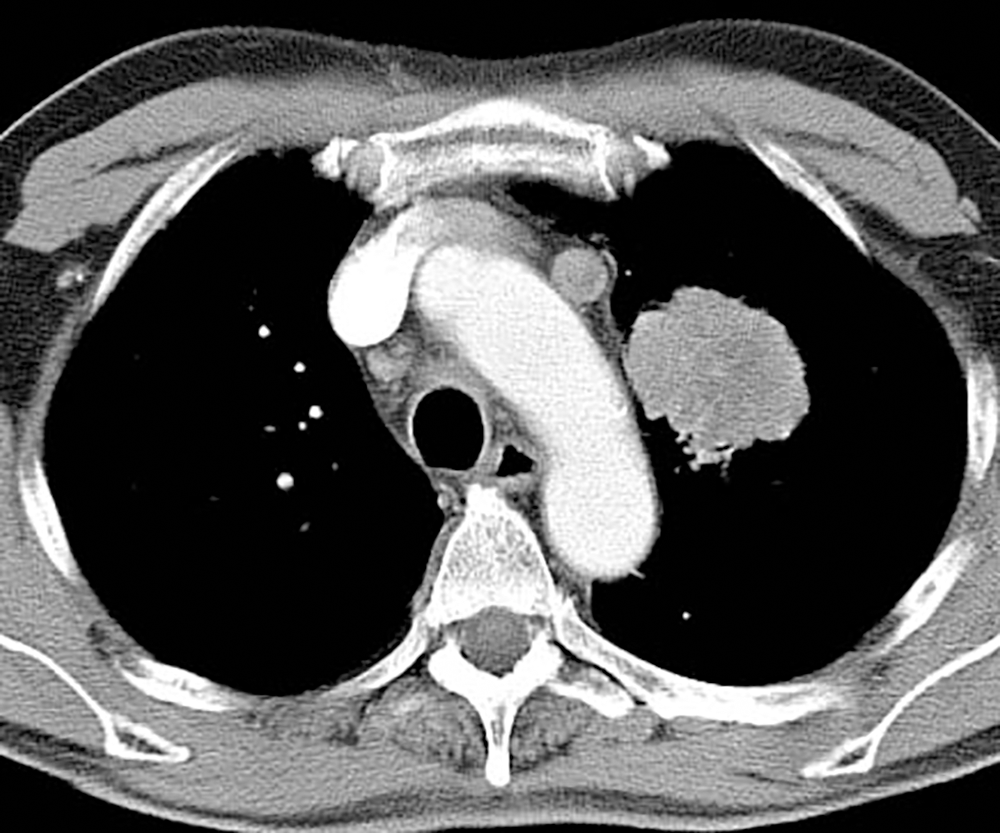
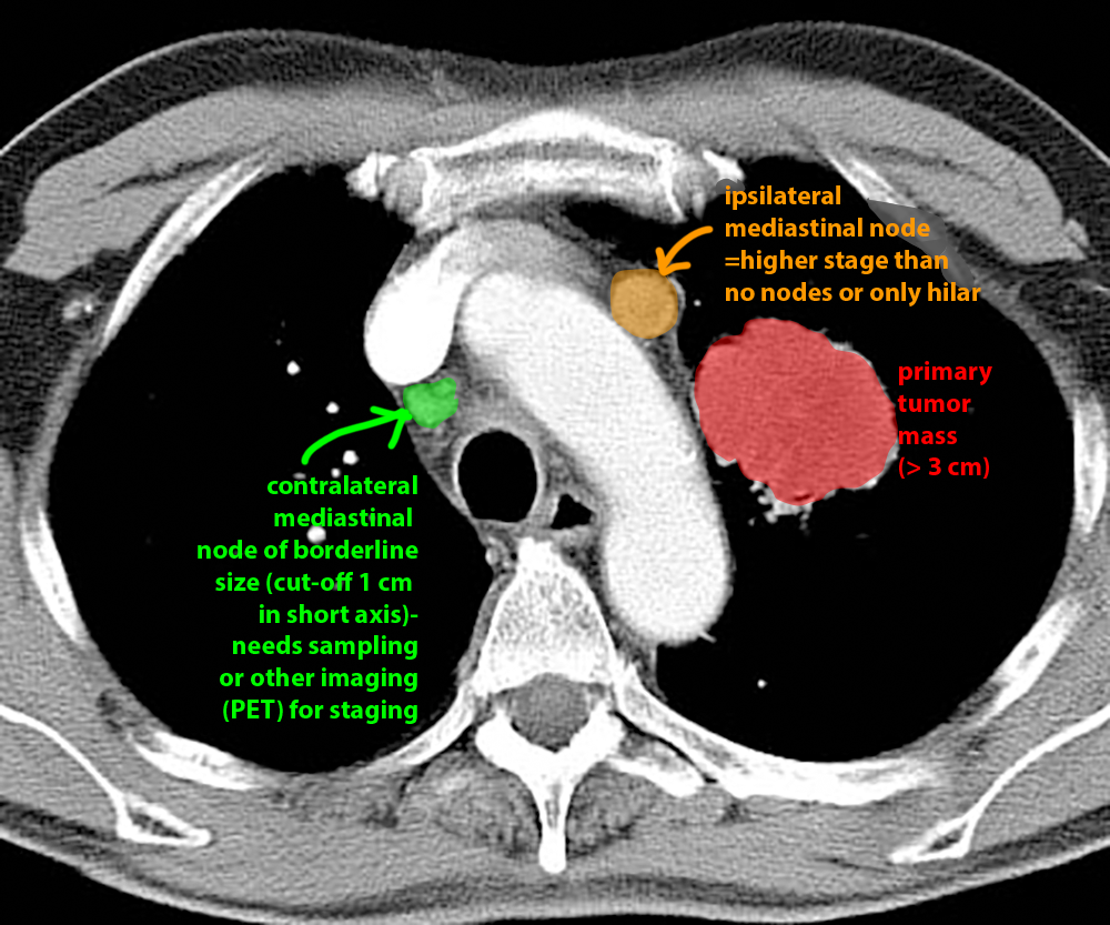
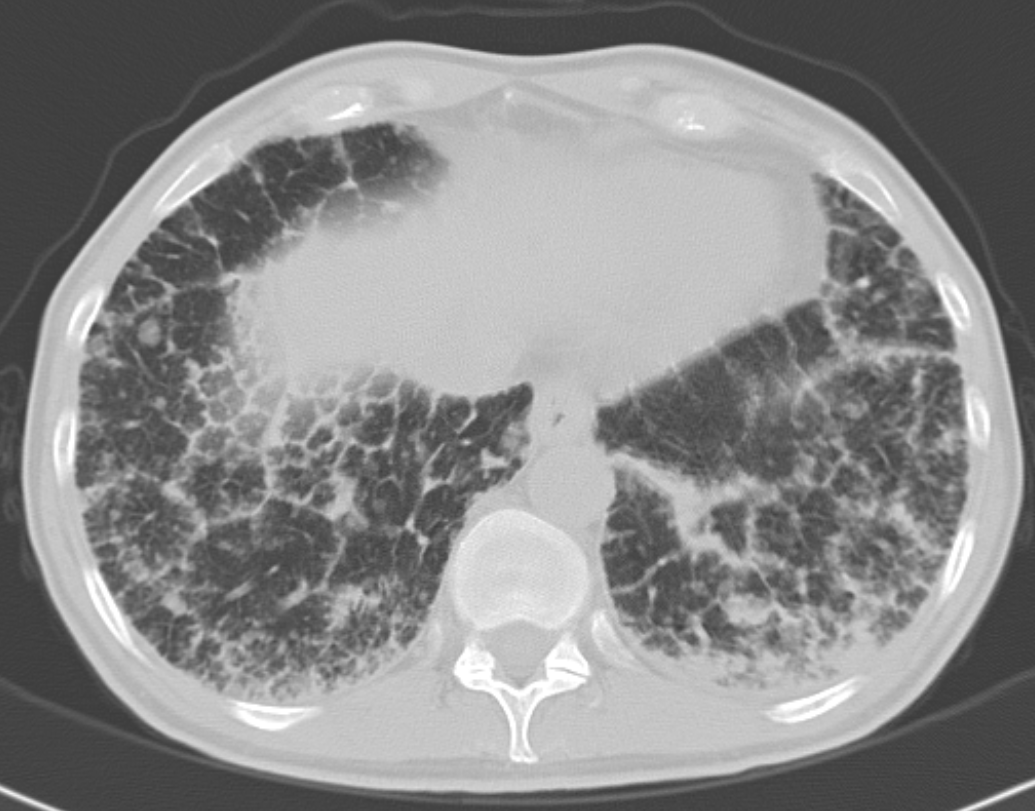
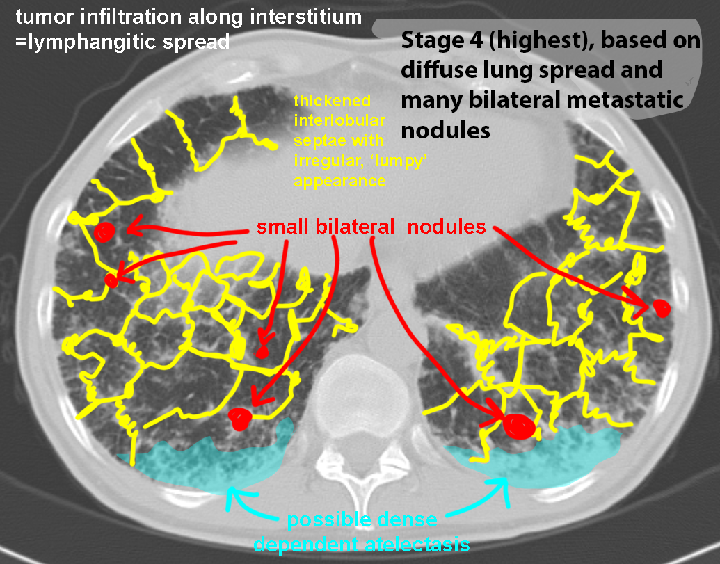
The labeled CT scans for the previous patients are shown here, along with other examples of the appearance of lung cancer on CT. Spread in lung cancer tends to be from a primary site to the hilum, then the ipsilateral mediastinum, then the contralateral mediastinum, and then systemic, although not all tumors follow this orderly path. But the farther the tumor has spread in the nodes, the higher the stage.
Further Explanation:
Case 1-lung cancer
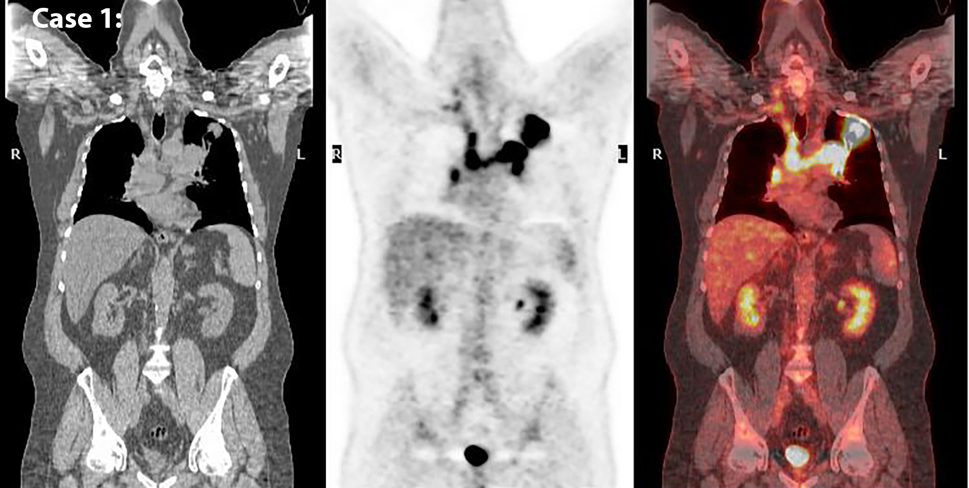
Radiography and CT are limited to anatomic information about tumors, which is not definite in terms of stage. For accurate staging of lung cancer, either biopsy or imaging that is more specific is needed. PET is a nuclear medicine modality with good specificity for many tumors, including lung cancer.
Further Explanation:
