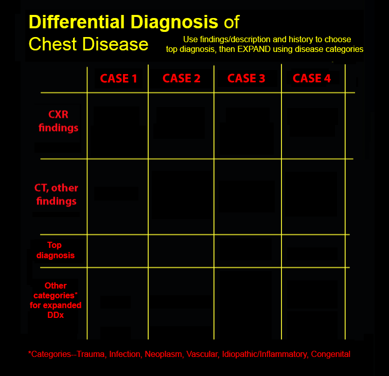
In this session, you will integrate your knowledge of chest imaging findings, including appearance of alveolar vs interstitial disease, with history to arrive at your top diagnosis for each case. Then, use general categories of disease to EXPAND your differential to include other possibilities. (In some rare cases, there is really only one reasonable diagnosis to consider).
After completion of this self-directed learning module on Imaging and Differential Diagnosis of Lung Disease, students will be able to:
- Predict the likeliest pathogens from among the following: community-acquired pneumonia, COVID, TB, atypical pneumonia, Legionella, based on imaging findings, and consider any alternative diagnoses
- Predict the likeliest diagnosis from among the following: idiopathic pulmonary fibrosis, scleroderma, asbestosis, silicosis and sarcoidosis, based on imaging findings, and consider any alternative diagnoses
- For patients with likely lung tumor on imaging, identify and describe the significance of adenopathy and calcification, and consider any alternative diagnoses
- Recognize and describe the significance of the following imaging findings: enlarged pulmonary artery (central vs peripheral), pseudo tumor appearance on radiography, air-trapping and ground glass opacity
