


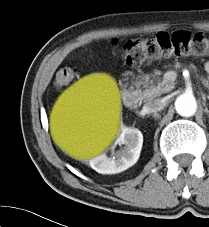
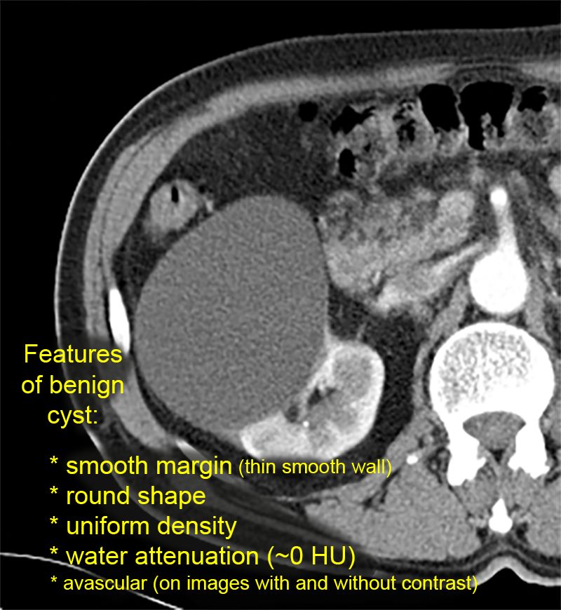
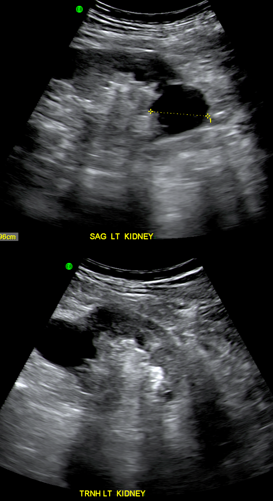
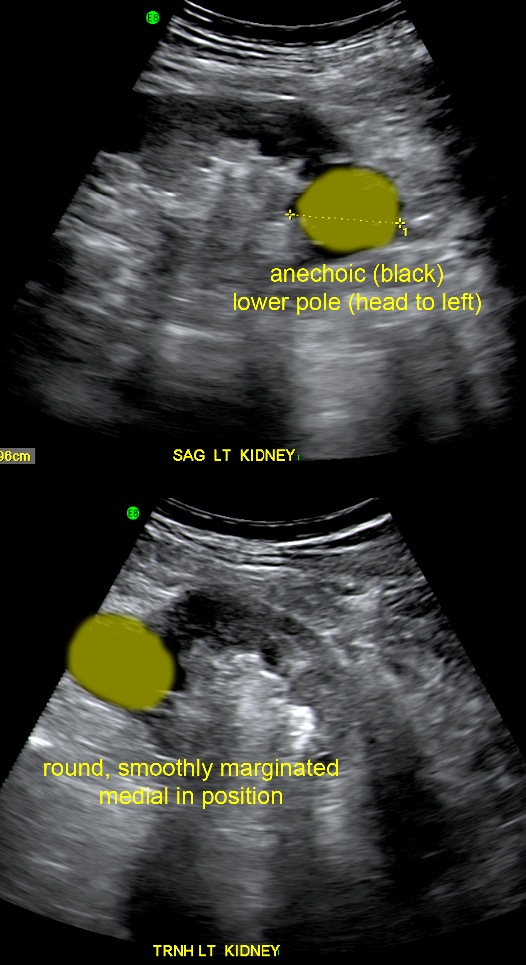
Stones and Cysts
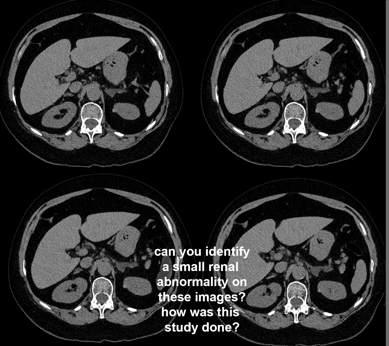
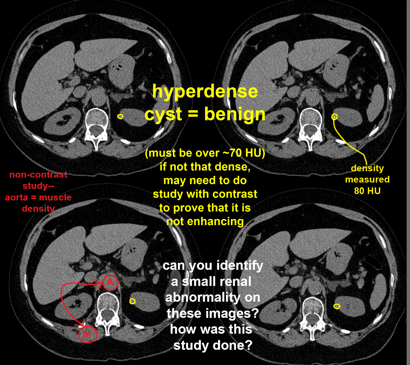
This set of images demonstrate features of a very common finding on radiologic studies of the kidneys: renal cysts. They are present in about 5% of the general population and about 30% of patients over the age of 70. It is important to recognize their typical imaging features, since they do not need further workup in most cases.
Further Explanation:
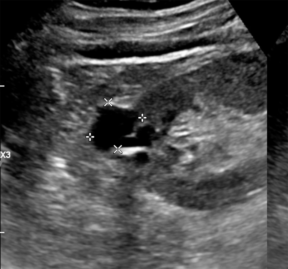
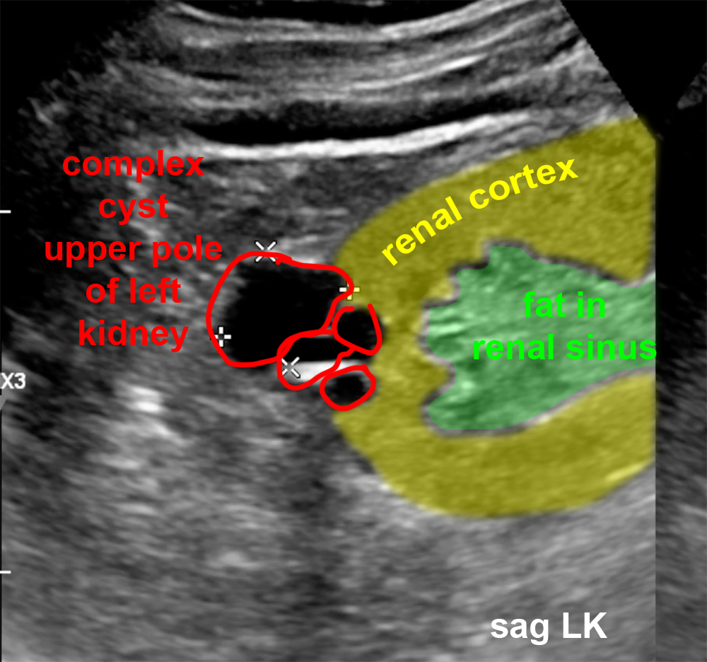
Stones and Cysts
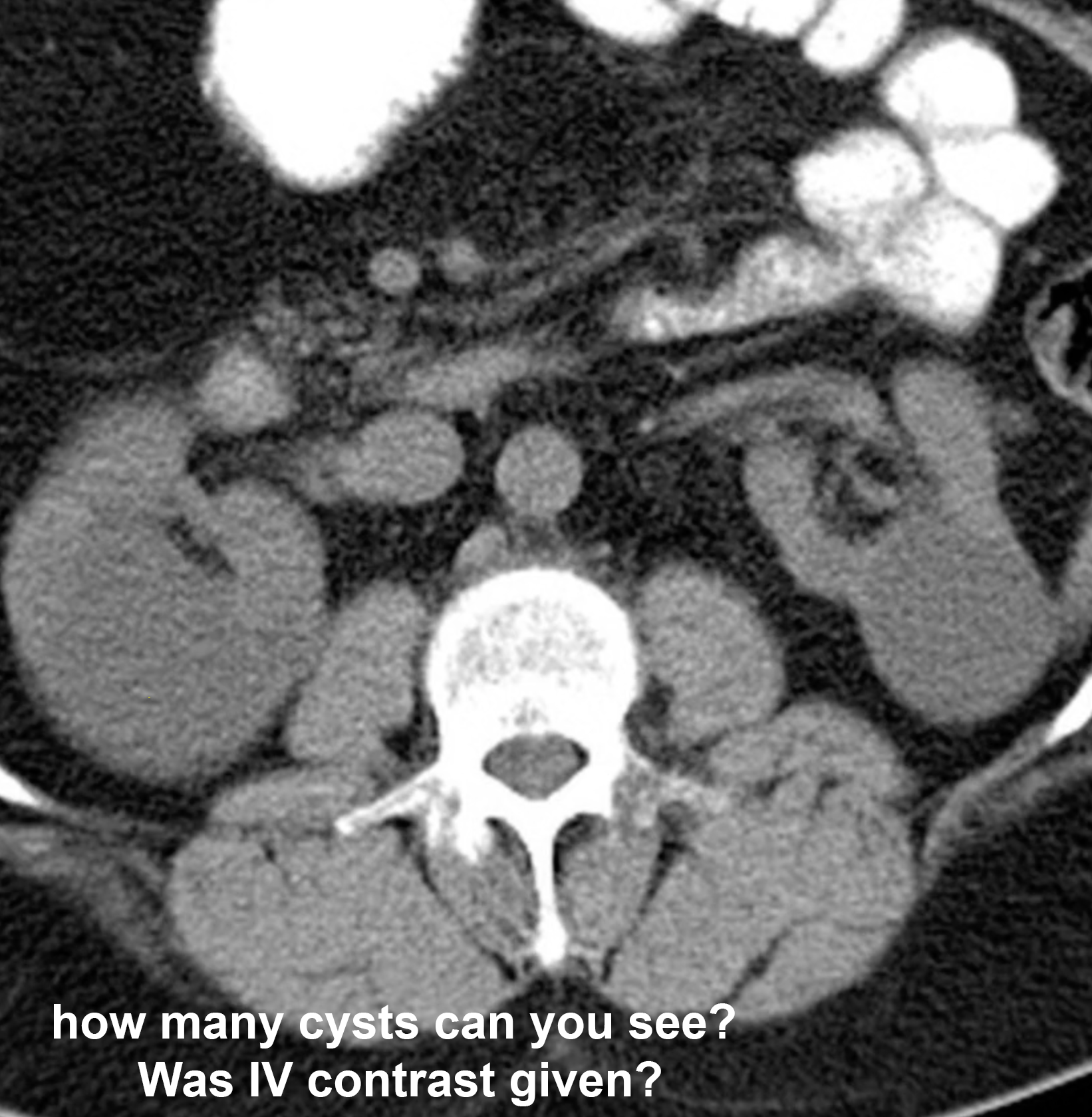
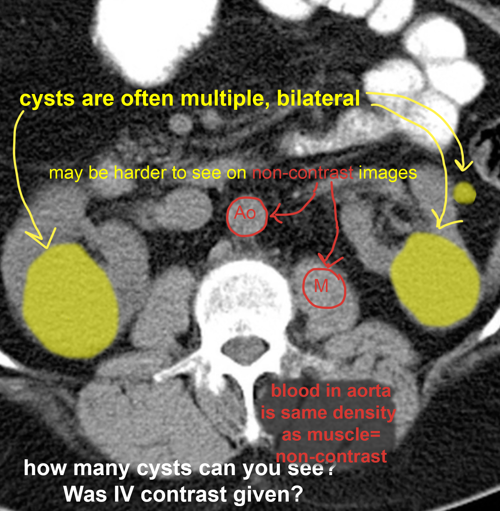
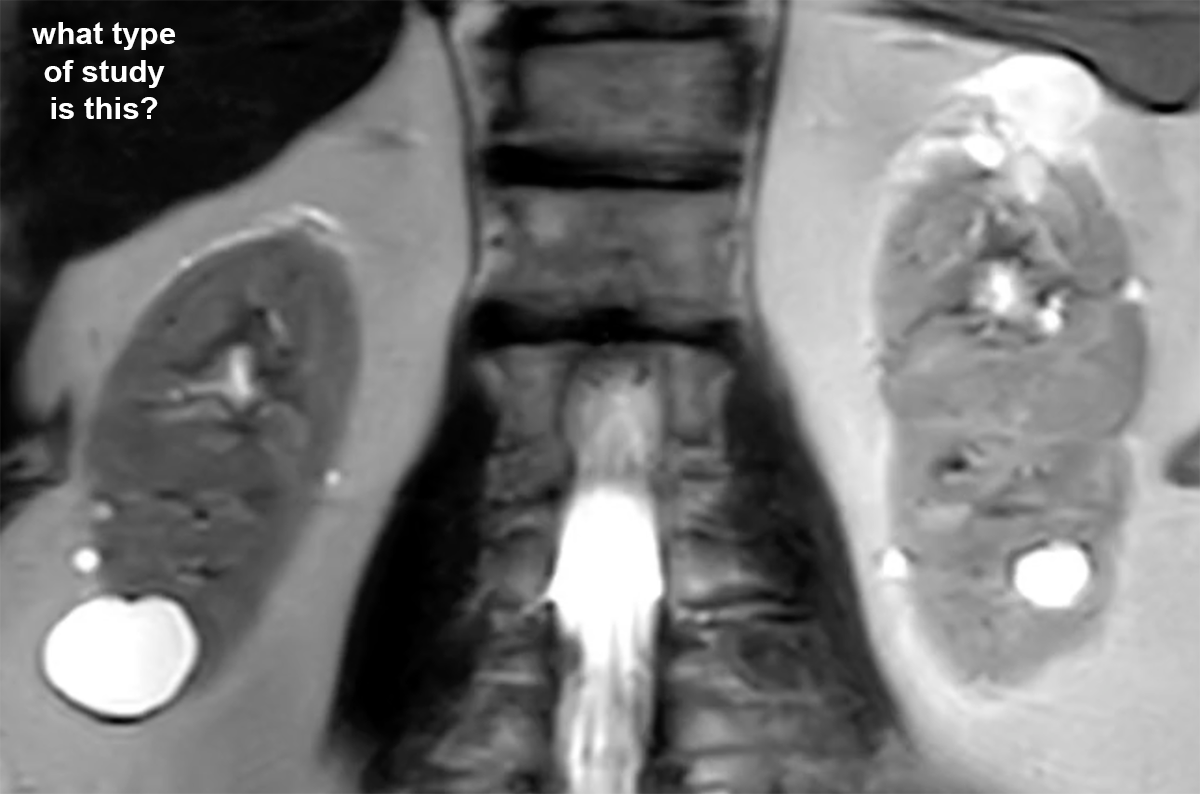
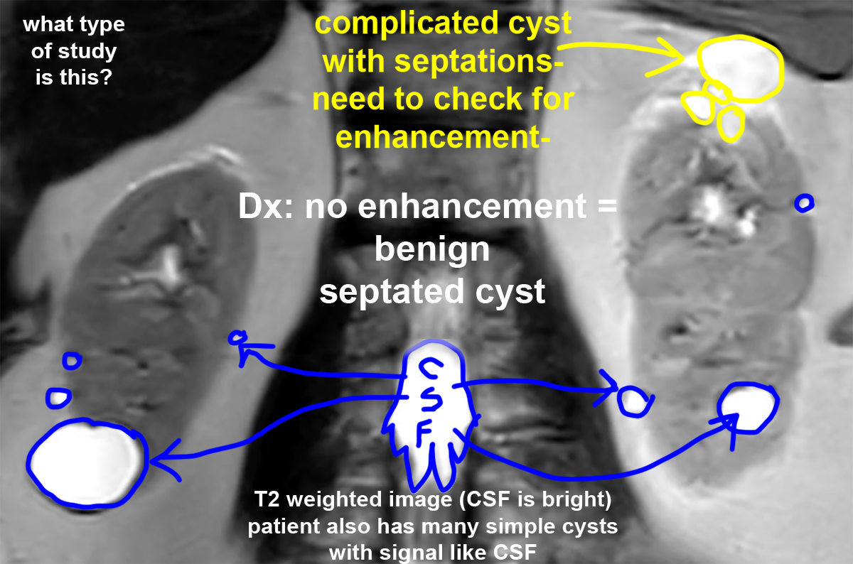
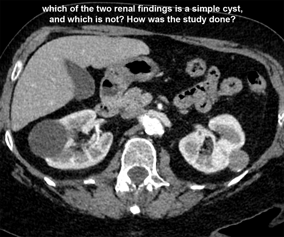
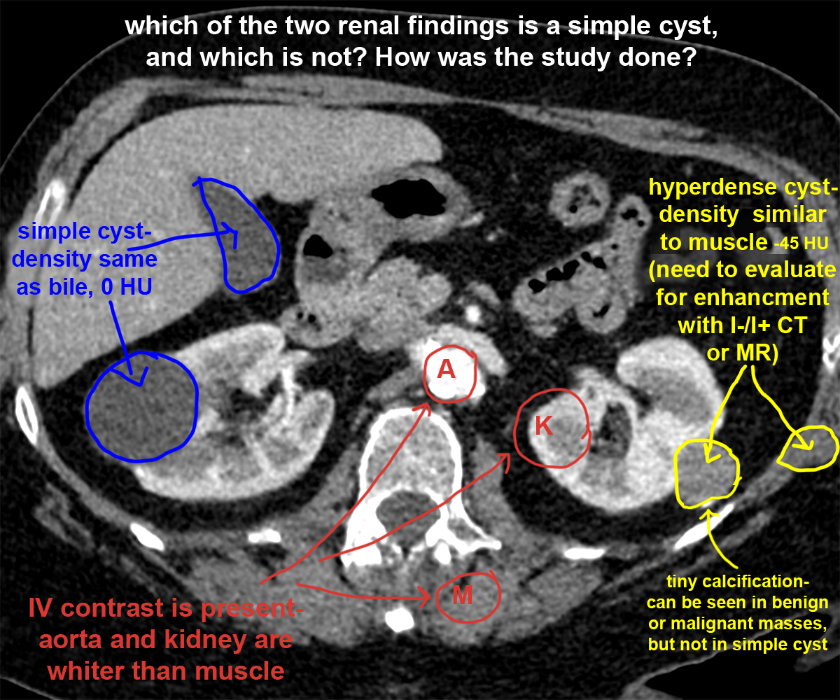
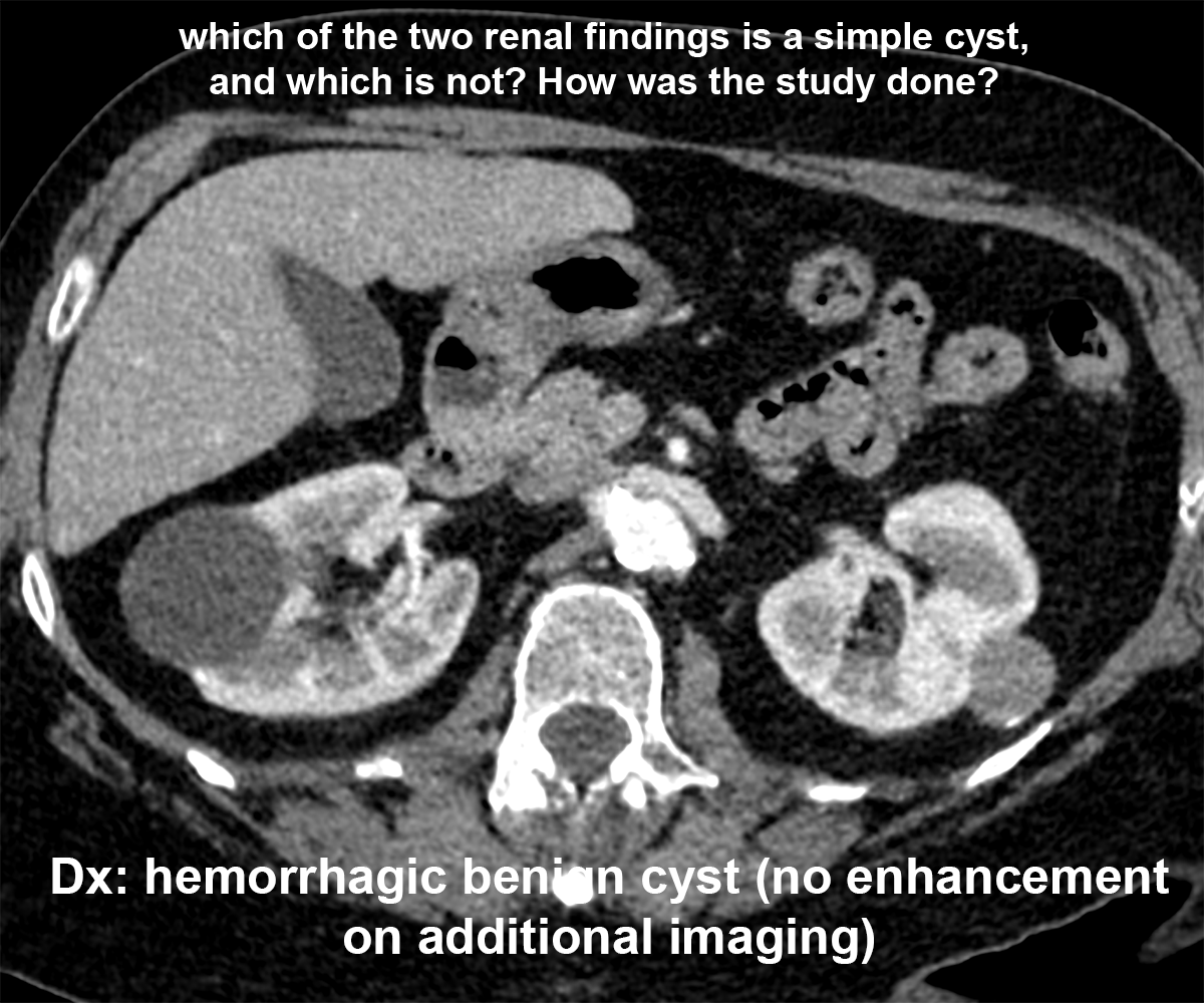
Sometimes things are not that simple. Some patients have renal abnormalities that have some features of simple cysts but are not perfectly round and homogeneous. This page demonstrates imaging features of complex cystic lesions of the kidney, which generally need additional imaging to determine whether they require biopsy, followup, or can be considered benign.
Further Explanation:
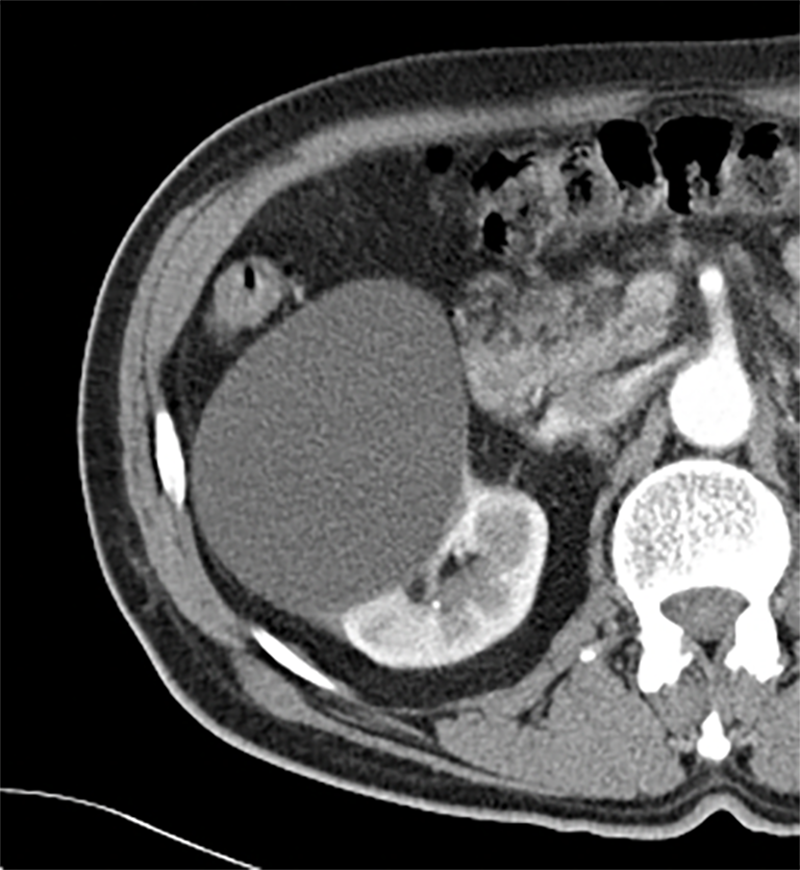
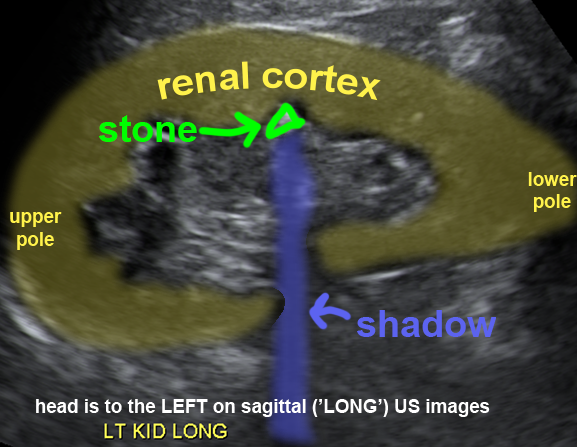
Stones and Cysts
Nephrolithiasis is a common indication for various types of imaging, most often CT and ultrasound. Imaging can influence management, since small stones may pass with conservative treatment (IV fluids and pain relief), while larger stones may require various types of intervention.
Further Explanation:
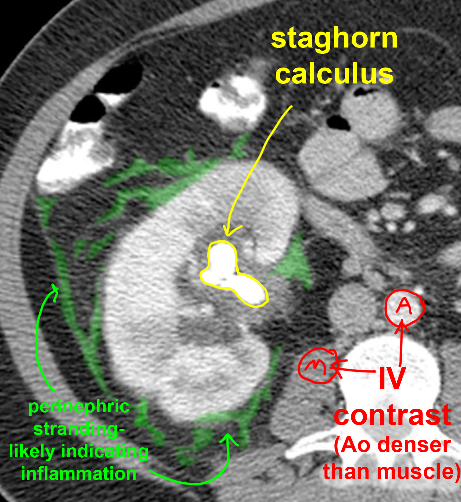
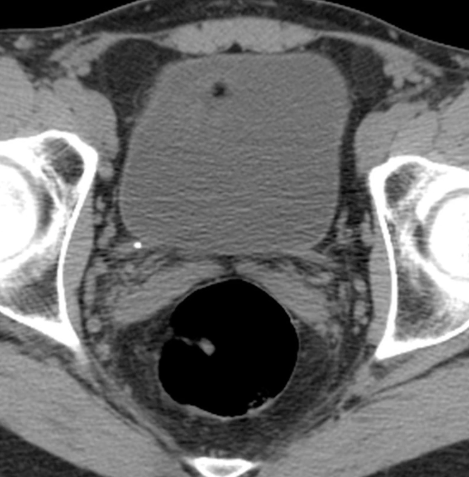
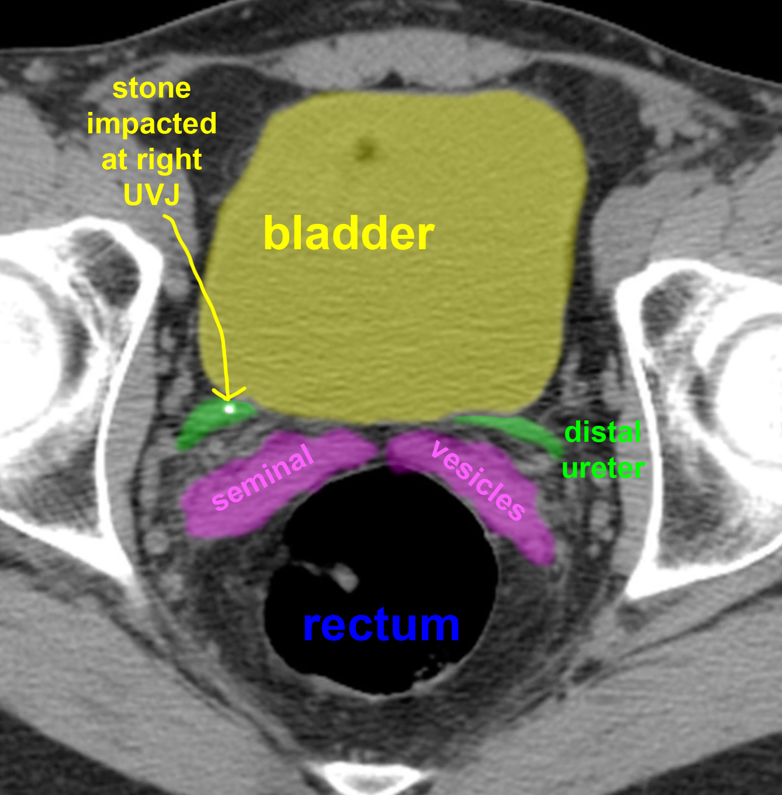
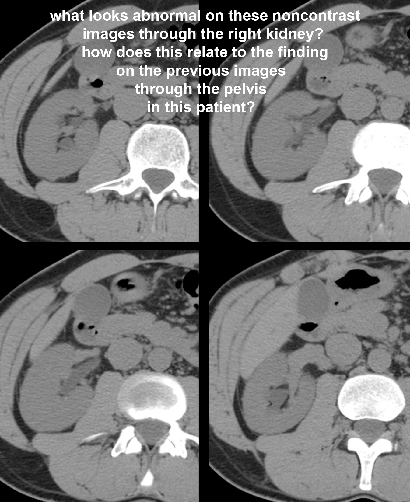
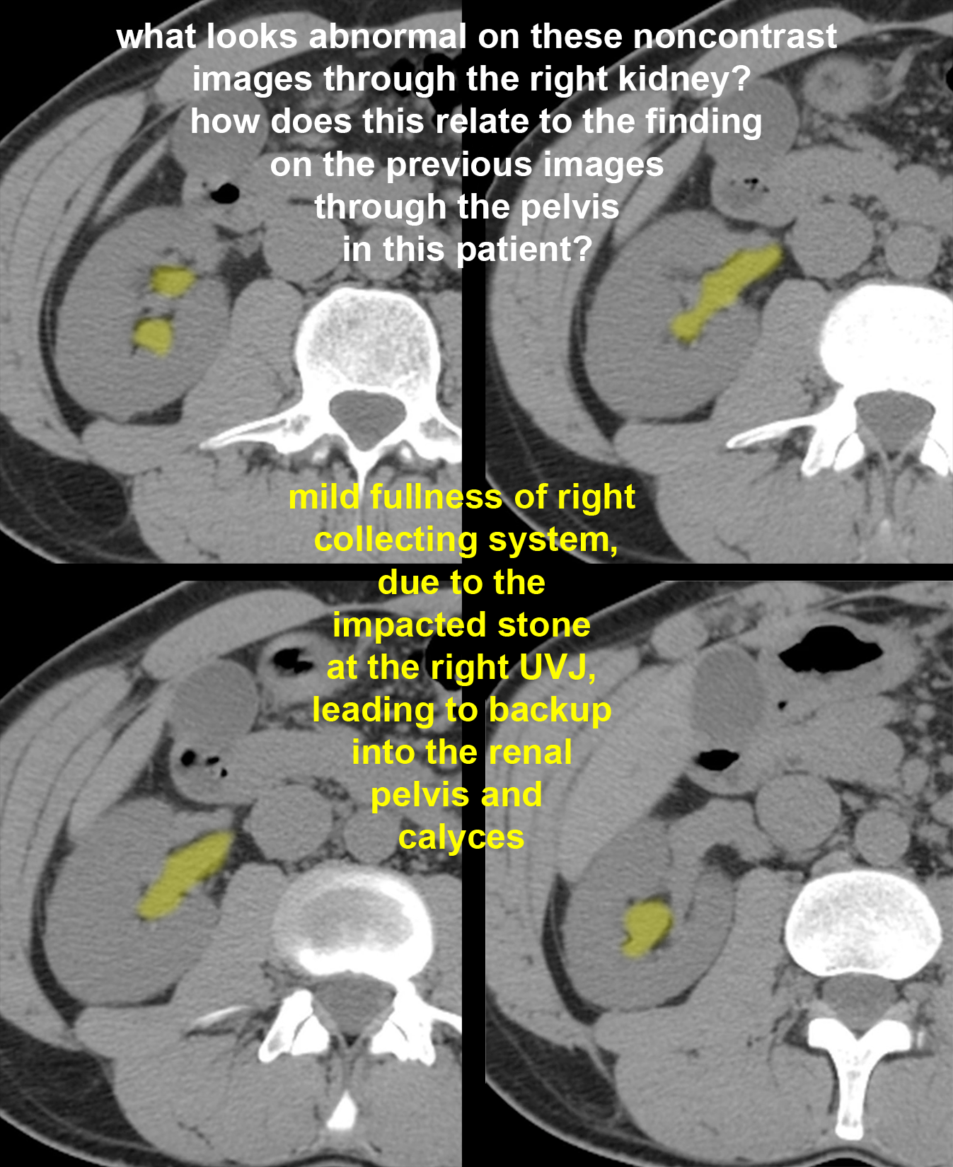
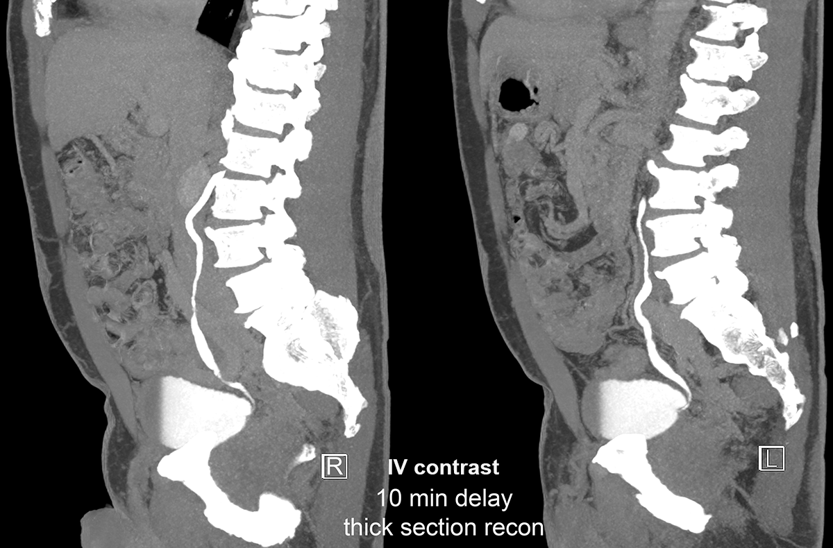
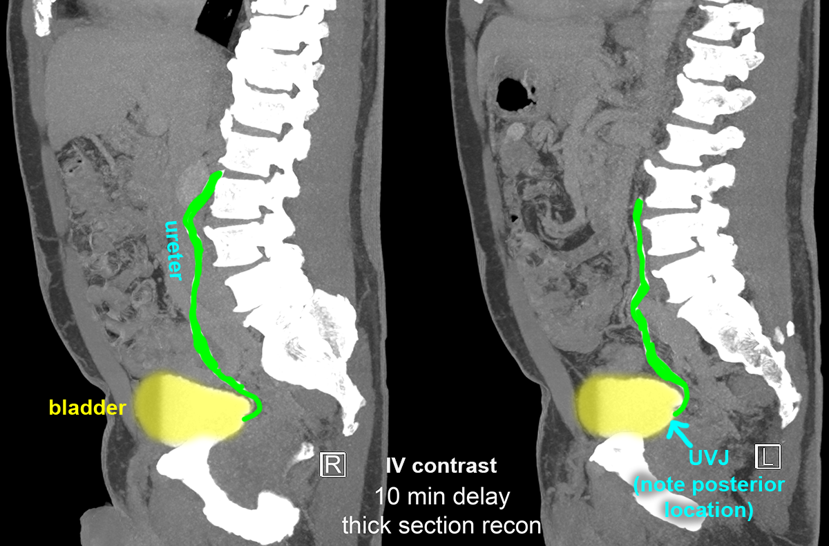
Stones and Cysts
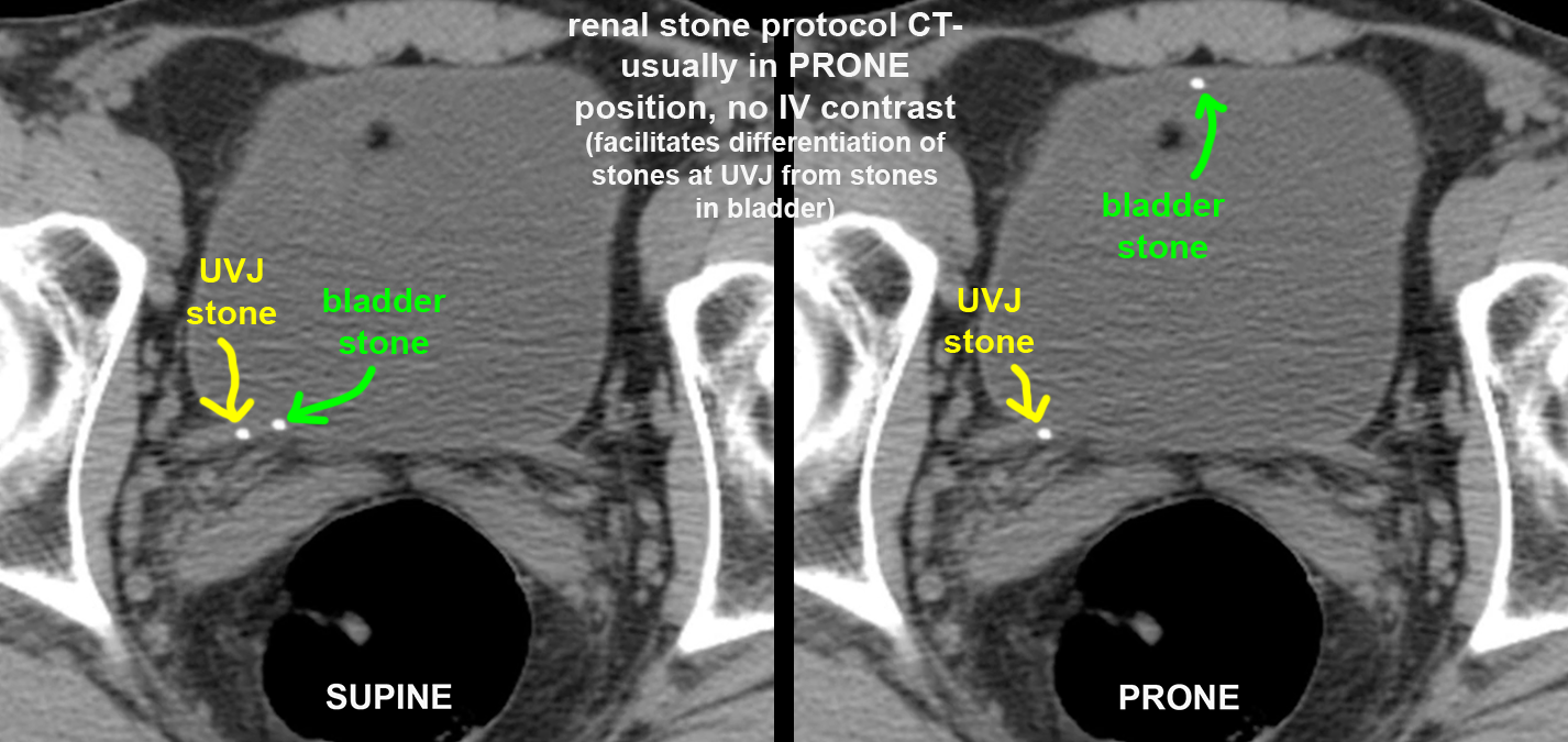
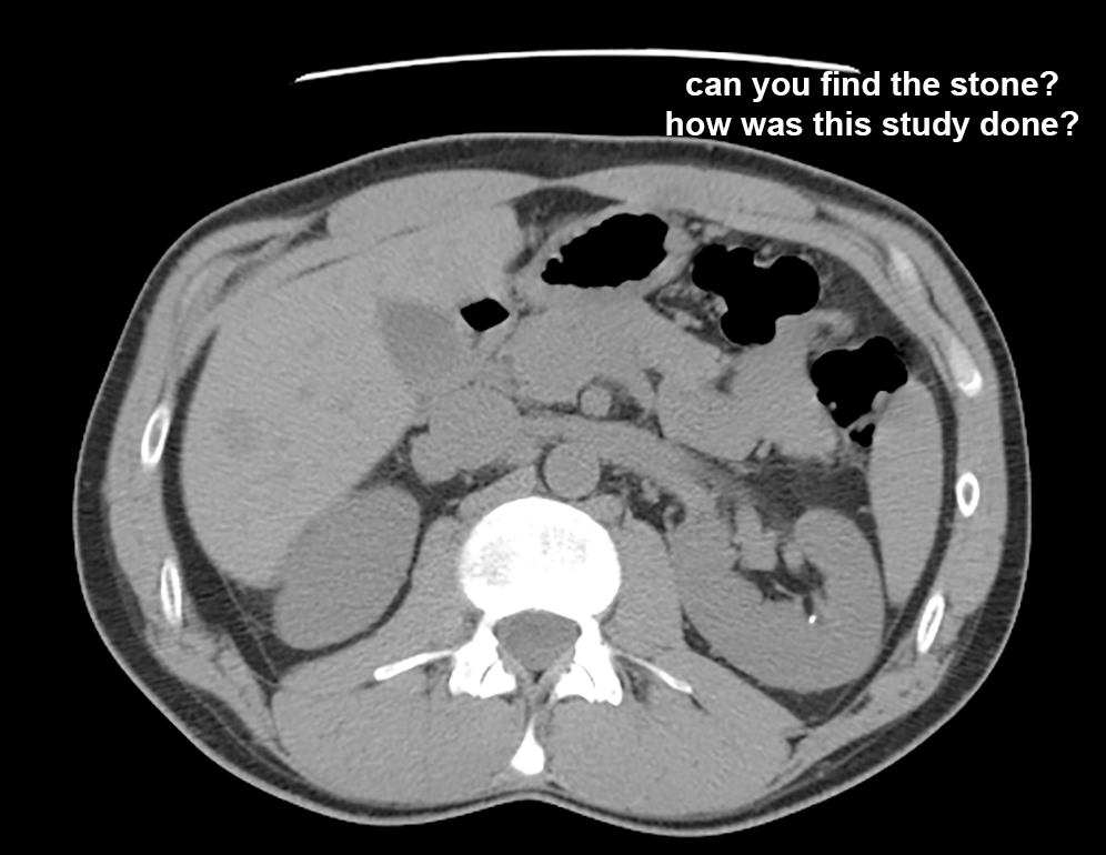
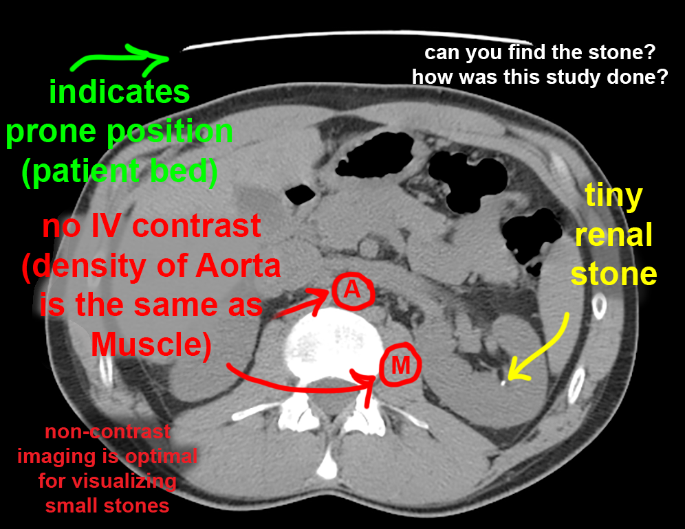
Since renal stones are a common indication for CT imaging (visualizing the ureters better in most cases than with ultrasound), it is important to understand how an optimal CT should be done in this setting. In fact, most radiology departments have a defined 'renal stone protocol CT' for this clinical setting. The cases on this page illustrate a common set of parameters for this protocol. Start by trying to identify the stone on the CT images through the pelvis.
Further Explanation: