

Imaging of Renal Masses
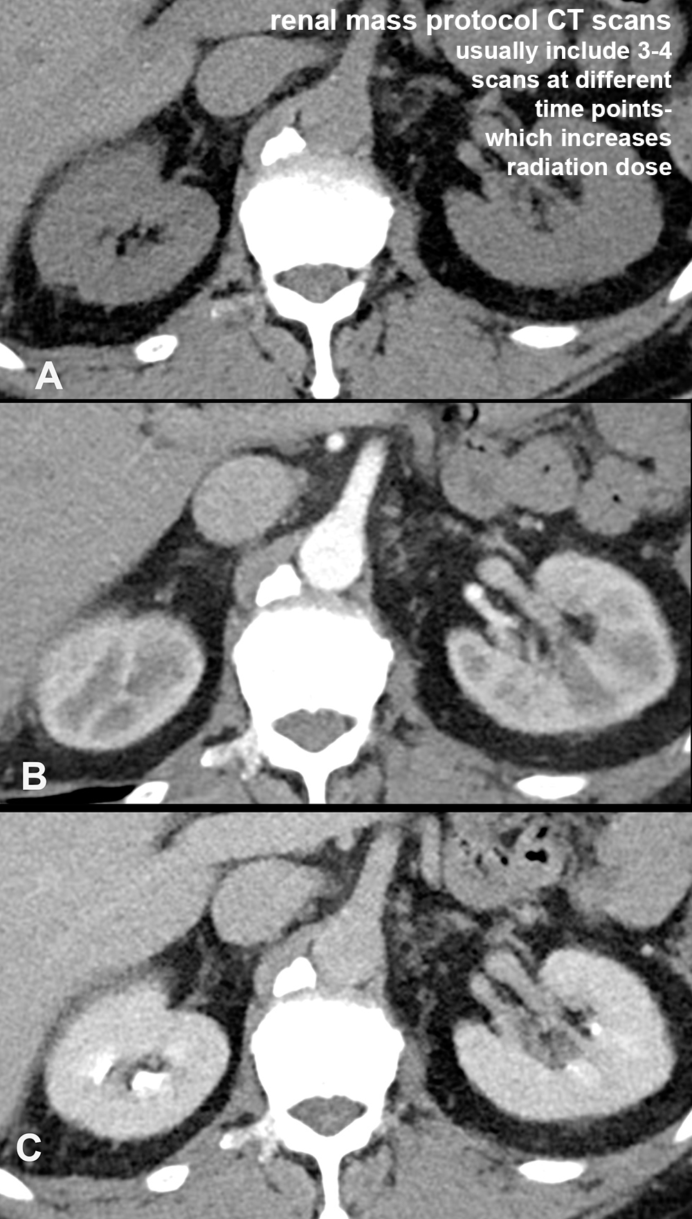
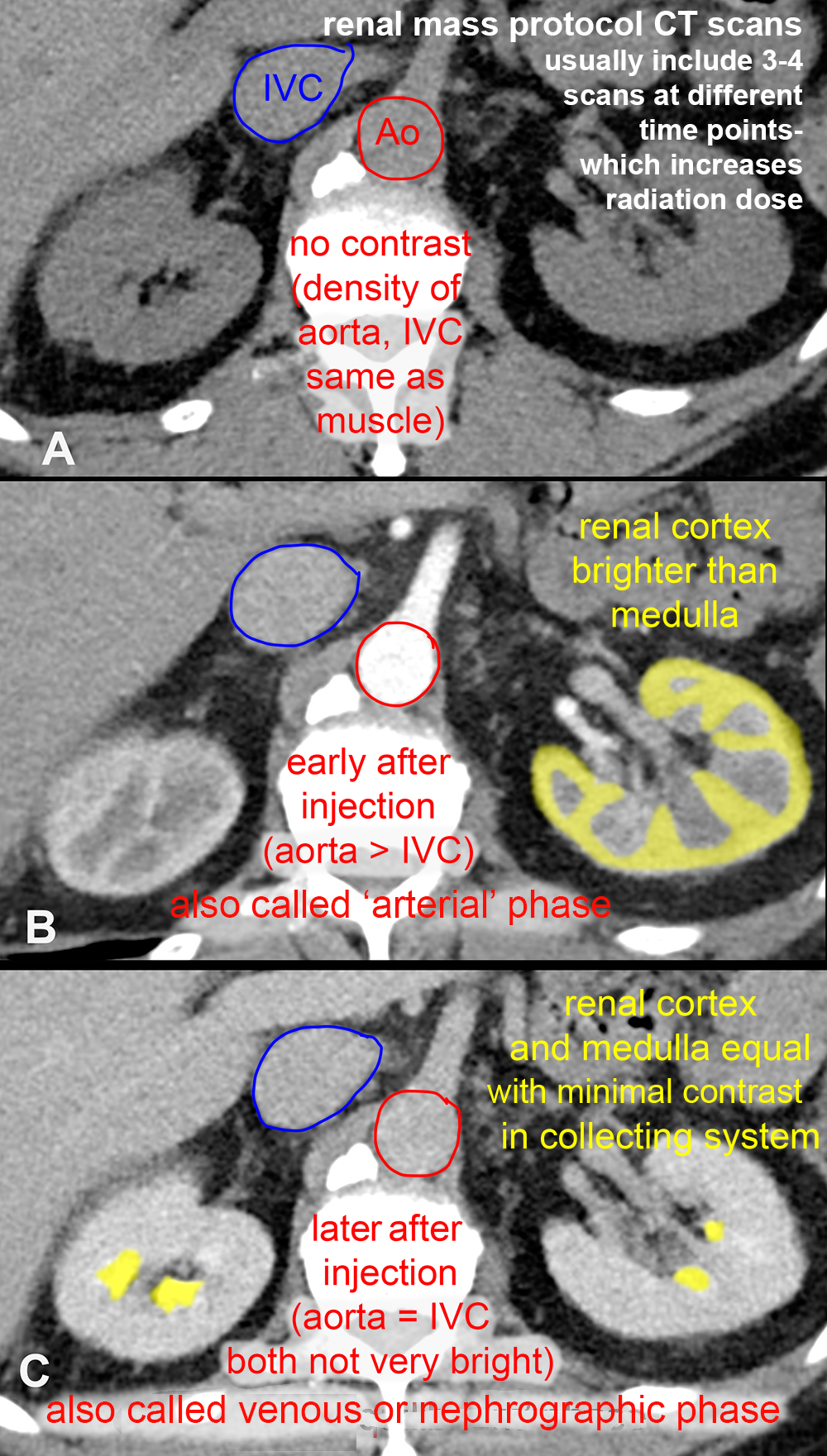
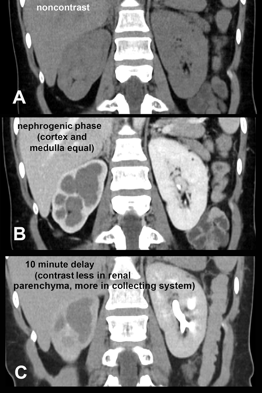
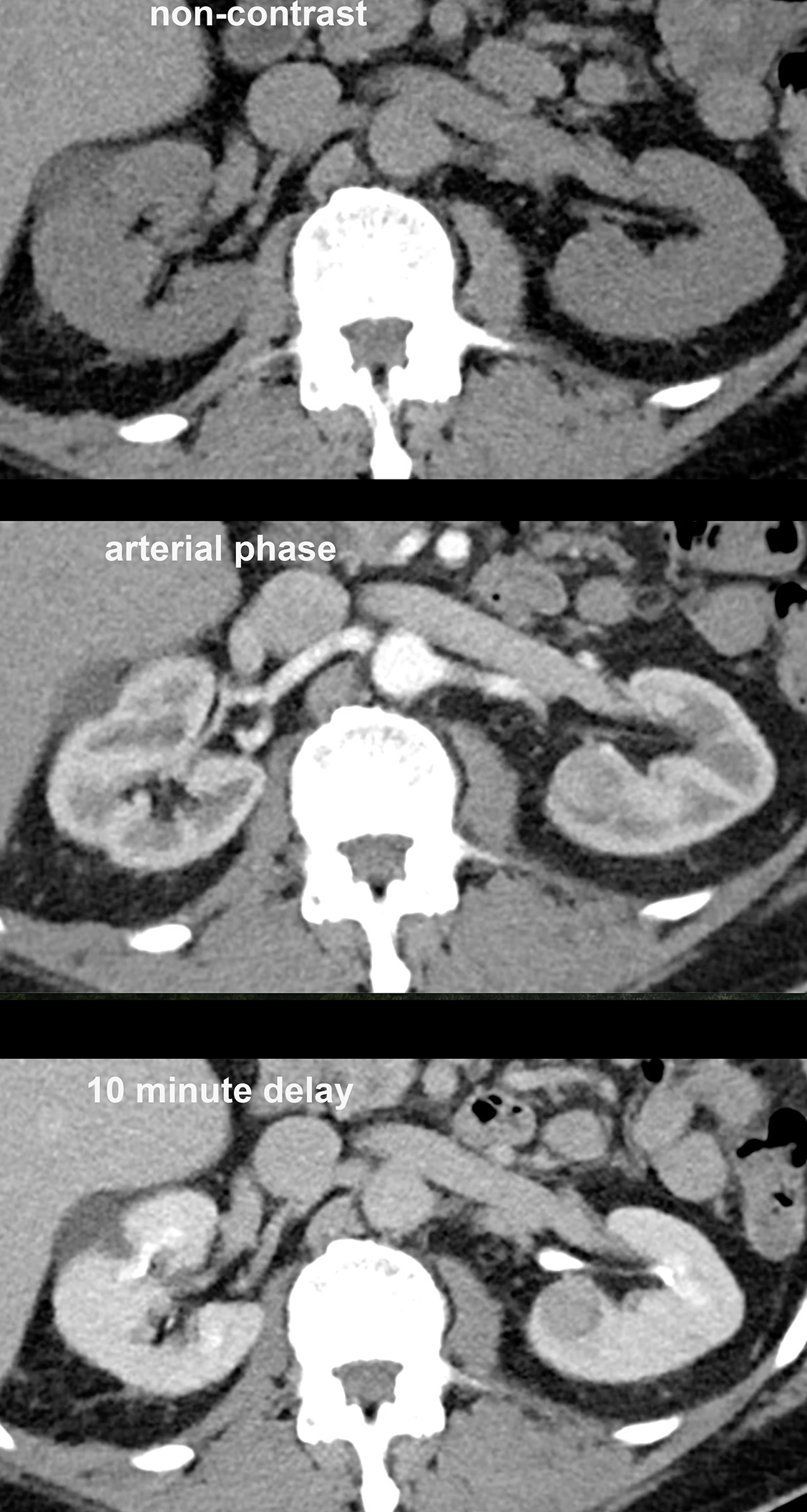
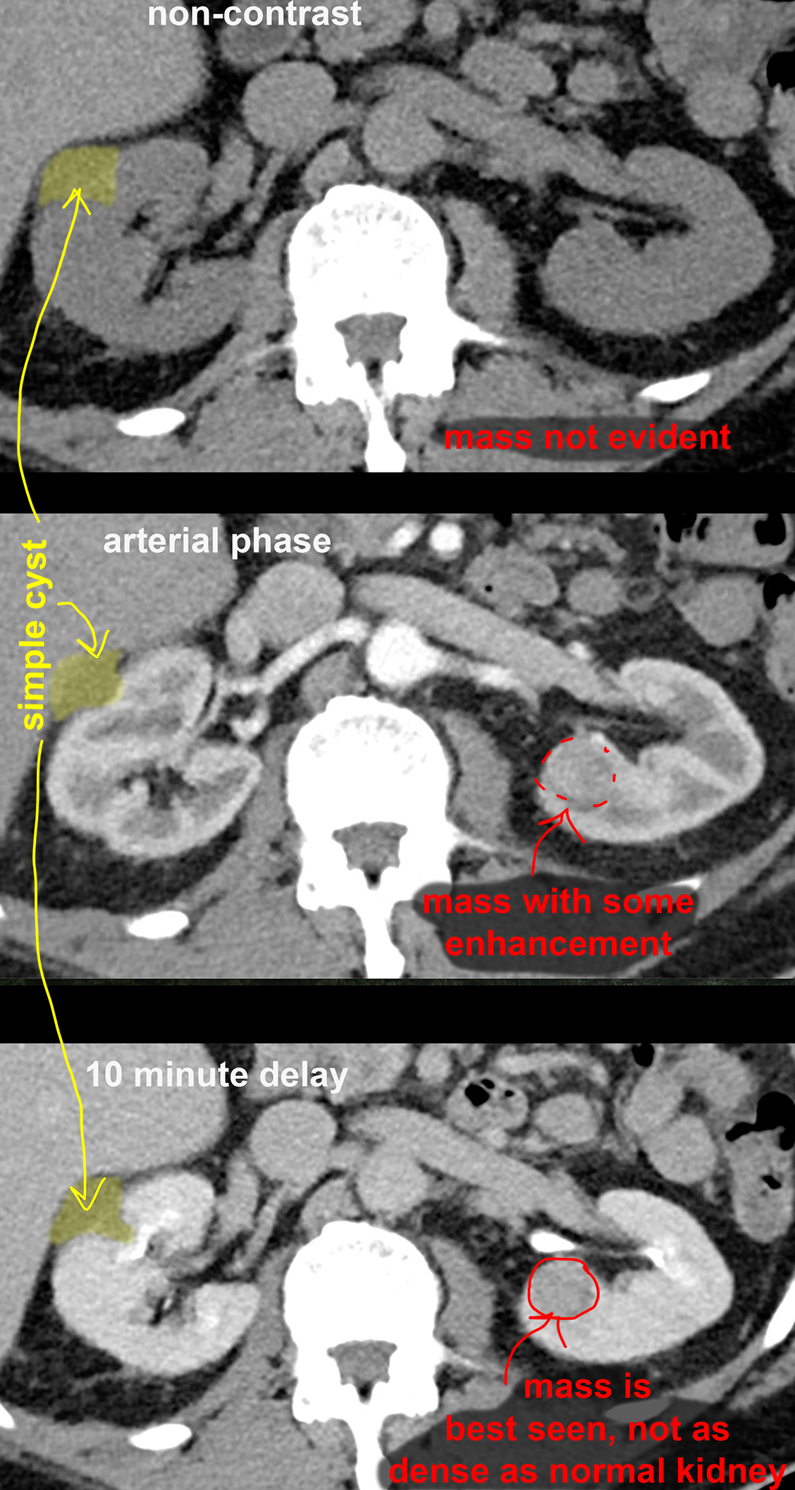
One of the distinguishing features of many renal masses is their vascularity, particularly when compared to avascular processes like cysts. So imaging of renal masses often includes an analysis of the vascular flow in the mass, and often requires imaging at different time points after administration of IV contrast. The first case on this page illustrates how the normal kidney looks at various time points after contrast injection. Many of the imaging studies shown also have oral contrast present, but this is not necessary if the study is being done specifically to evaluate a renal mass. Many of these studies were done for other reasons, and so may show GI contrast.
Further Explanation:
Imaging of Renal Masses
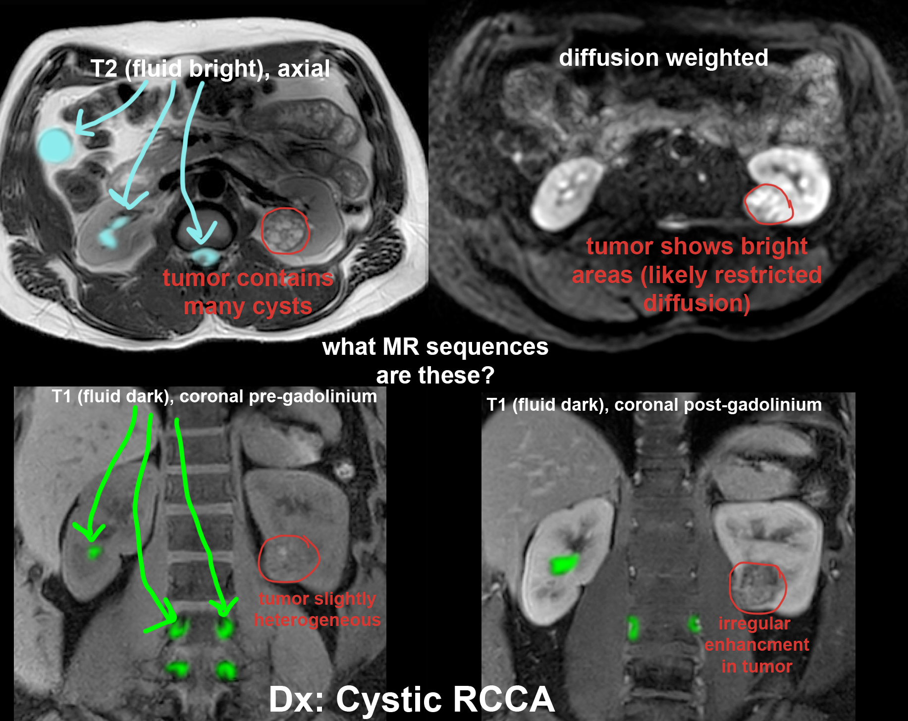
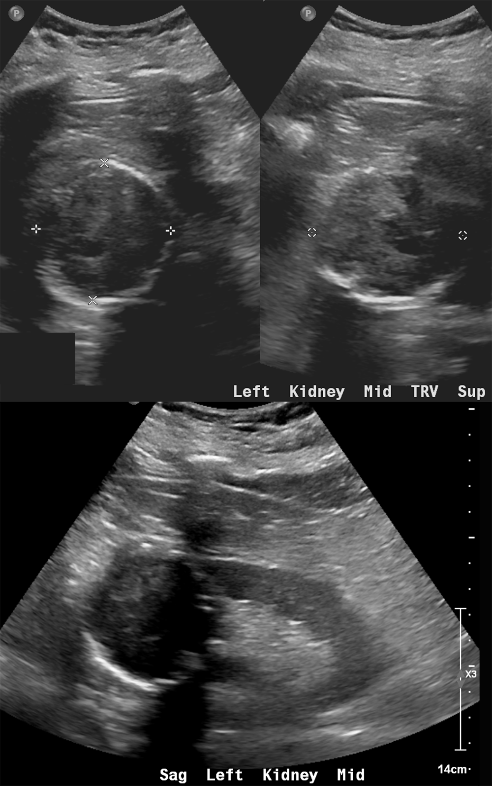
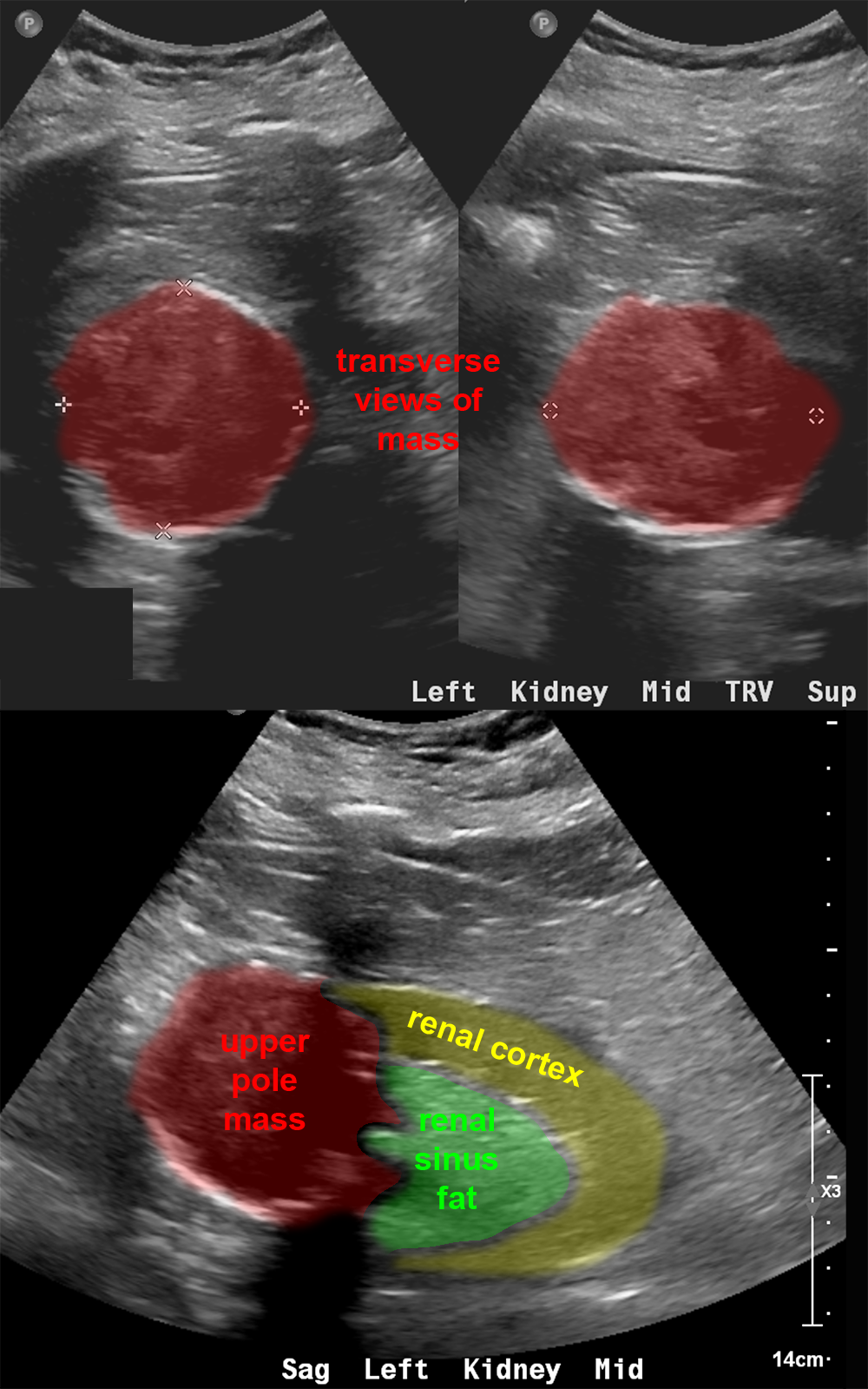
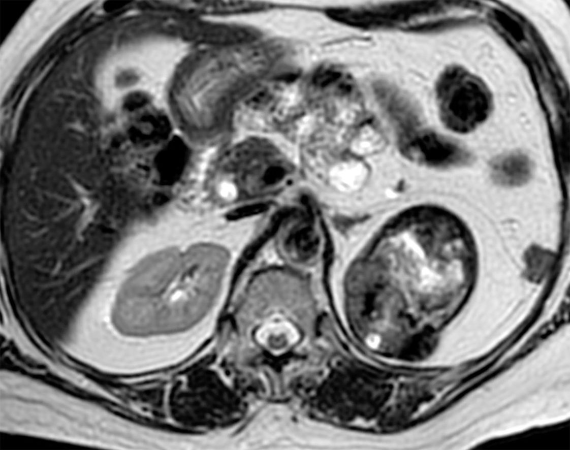
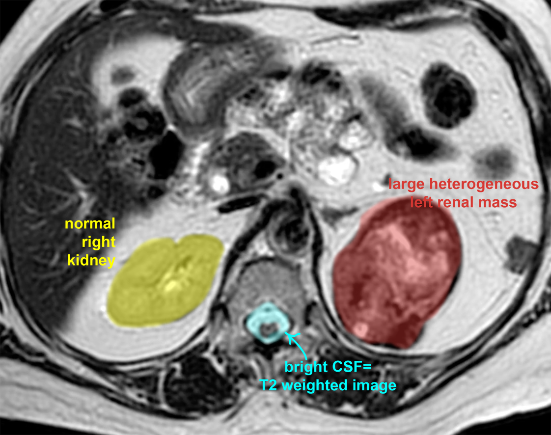
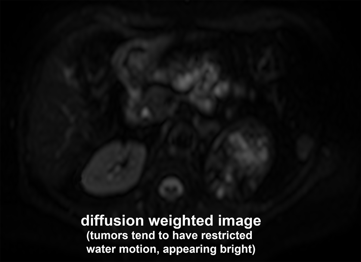
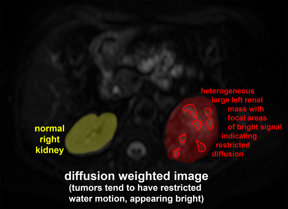
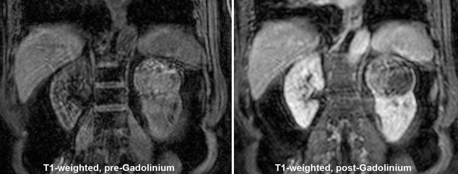
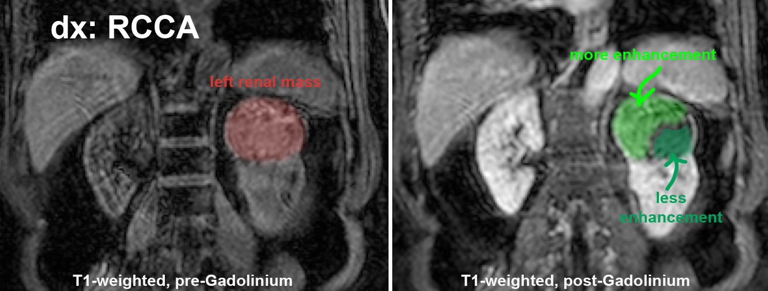
While imaging of renal masses often starts with CT, other imaging modalities can play a role, particularly in complicated cases. Renal masses may be incidental findings on US or CT studies done for other reasons. MRI can be helpful in several ways, including detection of small amounts of fat that may not be evident on CT, since macroscopic fat often indicates a benign diagnosis. Vascularity can also be assessed with MR, as well as diffusion of water through tissues (diffusion weighted imaging or DWI). Many tumors show restricted water diffusion due to the complexity of the tumor microscopic structure.
Further Explanation:
Imaging of Renal Masses
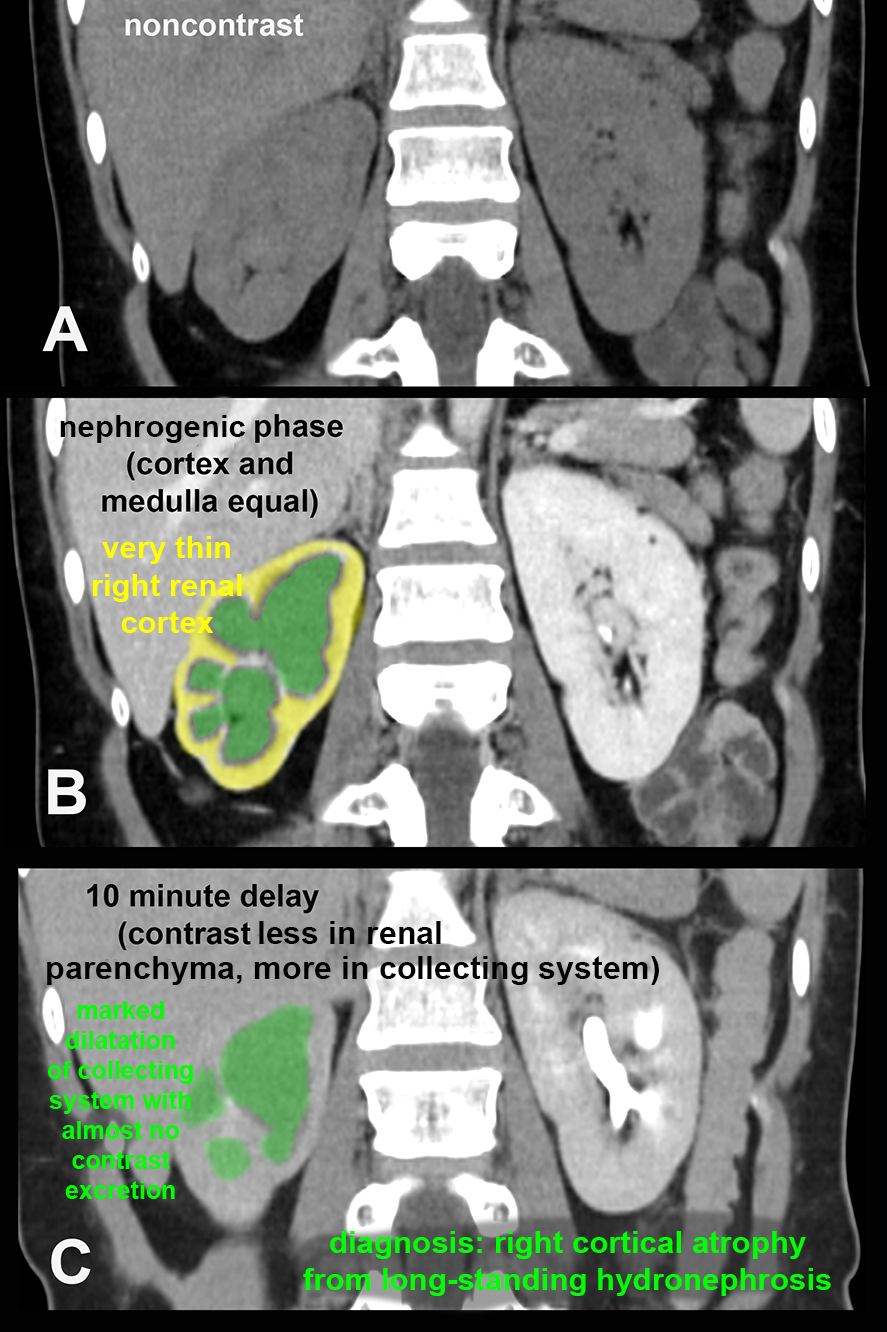
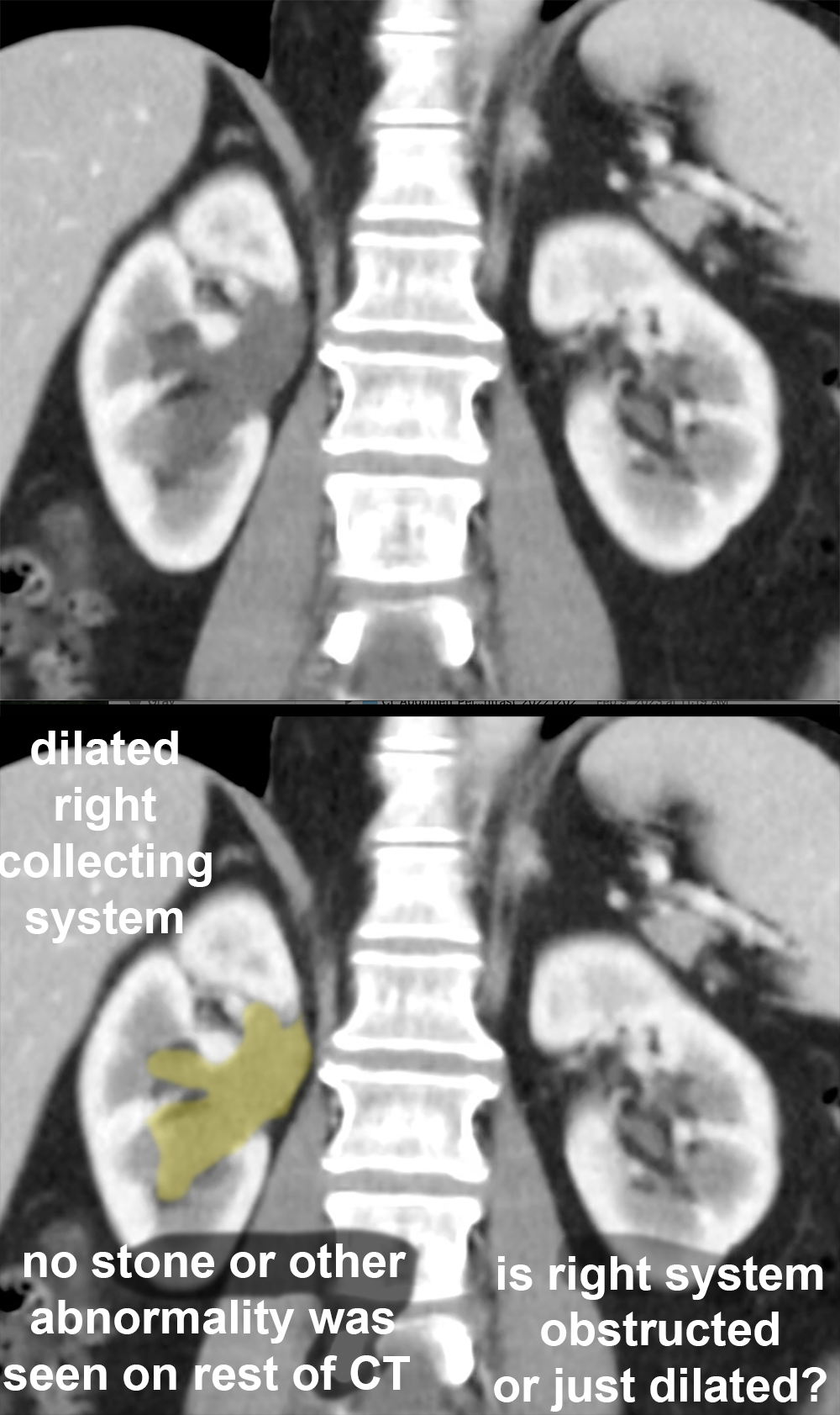
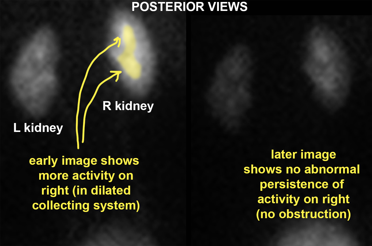
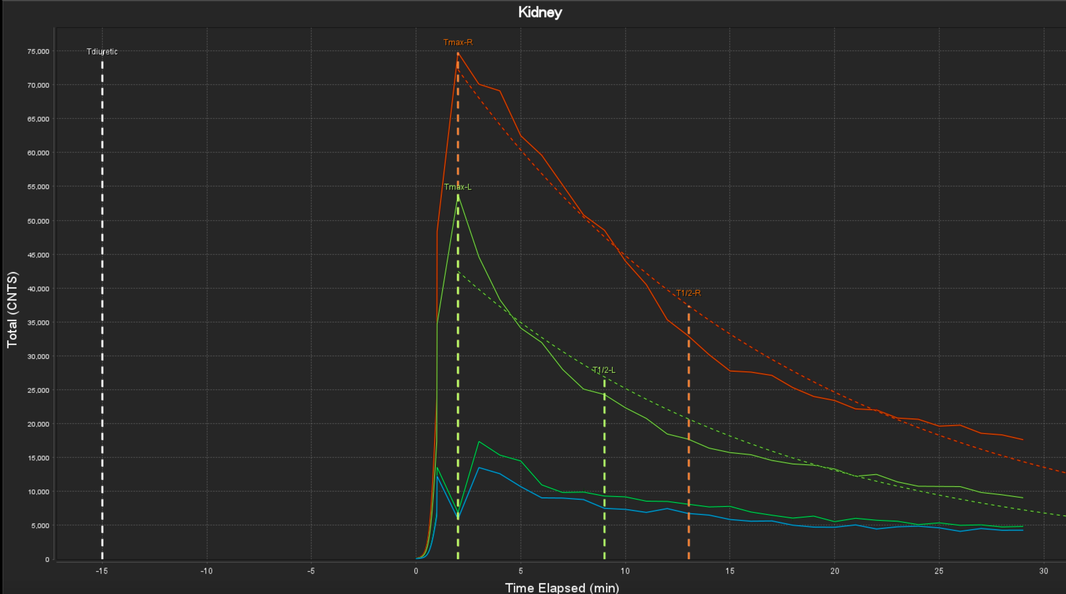
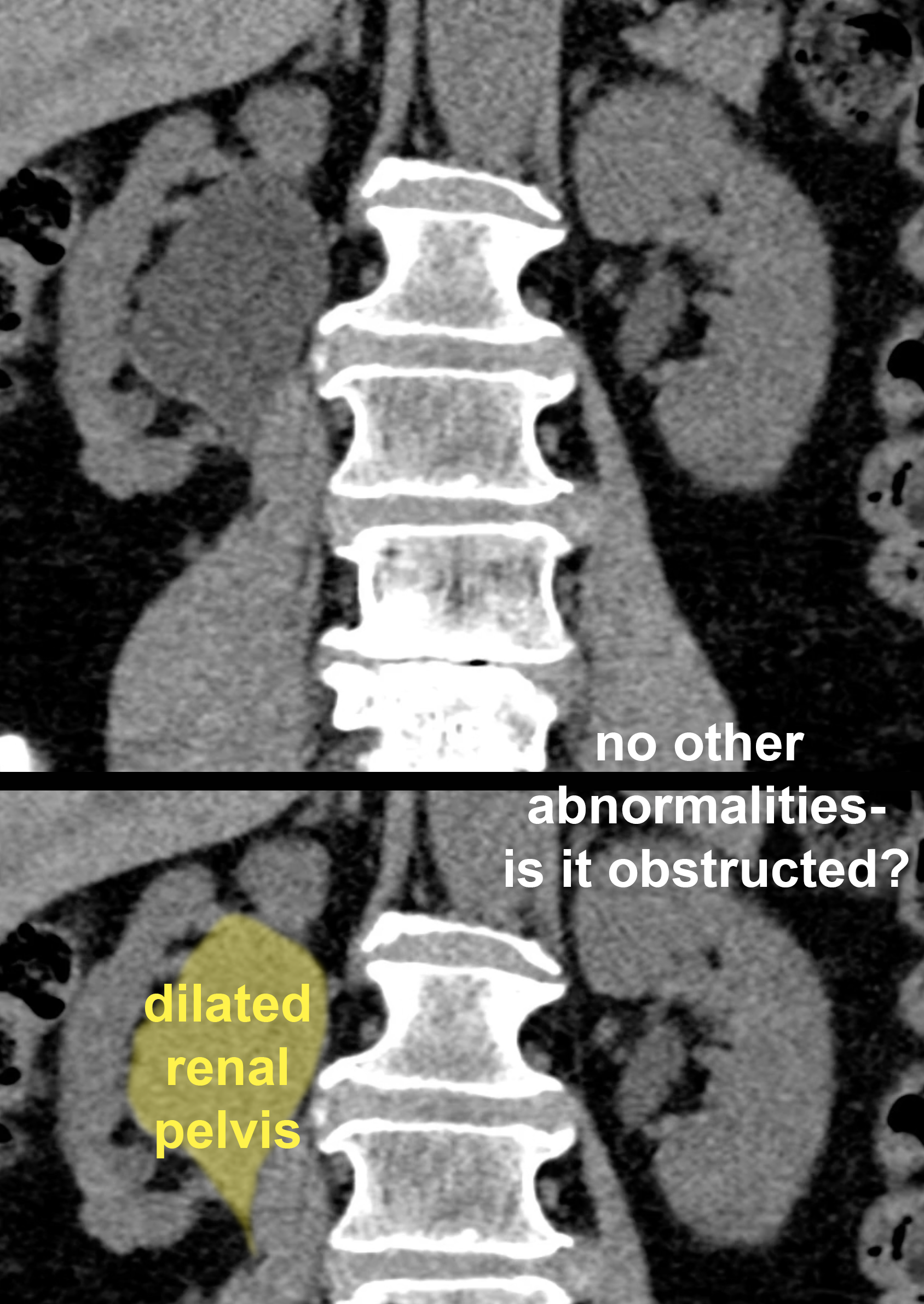
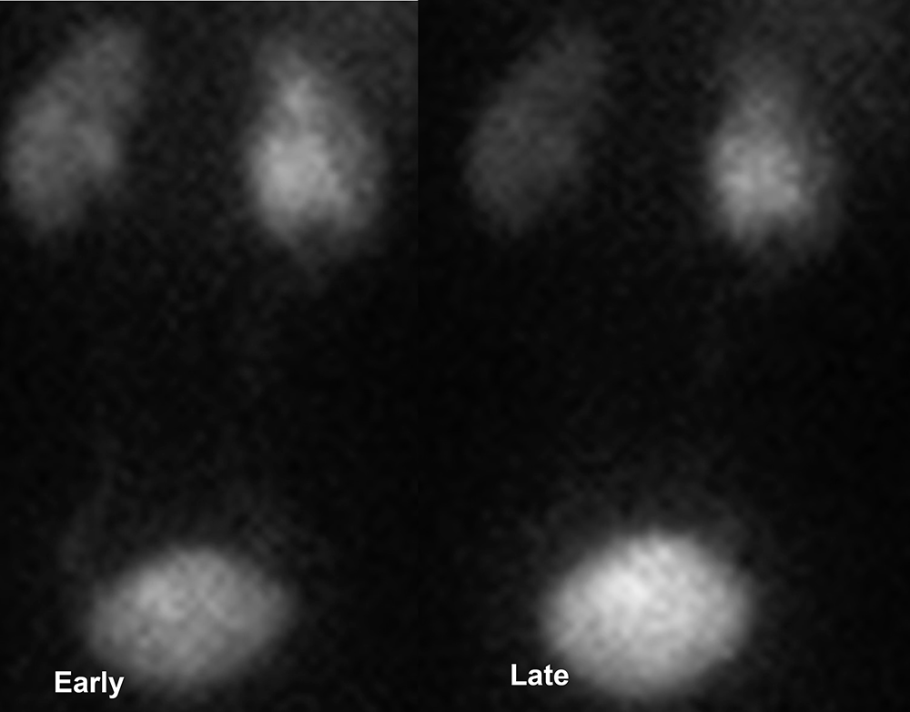
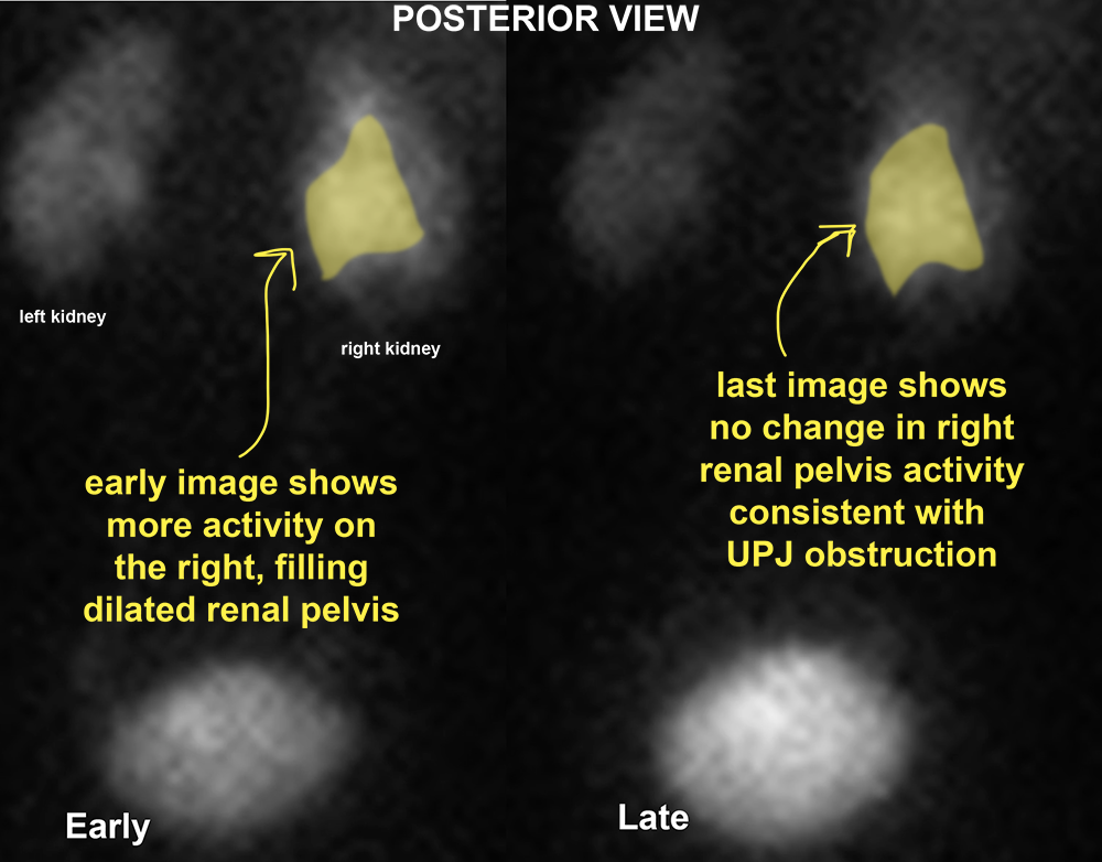
CT and to a lesser extent MR are most often used in evaluation of renal masses, but nuclear medicine imaging can also play an important role, particularly in quantifying renal function on each side (sometimes needed for surgical planning), and for determining whether a dilated collecting system is obstructed, or just distended (likely related to prior obstruction). While the anatomic detail of the images is not high, the value of quantitative data on function and flow can be very helpful. For example, if one kidney is small (atrophic), a nuclear medicine scan can determine how much function is present on that side compared to the other side. The first case shows CT findings of dilated collecting system on the right.
Further Explanation:
Imaging of Renal Masses
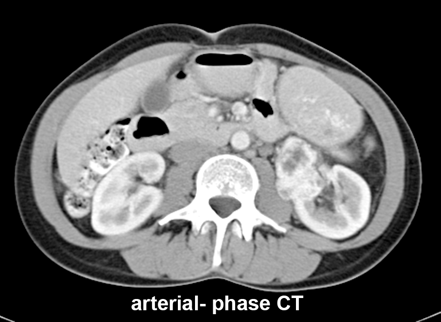
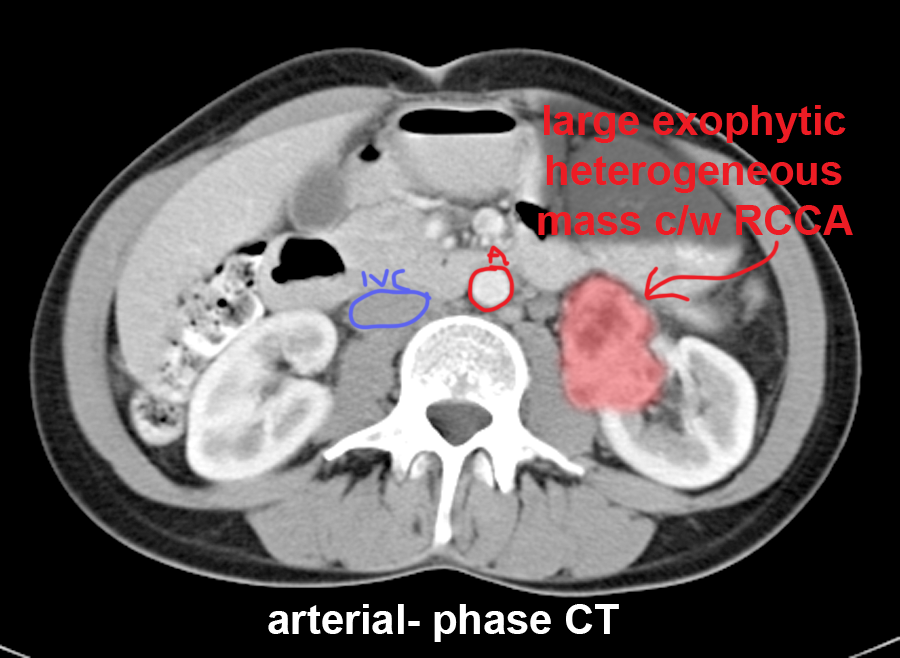
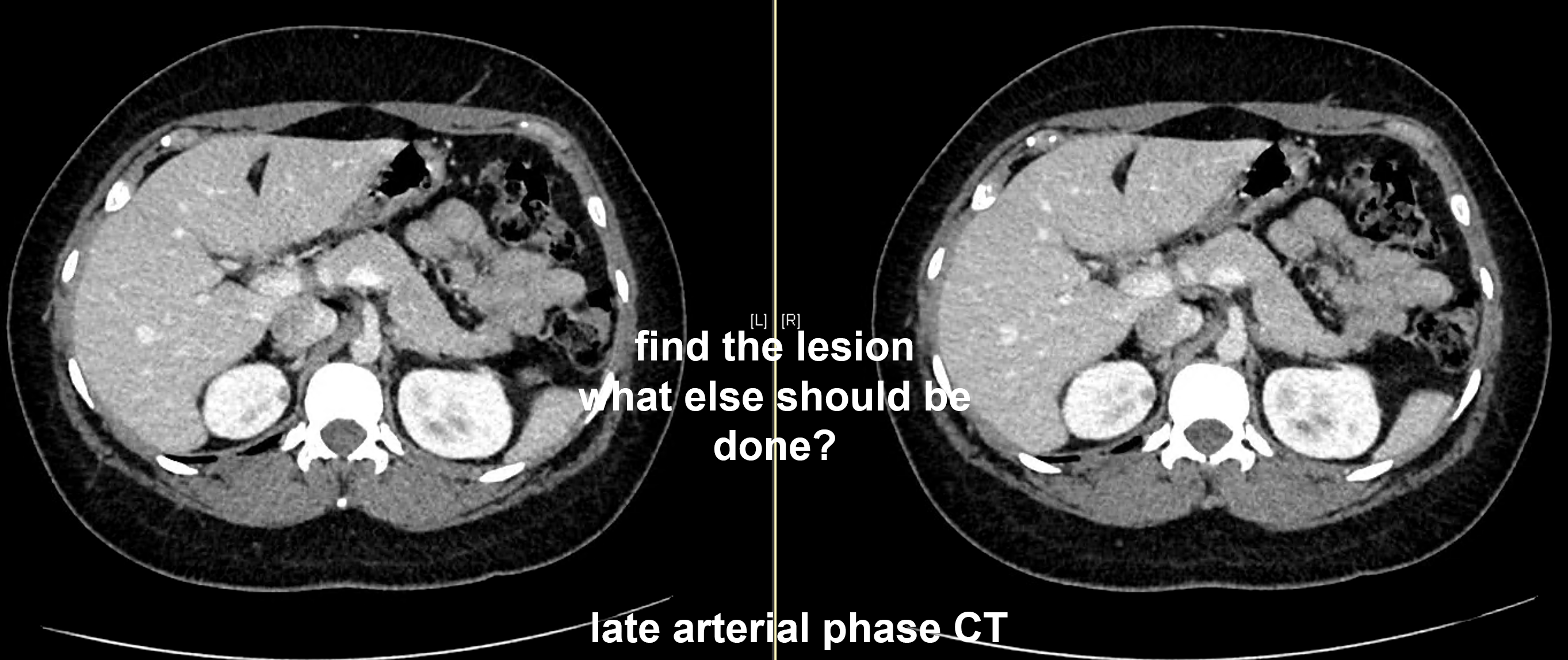
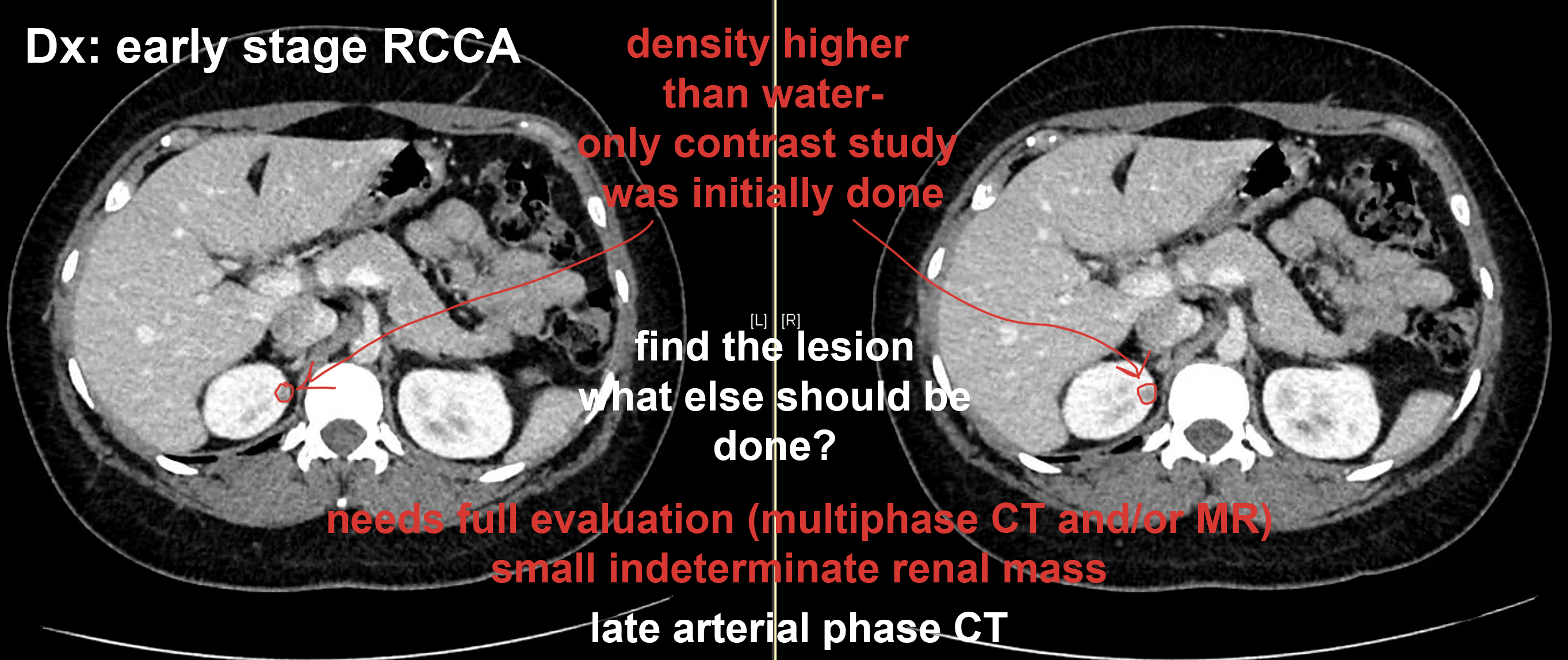
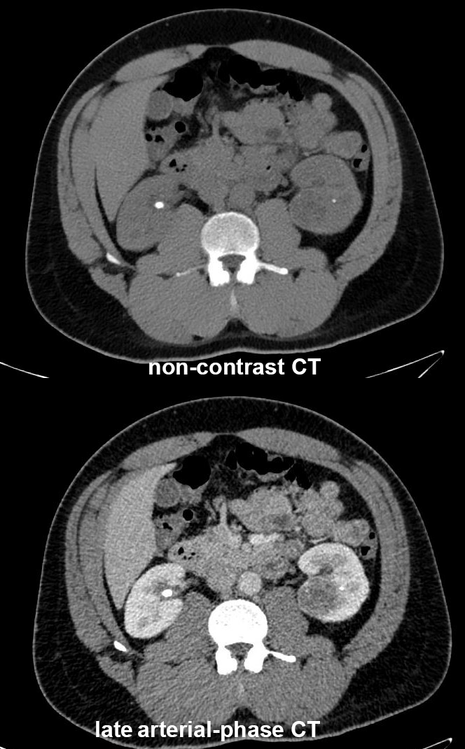
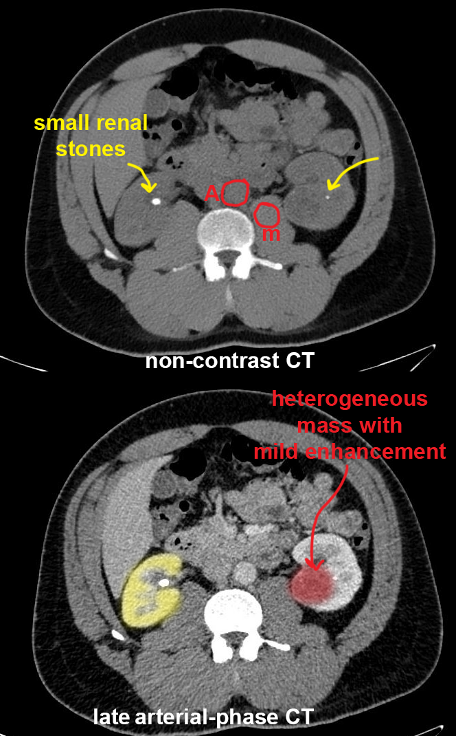
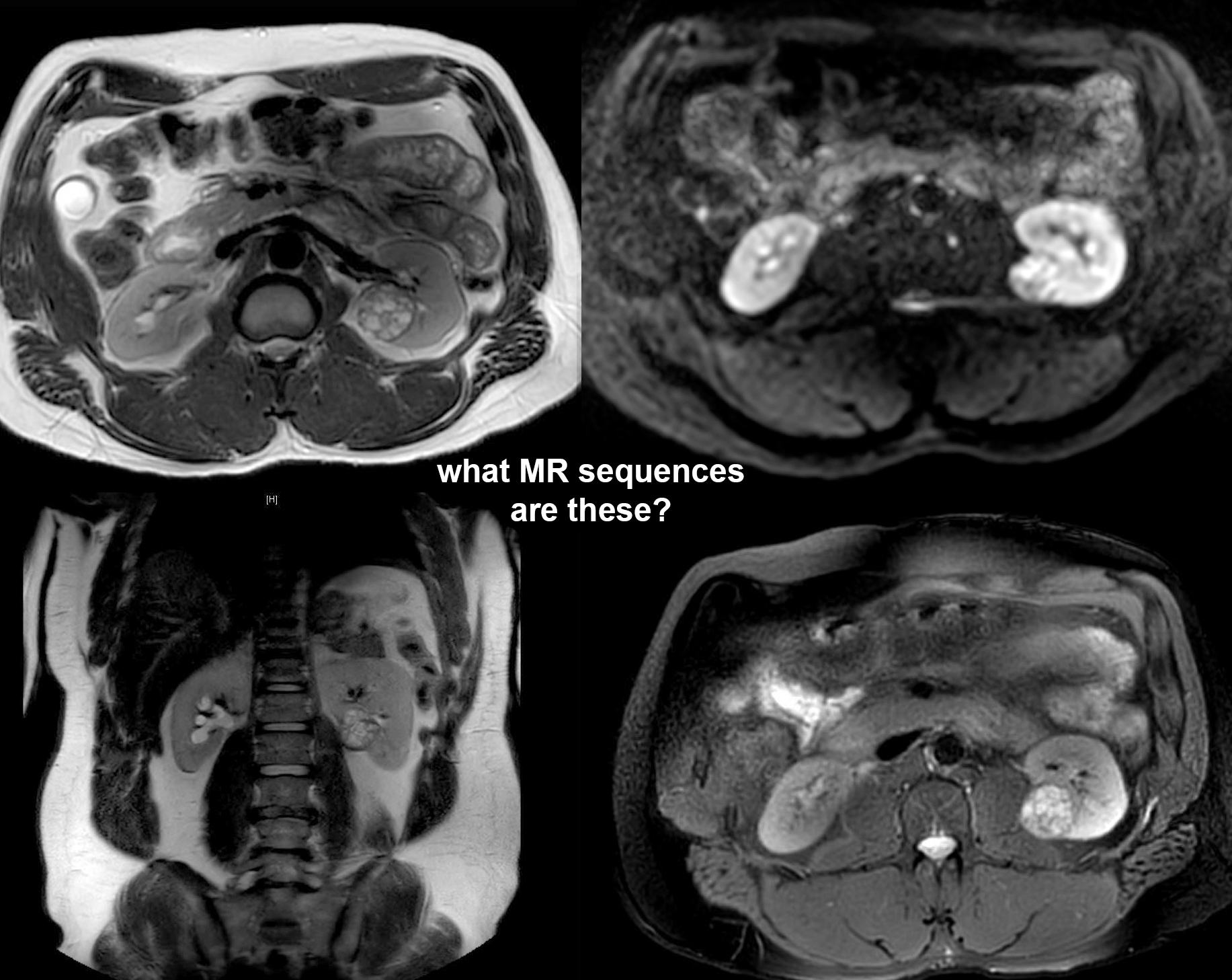
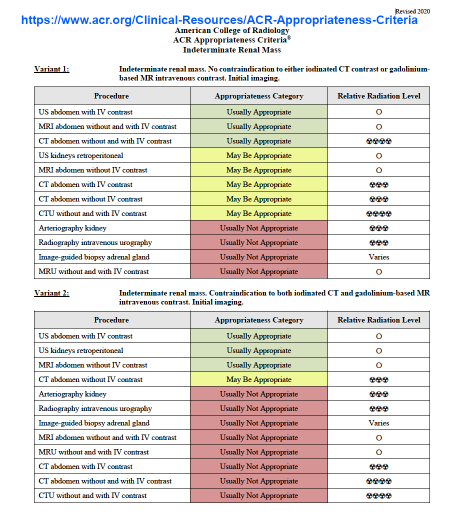
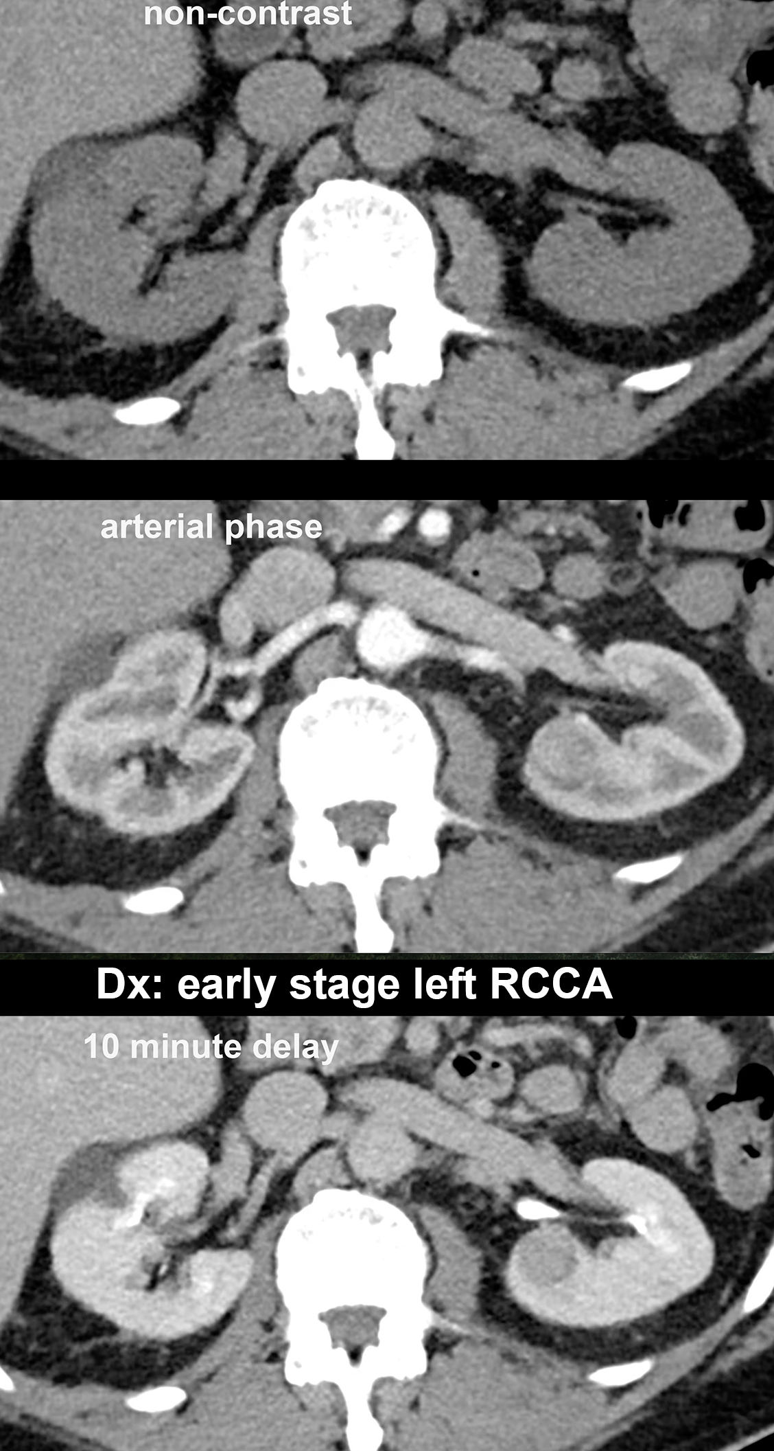
The imaging of renal masses with CT is extremely complex, since there are many different tumors that can occur in this region, and there is considerable overlap among imaging features for different tumors. These cases show the range of appearance for RCCA on CT, with one more MRI example. A useful resource in deciding what imaging is appropriate in a given setting is the American College of Radiology Appropriateness criteria. An example of a page related to renal masses is included and this information is freely available to all online.
Further Explanation:
