

Questions on Imaging Cysts and Stones
Questions on Imaging Cysts and Stones
Question 1:
a) What are imaging features of simple renal cysts?
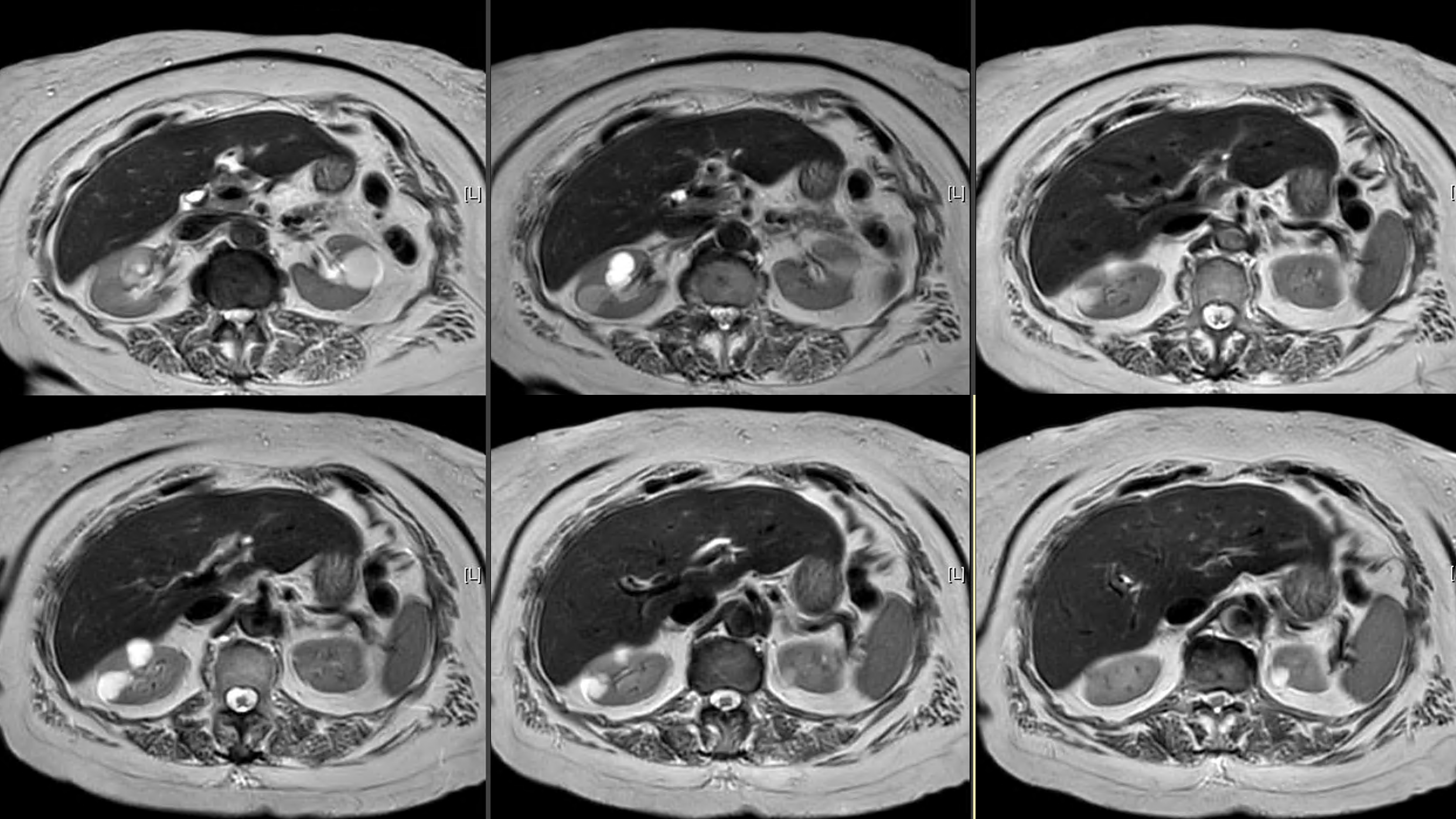
A simple cyst of the kidney is defined as having a round shape, smooth thin wall, uniform density of water (around 0 HU on CT, and bright on T2-weighted MR), and no enhancement on contrast CT or MR studies with no nodularity, septations or calcifications.
b) What is the study shown here?
This is a T2-weighted MR showing multiple bright rounded lesions in both kidneys consistent with simple cysts. You can see bright CSF, which indicates a T2-weighted sequence. In fact, there are special types of T2-weighted sequences used in neuroimaging that have LOW signal in CSF, called FLAIR imaging, but these types of images are not used for evaluation of the kidneys. If the CSF is bright, it is a T2-weighted sequence, but if it is dark, it might be a FLAIR or a T1-weighted sequence.
c) What other kinds of studies can be used to evaluate renal cysts?
Renal cysts are a common incidental finding on CT or US exams done for other reasons. The cysts seen on this MR were also incidental. MR is not the typical imaging modality for initial evaluation of simple cysts.

Questions on Imaging Cysts and Stones
Questions on Imaging Cysts and Stones
Question 2:
a) What are technical parameters for this study?
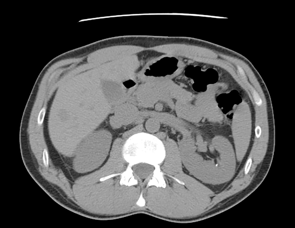
This is an axial CT with no oral or intravenous contrast, done in the prone position (look at the table at the top of the image). This is a renal stone protocol study.
b) How would you describe the abnormality on this study?
There is a tiny high attenuation lesion in the left kidney, most consistent with a renal stone. There is no sign of dilation of the collecting system on this single image.
c) Why is a renal stone protocol done with the patient in prone position?
A stone protocol CT is performed in the prone position because small stones may become lodged in the uretero-vesicle junction (UVJ), and the prone position will separate those stones from stones that are free in the bladder. In the supine position, stones in the UVJ and bladder may be hard to tell apart, but in the prone position they are on opposite sides of the bladder.


