

Questions on Imaging Renal Masses
Questions on Imaging Renal Masses
Question 1:
a) What is this study and how was it done?
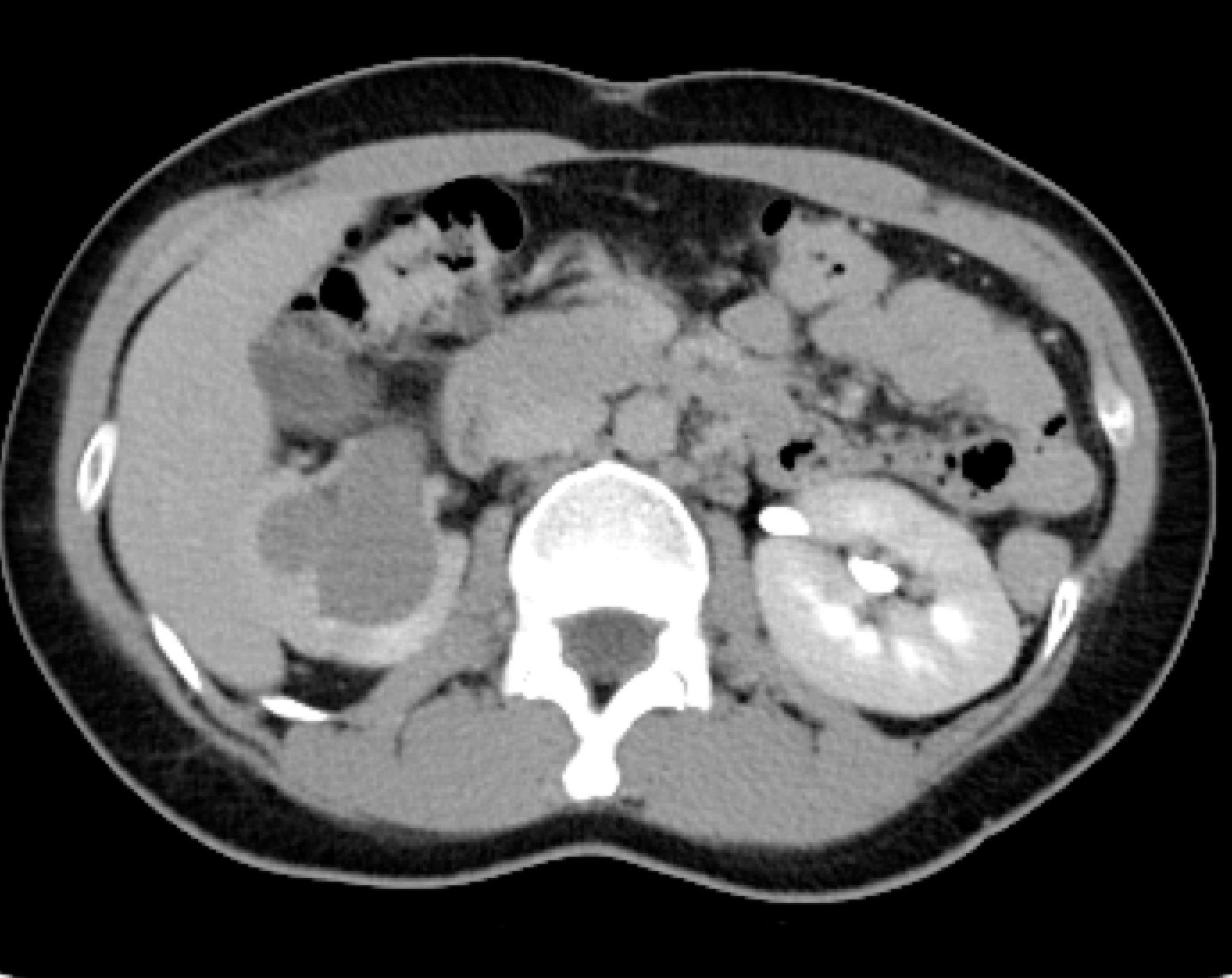
This is an axial CT image in the delayed phase. Most of the visible contrast is in the left renal collecting system, and the aorta appears similar to the density of muscle, so most of the IV contrast has been or is being filtered out of the blood by the kidney.
b) What is the abnormality?
There is marked right hydronephrosis (very large renal pelvis), with thinning of the cortex of the kidney consistent with atrophy. It is not possible to tell from this one image whether the right collecting system is currently obstructed or just dilated, related to prior obstruction.
c) What type of study could give information about how much renal function remains on the right in this patient?
A nuclear medicine study could give numeric information about relative function on the right vs the left in this setting.

Questions on Imaging Renal Masses
Questions on Imaging Renal Masses
Question 2:
a) What are these studies?
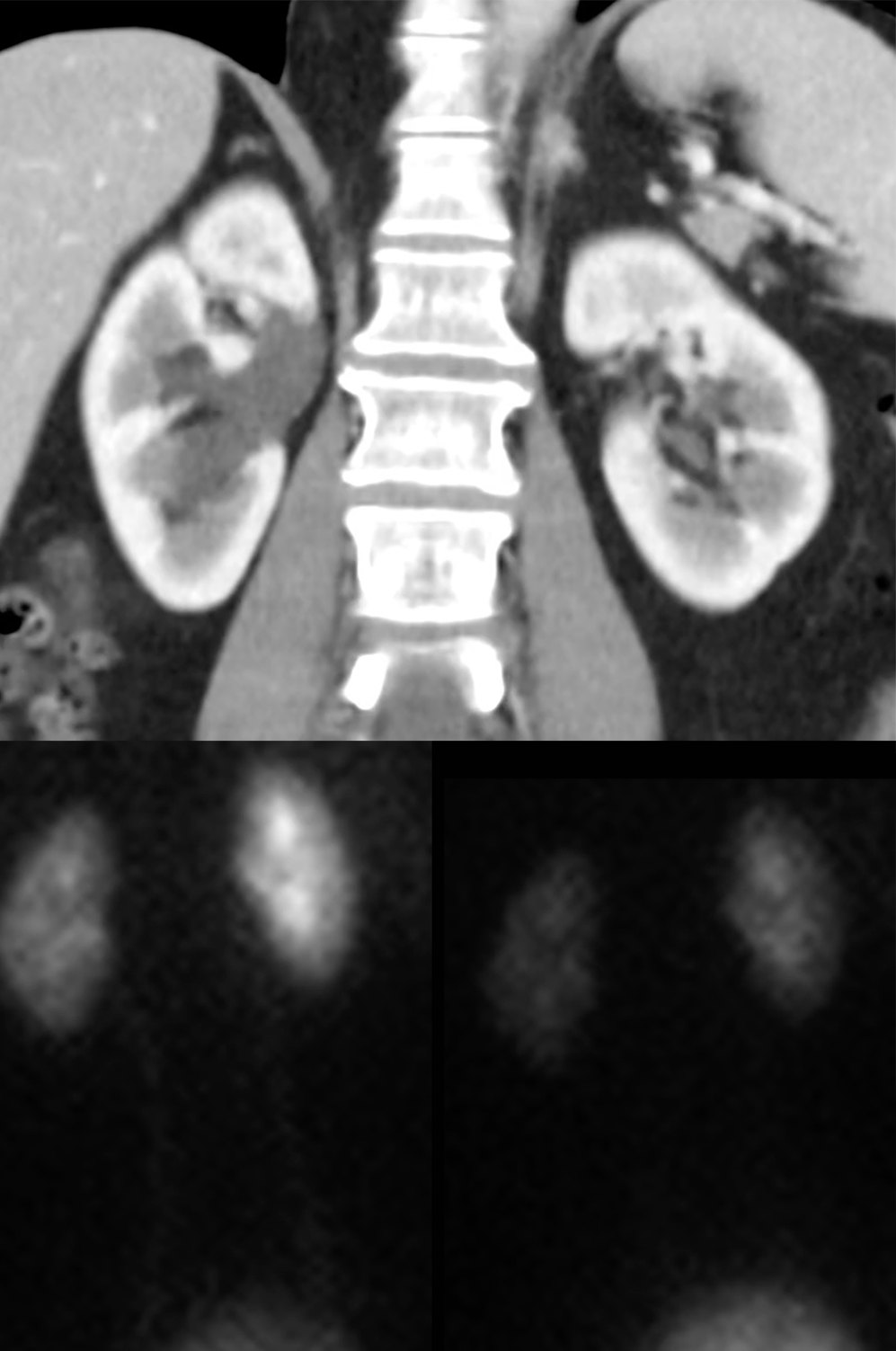
The top image is a CT reconstructed in the coronal plane with IV contrast, scanned in the arterial phase (cortex and medulla appear different in density). The bottom images are from a nuclear medicine study to check for obstruction of the collecting systems.
b) What is abnormal on the top image?
There is dilatation of the right renal pelvis. This could be due to current obstruction or prior obstruction.
c) What do the two bottom images show?
The early image on the left shows asymmetry of activity, with more on the patient's right (these are posterior images, so the right kidney is shown on the right side of the image) than on the left, due to accumulation of tracer in the dilated right renal pelvis. On the later image on the right, the two sides look more symmetric, indicating that there is no current obstruction--the activity moves out of the collecting system and into the bladder.


