

Questions on Imaging RCCA
Questions on Imaging RCCA
Question 1:
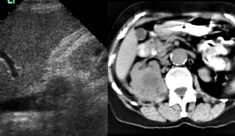
a) What are these two studies?
The image on the left is a sagittal US image through the IVC. The image on the right is an axial CT at the level of the right kidney, with oral but without IV contrast.
b) What do the two studies show?
The US image shows tumor in the lumen of the IVC, below the level of the liver. This is highly suggestive of renal cell carcinoma. The CT image shows an irregular mass in the right kidney.
c) What are common and uncommon sites of metastatic disease from renal cell carcinoma?
The commonest sites for RCCA metastasis are lung, lymph nodes, bone and liver. There are many unusual sites of metastasis that have been reported for RCCA including pancreas, thyroid, breast, skeletal muscle and endobronchial.

Questions on Imaging RCCA
Questions on Imaging RCCA
Question 2:
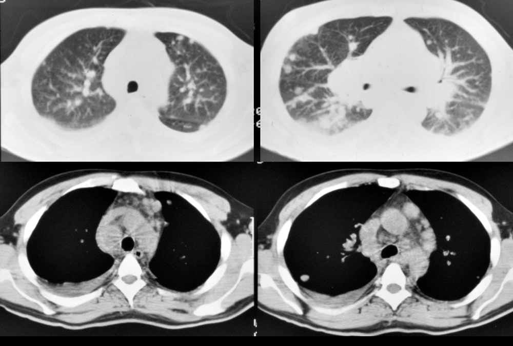
a) What are technical parameters for this study?
These are images from a chest CT without intravenous contrast. The top two images are displayed with lung windows and the bottom two with mediastinal windows.
b) What abnormalities are present?
There are many small round lung nodules bilaterally, worrisome for metastatic disease. There are also enlarged mediastinal nodes.
c) What diagnoses would you consider in this case?
Nodules and nodes could be due to lymphoma but another consideration is renal cell carcinoma with a 'pseudosarcoidosis' appearance. RCCA often spreads to retroperitoneal nodes at the level of the kidneys, and may then climb up the lymphatic chain to involve the mediastinum and hila of the lungs. Most other abdominal tumors do not spread in this fashion. Many other malignancies could also be considered, since nodal disease and lung metastases are very common in a variety of tumors, such as melanoma, breast cancer or sarcomas.


