

Questions on Other Renal Masses
Questions on Other Renal Masses
Question 1:
a) What are technical parameters of this study?
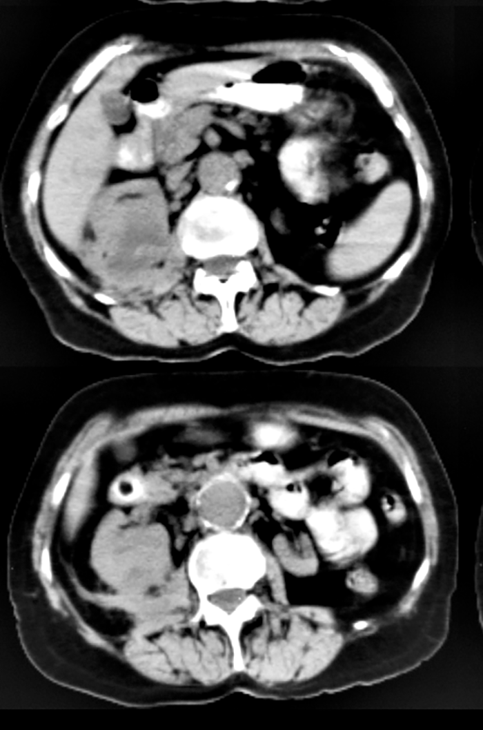
This is an axial CT at the level of the right kidney with oral but without intravenous contrast. GI contrast is not generally needed for imaging of renal tumors, but this study was done for vague symptoms, and the renal mass was unexpected. When doing a CT without a specific diagnosis in mind, oral contrast is often helpful.
b) What are differential possibilities for the abnormality seen in the right kidney?
The most worrisome possibility is renal cell carcinoma. Other considerations would include pyelonephritis or metastatic disease.
c) What features on this image would favor infectious etiology for the finding?
There is extensive fat stranding posterior to the mass. While this could represent tumor invasion, it could also be a sign of infection. This patient had a fever, flank pain and an elevated white blood cell count as well as a history of right renal stones. The diagnosis was chronic pyelonephritis. Of note, there is also a small aneurysm of the abdominal aorta.

Questions on Other Renal Masses
Questions on Other Renal Masses
Question 2:
a) What are technical parameters for this study?
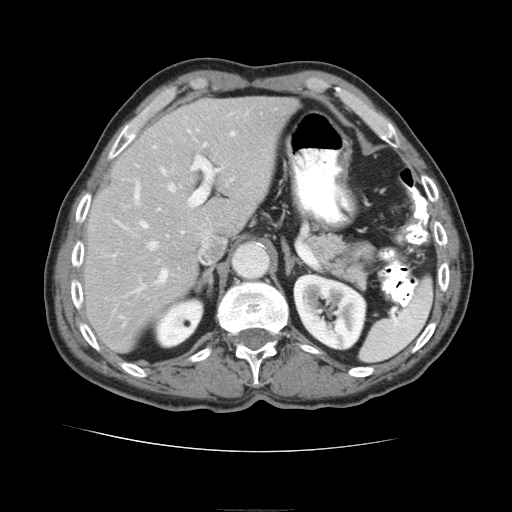
This is an axial CT through the upper abdomen with IV contrast (in the early or arterial phase, as the renal cortex and medulla appear different in density) and with oral contrast.
b) What renal abnormality is present?
There is a very small low attenuation rounded lesion in the upper pole of the right kidney.
c) If the density of the lesion is measured at -40 HU, what is the likely diagnosis?
A density of -40 HU indicates the presence of macroscopic fat. This would indicate that this is a small benign angiomyolipoma. This would not require further imaging or followup. Since it is so small, it might be difficult to prove on CT that it contains fat, due to averaging with adjacent bright renal tissue, which might yield higher density measurements. In that case, evaluation with ultrasound (to show an echogenic appearance of the lesion consistent with fat), or MRI (to do special sequences that show presence of fat, such as fat suppression) might be helpful.
This completes the Knowledge Assessment questions for the module on Renal Imaging. Click here to return to the list of learning objectives.


