

Radiography (XR)
Introductory radiograph image
Question 1:
What is the imaging modality depicted?
A PA (posterior-anterior) chest radiograph is depicted. This is also referred to as a plain film of the chest. It may also be abbreviated as “CXR”.
Question 2:
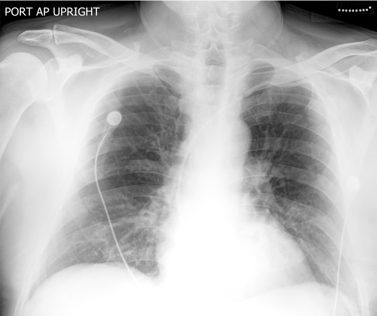
What appears “black” , “white” or “grey” in this modality?
White: densely radiopaque structures (eg. Bone)
Black: air-filled low density structures (eg. Lungs)
Grey: intermediate to low density structures (eg. Fat)
Question 3:
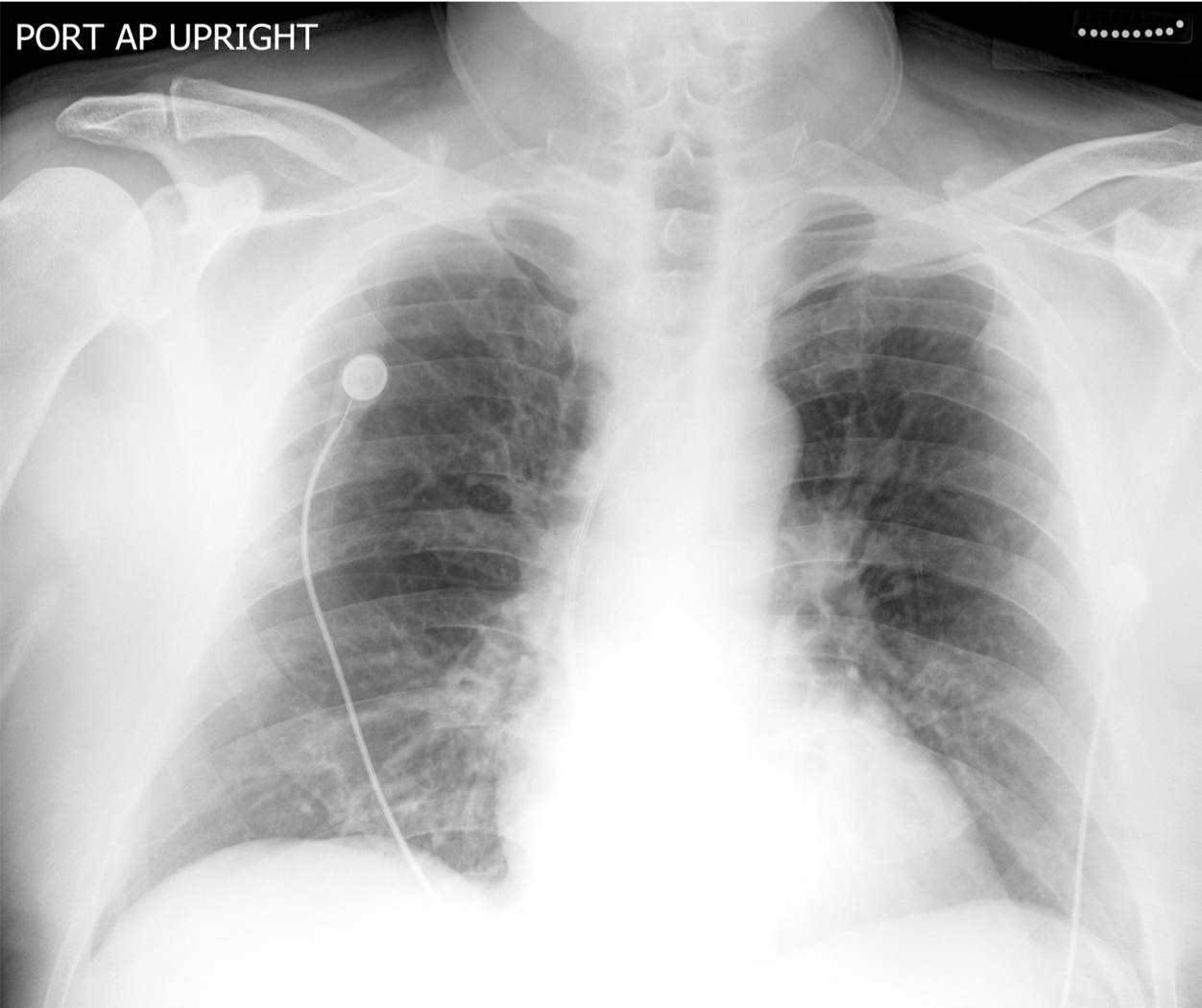
What vascular access device has the patient received prior to obtaining this image?
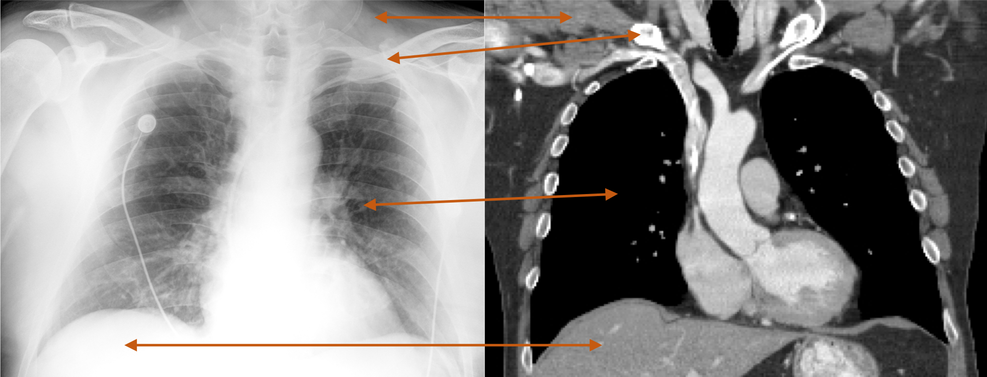
A port is depicted in the left chest. It consists of a reservoir and an attached catheter, which is seen coursing from the reservoir to the right cardiac atrium.
Further Explanation:
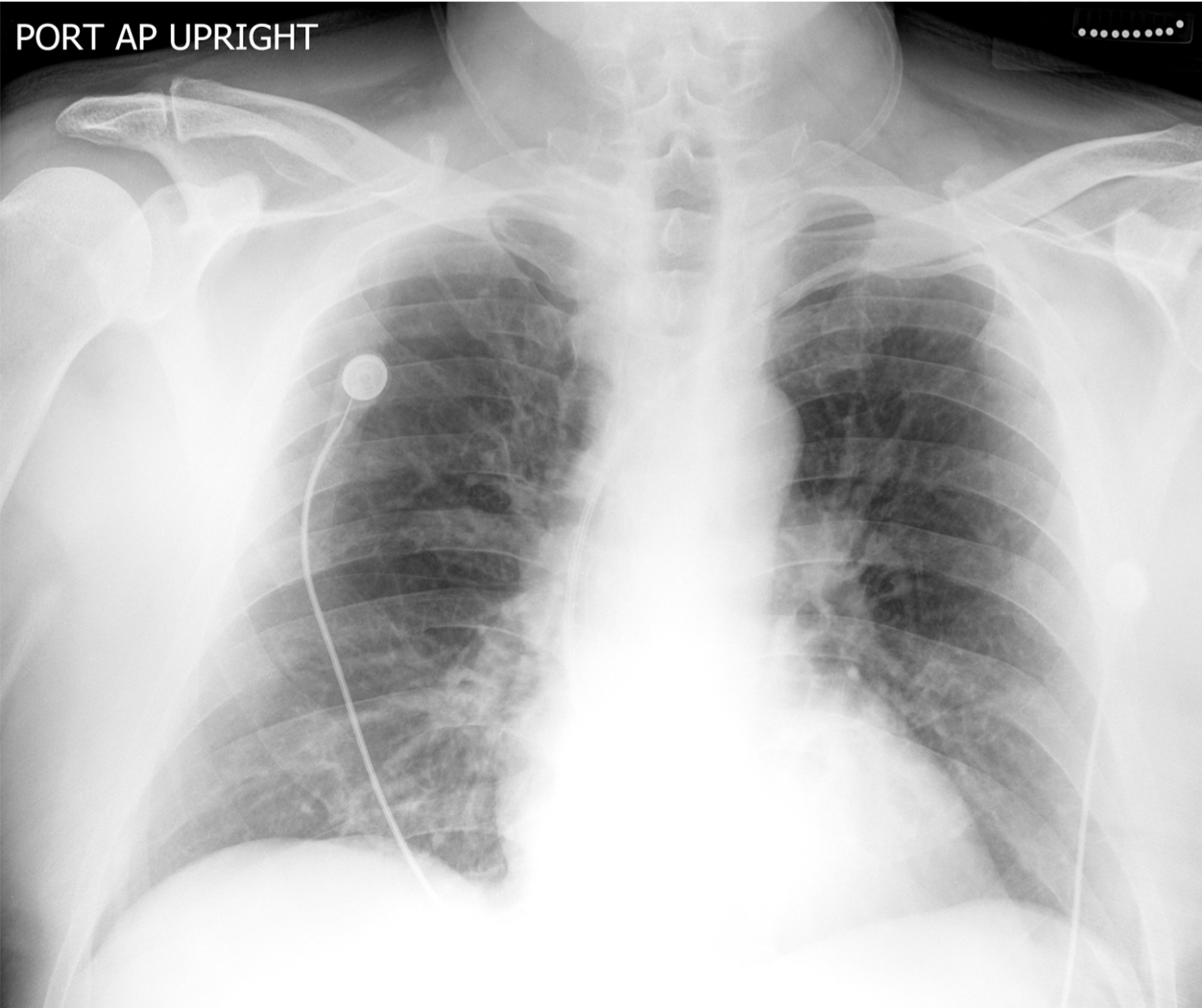
Notice how the Computed Tomography (CT) scan of the chest in the same patient illustrates the corresponding structures in question.
Question 4:
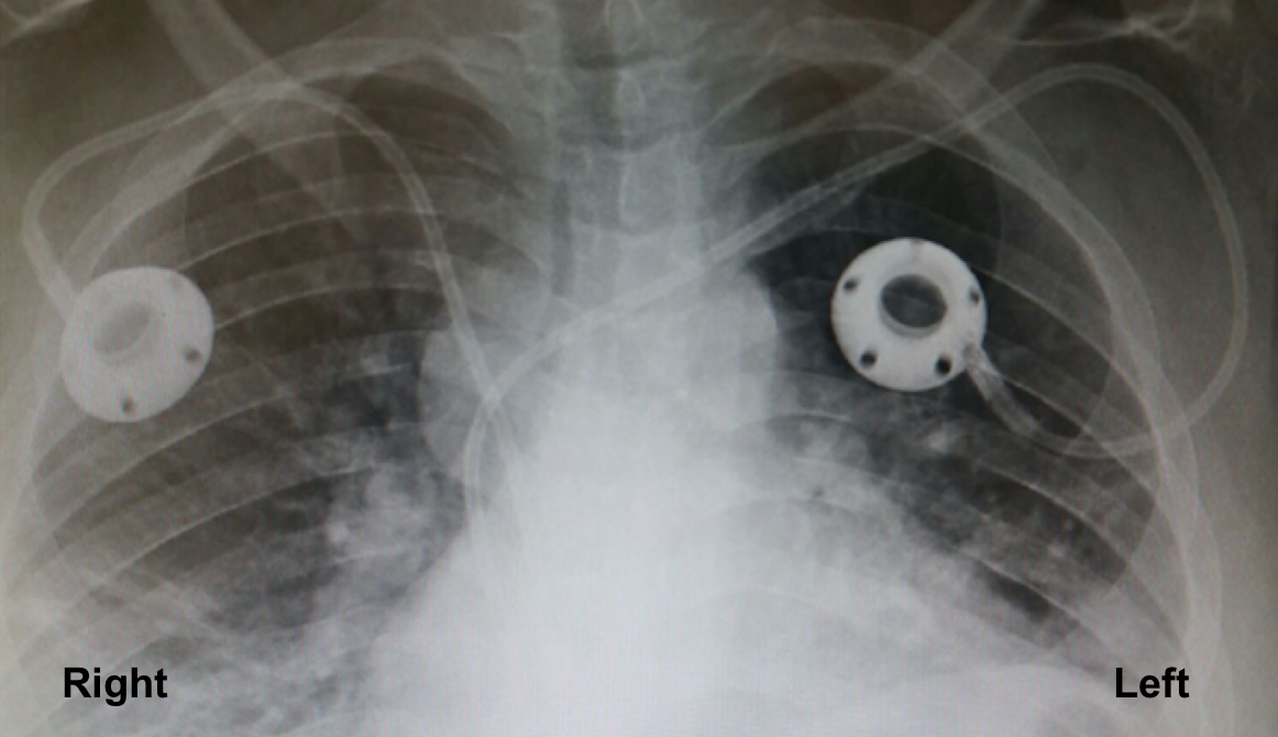
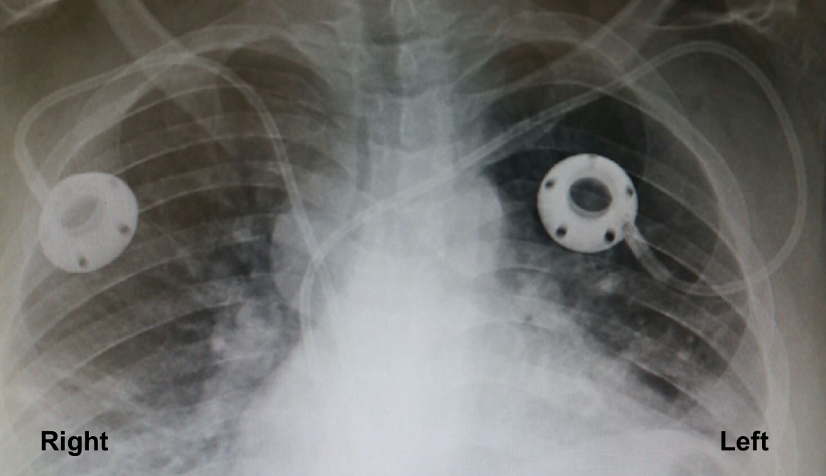
What are the devices depicted in this frontal chest radiograph?
The devices depicted are bilateral port-a-caths. These are venous access devices for long term venous access, and used primarily for administration of chemotherapy.
Question 5:
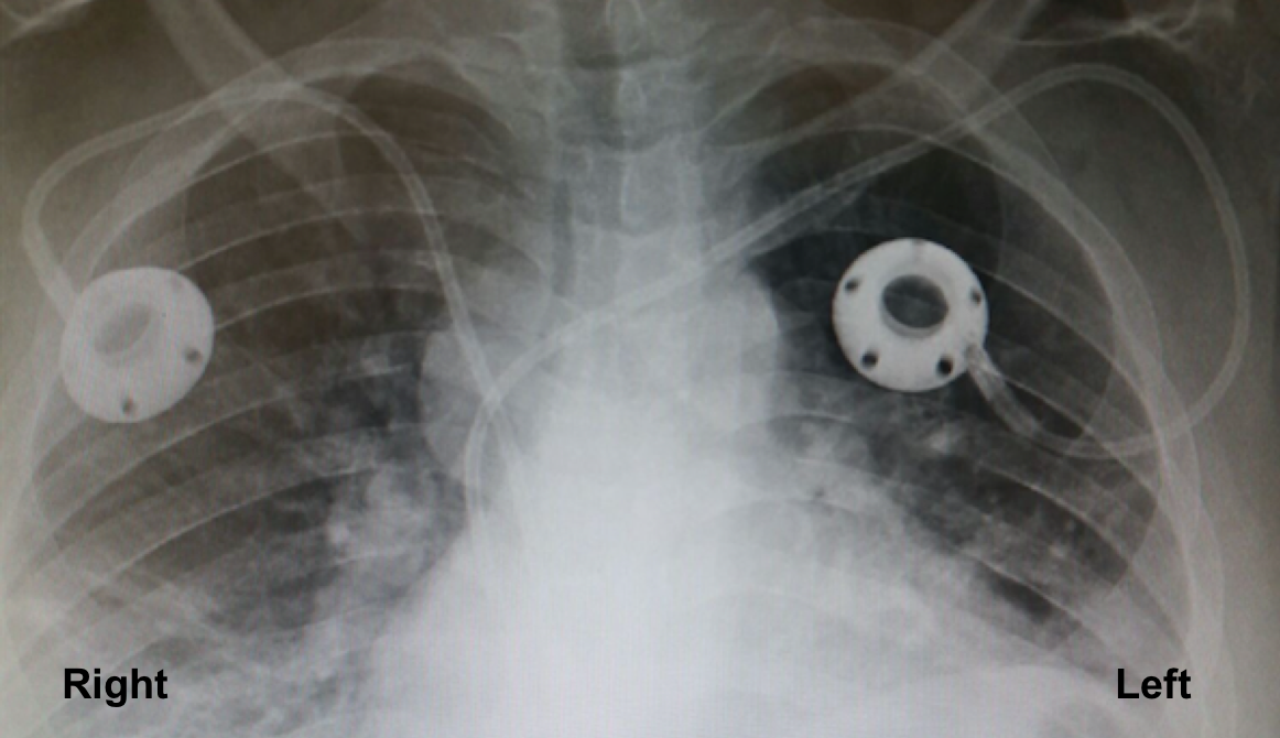
What is the difference in the route via which the ports were placed as depicted on this radiograph?
The port on the RIGHT was placed via the internal jugular vein. The port on the LEFT was placed via the subclavian vein.
Question 6:
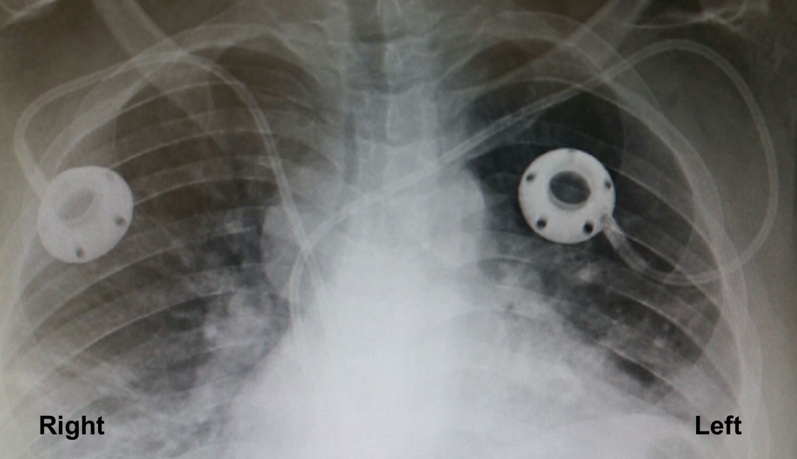
What is the recommended venous access route for ports?
The port on the RIGHT was placed via the recommended venous route, namely the internal jugular vein. This is the route for venous access device placement recommended by the Standards and Practice Committee of the Society of Interventional Radiology and the American College of Radiology’s Interventional Radiology Appropriateness Criteria expert panel. The port on the LEFT was placed via the subclavian vein.
Question 7:
What is the potential complication when ports are placed via the route depicted on the left?
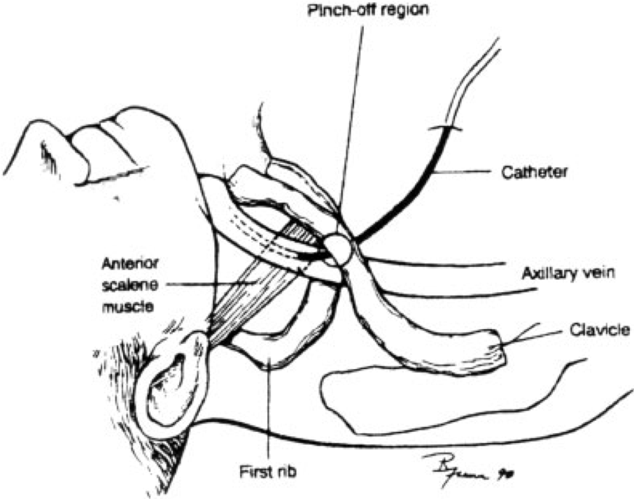
Pinch off syndrome is the primary risk to the patient when a port-a-cath is placed via the subclavian vein.
Further Explanation:
Pinch off syndrome is the primary risk to the patient when a port-a-cath is placed via the subclavian vein.
Schematic illustrates the pinch off phenomenon where a simulated right sided subclavian central venous catheter that through its initial course is compressed between the clavicle and 1st rib.
Jensen, M. O. (2008). Anatomical Basis of Central Venous Catheter Fracture. Clinical Anatomy , 21, 106-110.
Radiography (XR)
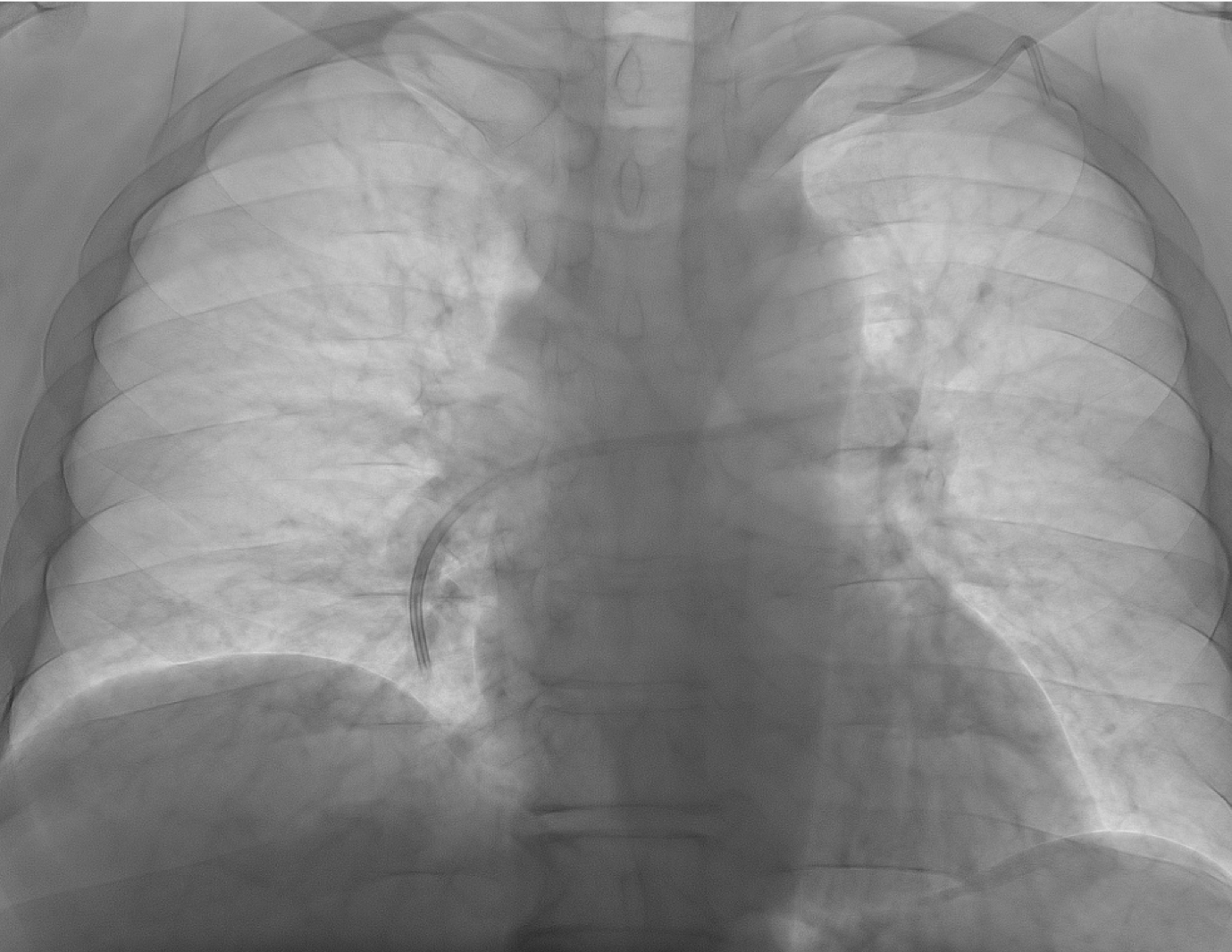
You are rotating through interventional radiology and see the following image of a patient who presents for port removal for completion of chemotherapy
Question 8:
What is the depicted imaging modality?
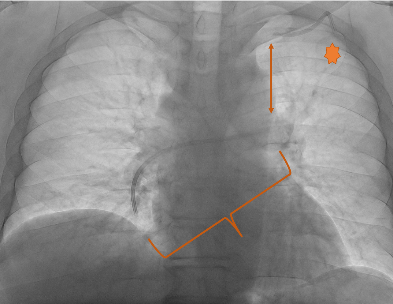
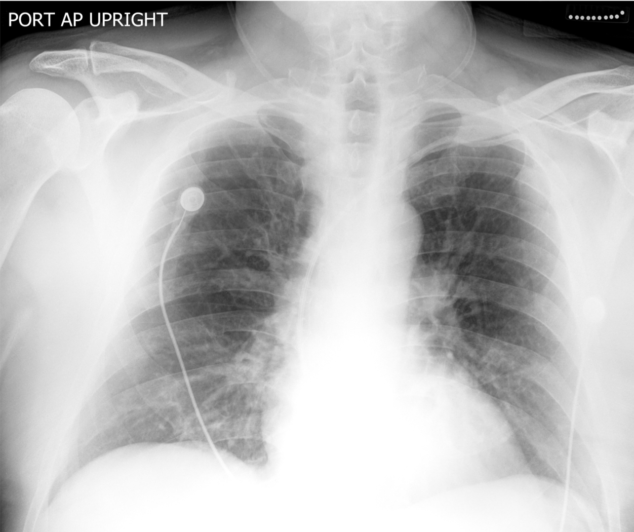
The depicted image is a fluoroscopic image. It resembles a CXR but has differing, contrasting densities. The density conferring “White” (and “Black“ are typically reversed in fluoroscopy when compared to a radiograph. This comparison is depicted well on the chest radiograph in the same patient shown above.
Further Explanation:
Chest radiography demonstrates the left sided chest port (star overlies port reservoir) placed via the subclavian vein (as evidenced by its course originating below the clavicle as opposed to above the clavicle as in the case of internal or external jugular venous accesses). The catheter is fractured at the point of its previous course between the clavicle and first rib.
The magnified radiograph depicts the fractured catheter from the left subclavian port. It is completely discontinuous from the proximal portion of the catheter arising from the port’s reservoir, which is fractured just inferior to the clavicle.
