

Ultrasound (US)
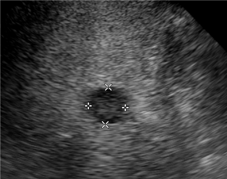
Introductory ultrasound image
Question 1:
What is the modality shown?
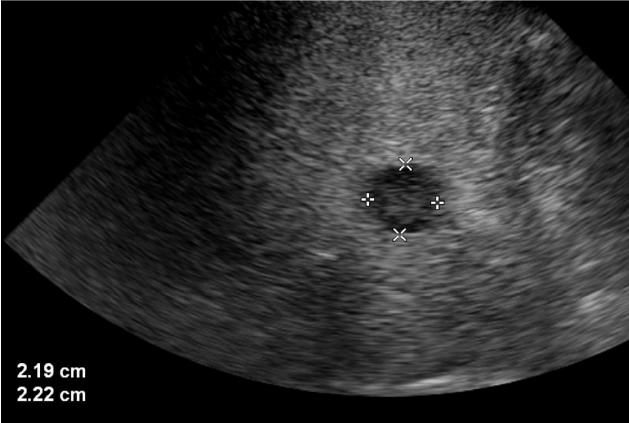
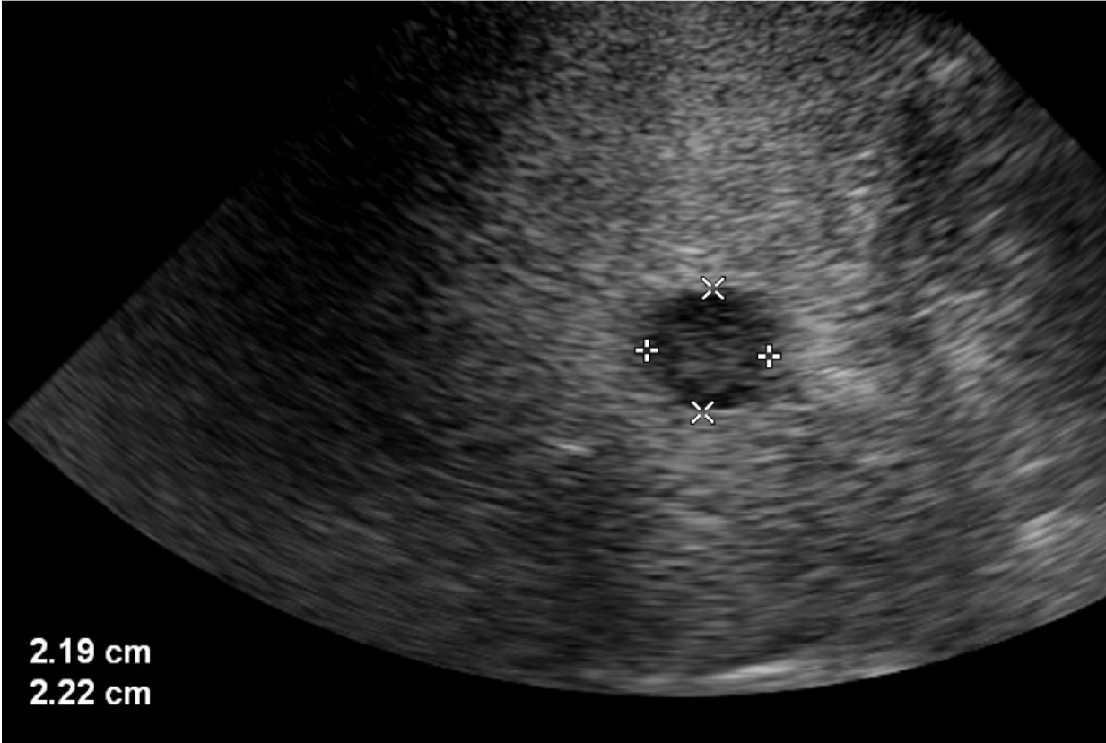
The image shown represents an ultrasound or sonogram of the liver. The echotexture marked by an alternating grayscale of hazy black, grey and white is characteristic. The conical pizza slice-like frame is dictated by the curvilinear transducer used here to image this patient’s abdomen. Curvilinear ultrasound transducers are best for imaging depth, such as anatomy in the abdomen.

Question 2:
The imaged patient has recently been found to have colonic adenocarcinoma. In this imaging evaluation of the patient’s liver, what do think the highlighted finding represents?
The finding which is also seen measured with calipers is a round well-circumscribed lesion that is centrally echogenic (“bright” in echotexture). It either represents a benign or malignant lesion. However, in a patient with known colonic adenocarcinoma, it is concerning for a site of metastasis.
Question 3:
What is the role of the interventional radiologist in such findings?
Interventional Radiologists subspecialize in interventional oncology, which is the 4th pillar of oncologic care after medical oncology, surgical oncology and radiation oncology. Locoregional therapies such as a thermal ablation are typical therapies offered for such lesions, particularly metastatic hepatic colorectal metastases <3cm in size.
Question 4:
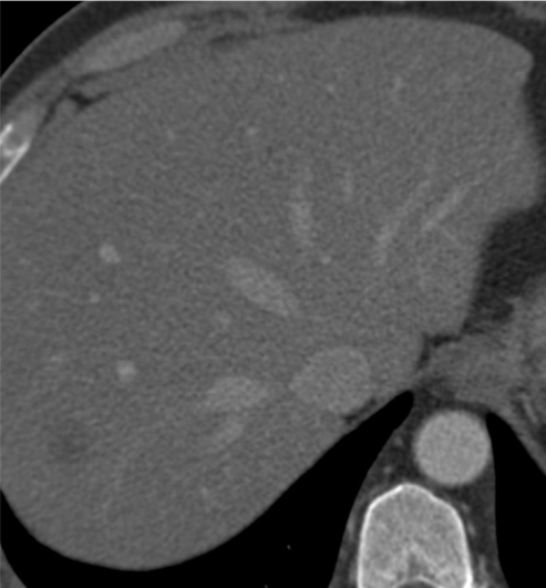
Another cross-sectional imaging modality is obtained in the same patient. What does it represent?
The image to the left is a contrast-enhanced computed tomography (CT) scan. The posterior right hepatic lobe lesion appears hypodense (ie. dark) and nonehancing (ie. no appreciable assimilation of contrast) on this CT scan. This finding on CT correlates well with the lesion seen rather well on ultrasound.


Question 5:
The imaged lesion is biopsied under ultrasound guidance by interventional radiology. 4 passes with a 20G coaxial biopsy needle are submitted in formalin to the pathologist and are found to represent metastatic adenocarcinoma and is presumed to be of colonic origin. What thermal ablation technique can the Interventional Radiologist use to treat this site of metastasis to the liver?
Thermal ablation includes heat-based technologies such as microwave and radiofrequency ablation or cool-based therapies such as cryoablation. To the left, the interventionalist is using ultrasound guidance to place a radiofrequency ablation probe percutaneously (ie. directly through the skin and soft tissues) into the concerning liver lesion. The schematic demonstrates the mechanism by which the probe is advanced and deployed into a similar lesion prior to heating and killing the tumor through heat-induced coagulative necrosis.

Question 6:
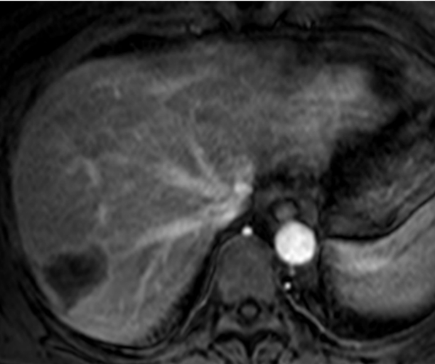
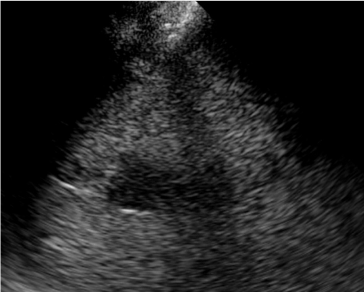
A percutaneous radiofrequency ablation of the site of liver metastasis is performed by interventional radiology. Post-procedure images are obtained. What are the relevant imaged findings?
A post-ablation triple-phase (noncontrast, arterial phase and portal venous phase) contrast-enhanced magnetic resonance image (MRI) obtained 1 month post procedure demonstrates absence of arterial enhancement within the ablation cavity, which is routinely larger than the treated lesion in an effort to prevent local recurrence from an incompletely treated tumor margin. The ultrasound obtained immediately post-ablation in the IR suite shows a well-circumscribed diffusely hypoechoic (dark in echotexture) focus enveloping the site of the treated lesion.

