

Case 1
Case 1 - 82 year old man with leg pain after walking one block.
Question 1:
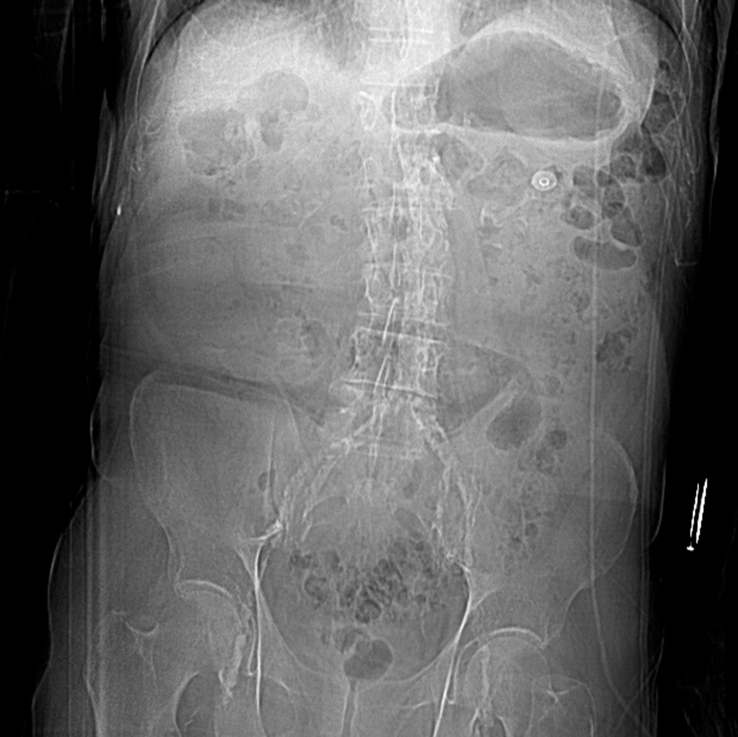
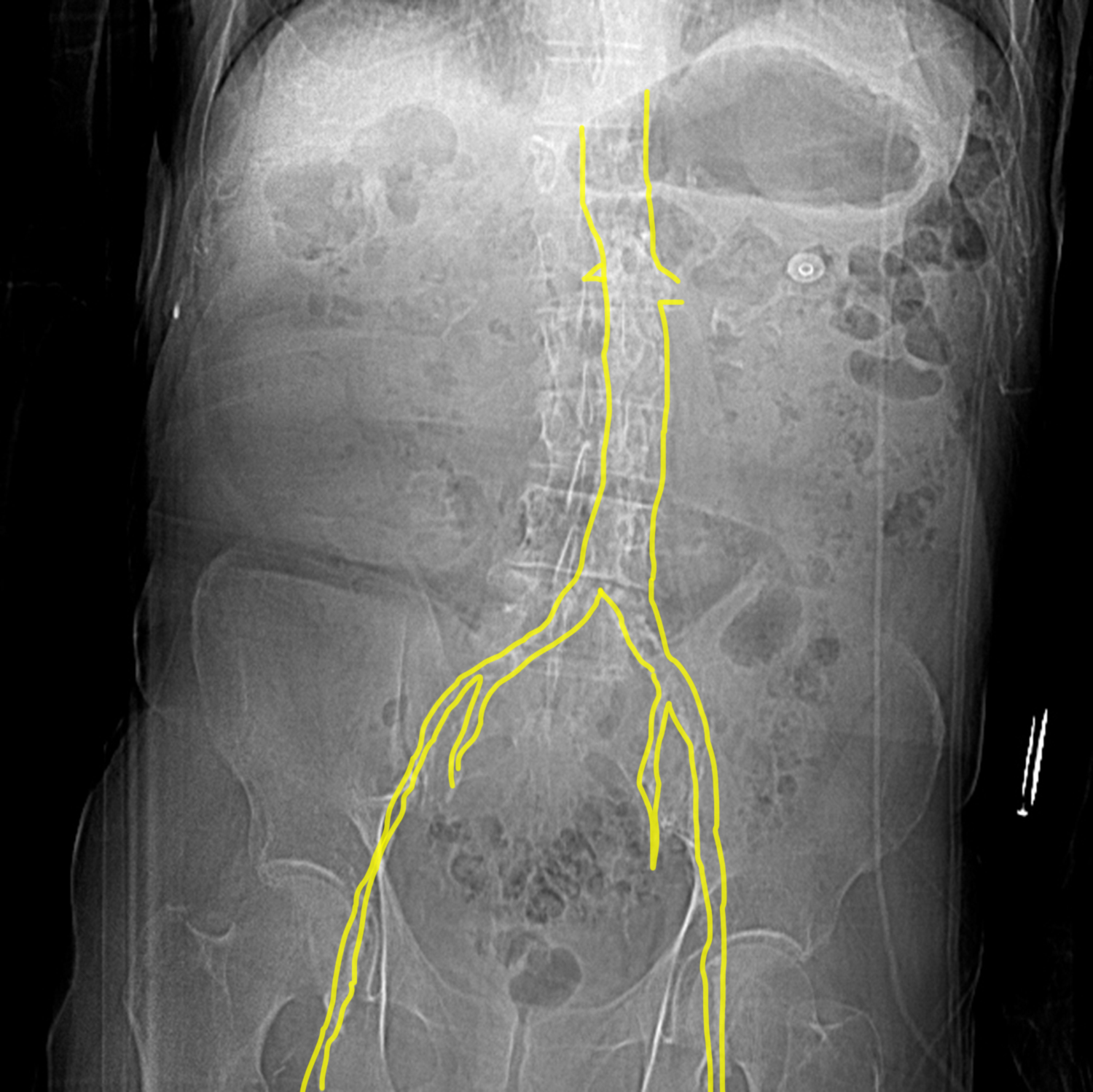
What type of image is this? Do you see anything abnormal that might relate to this patient's symptoms?
This is an abdominal radiograph. The hint below shows that you can see the abdominal aorta and iliac vessels outlined in white. You should NOT be able to see blood vessels on radiographs unless contrast has been injected into them. They blend with other soft tissues on x-ray images because their density is similar.
Question 2:
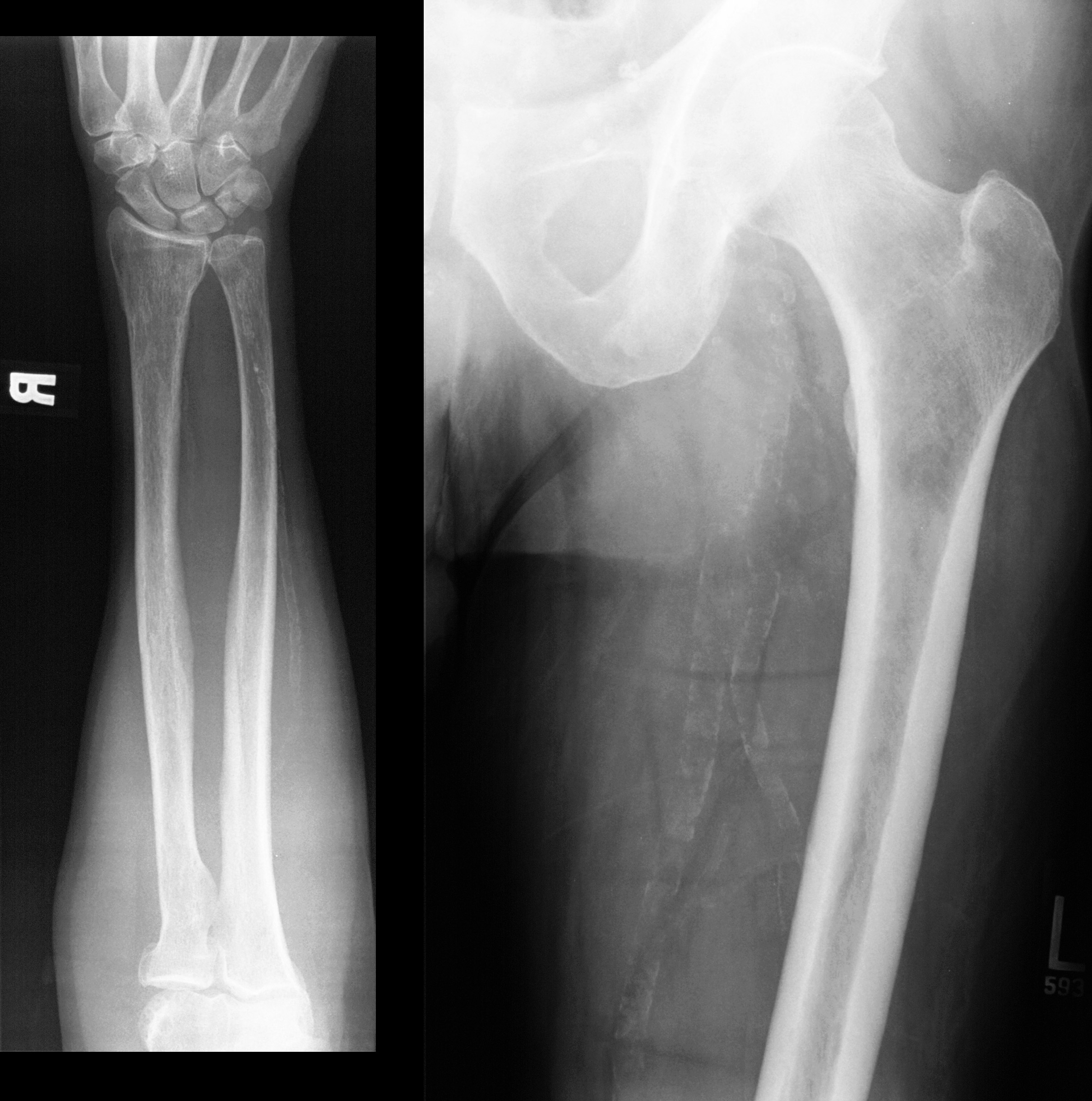
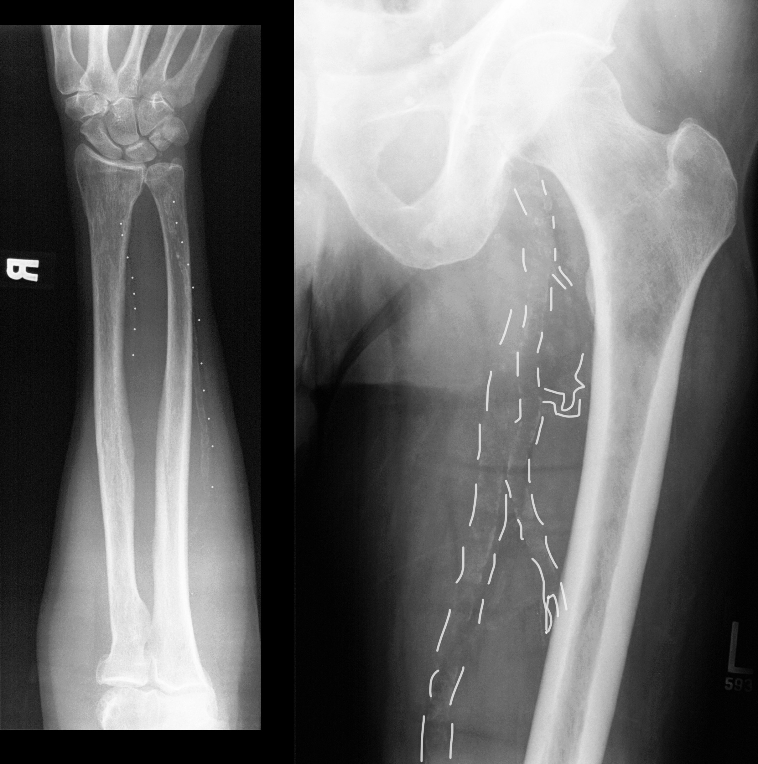
What do you see on these images? First identify the anatomy and then what is wrong.
These are AP radiographs of the femur and the forearm. Again, blood vessels are visible due to thin white lines along each side, indicating extensive atherosclerosis. The ulnar artery, the femoral artery and the profunda femoris artery are particularly densely calcified.
Question 3:
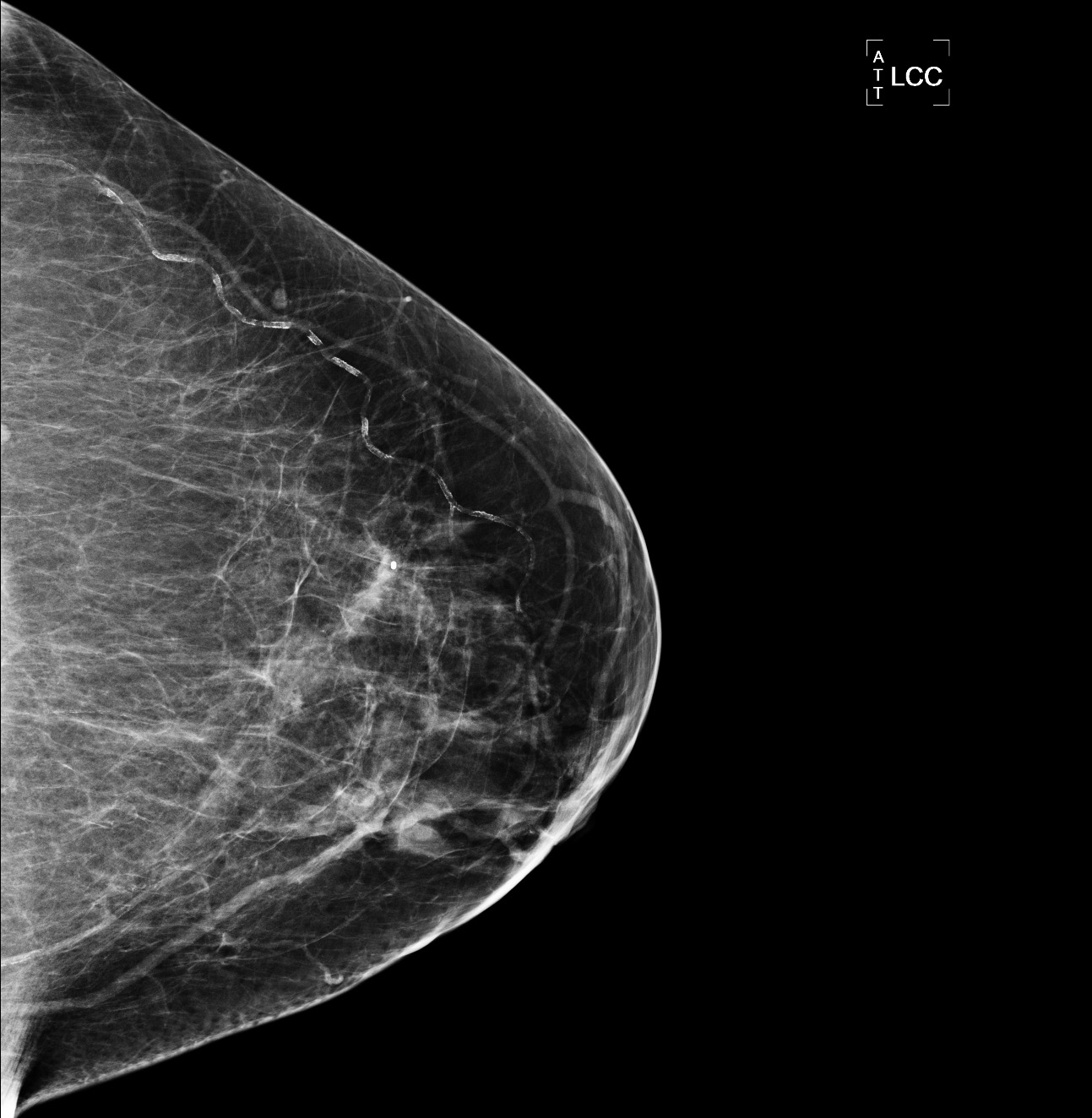
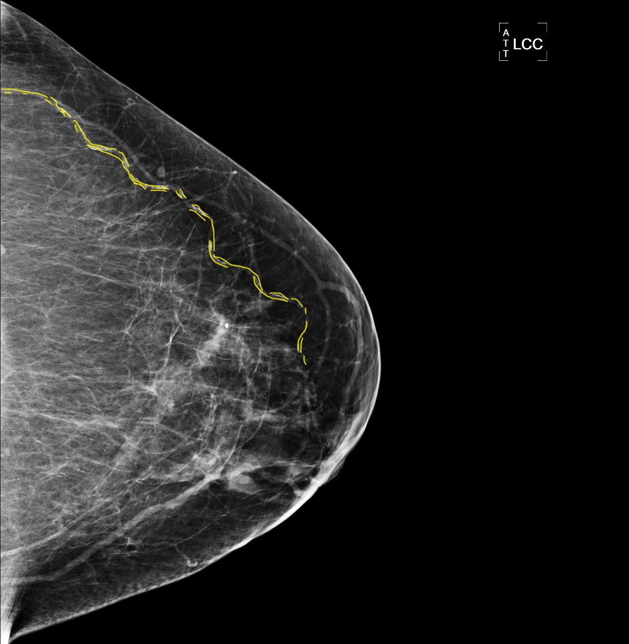
What is this study and what does it show?
These two images are mammograms, a special type of x-ray done of the breast with a VERY low strength (and low radiation dose). The breast must be compressed for the image to be useful, since it is so weak. Again, we see double lines indicating calcification of both walls of vessels, in this case branches to the breast from the internal mammary, lateral thoracic or pectoral arteries.
Question 4:
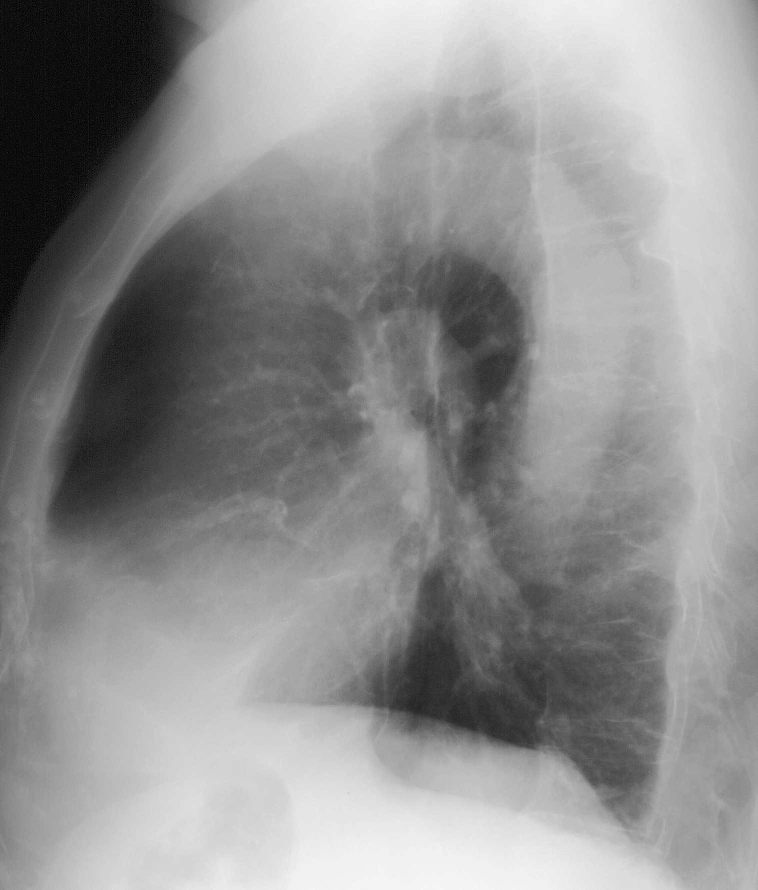
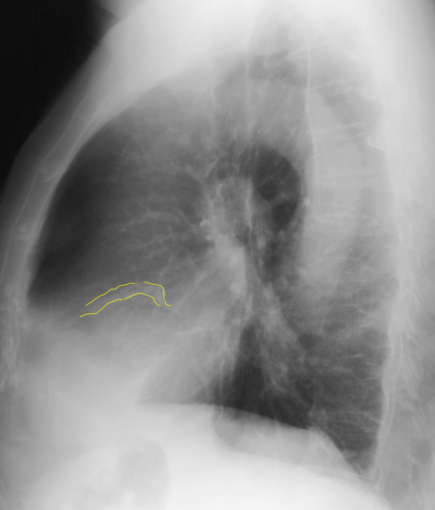
What is this image and what can we see in the region of the heart that relates to the topic of atherosclerosis?
This is a lateral chest radiograph. We do not expect to see the vessels on this type of image, but there are small double lines of white overlying the heart in the location of the coronaries. This pattern of double lines is sometimes called 'tram-tracking', as the lines look like tram tracks in the street.
Question 5:
What is this study? Give all pertinent technical details. What do you think of the vessels? Would intravenous contrast be helpful in evaluation of this finding?
This is a CT displayed in the axial plane, displayed with soft tissue windows, with no oral or intravenous contrast. When looking for vessel wall calcification in atherosclerosis, no IV contrast should be given since it could obscure the calcification. If the vessel diameter is being evaluated, rather than calcification, then IV contrast is helpful.
Question 6:
The previous case demonstrated such extensive calcifications in so many arteries that vascular surgeons sometimes refer to this condition with a term illustrated here. What is this?
This is a photo of a coral reef, and surgeons sometimes call very extensive calcification, particularly if it involves the aorta, as a 'coral reef' aorta. It is very difficult to perform vascular surgery on a coral reef aorta.

Case 1
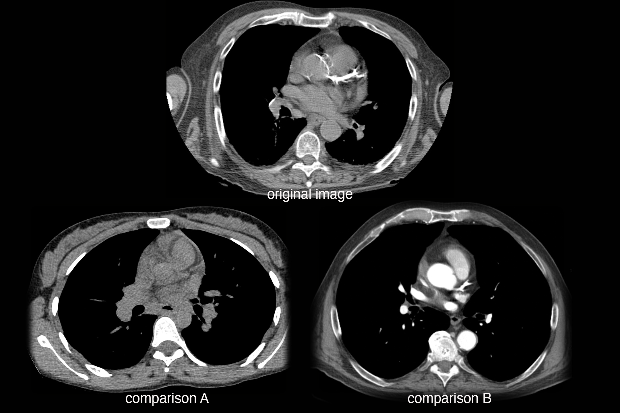
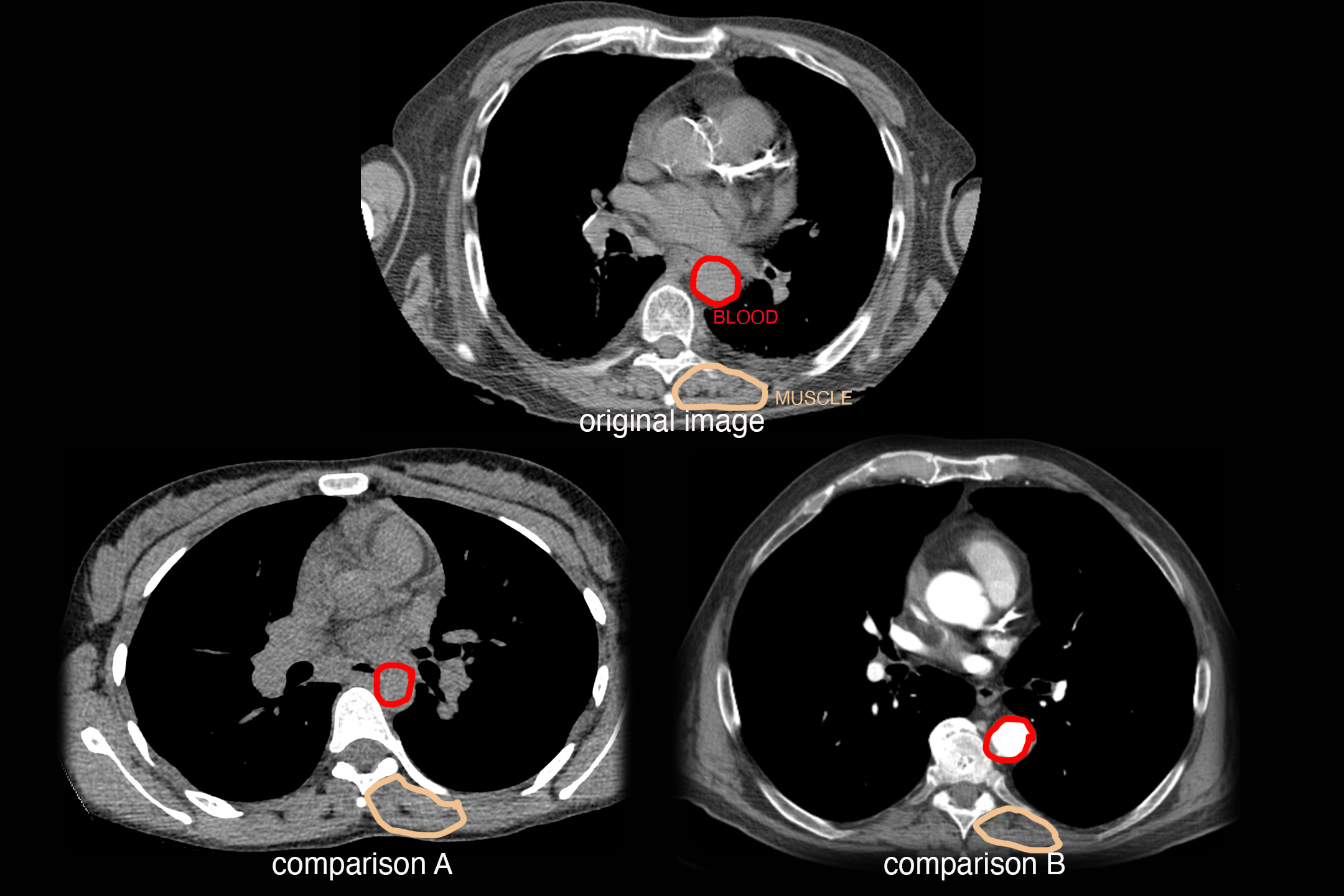
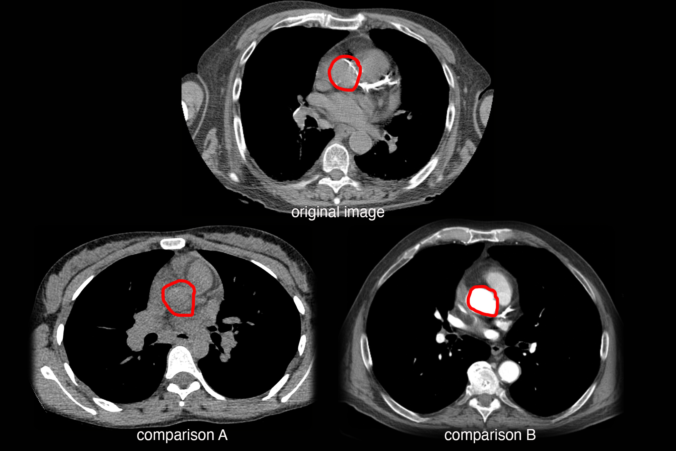
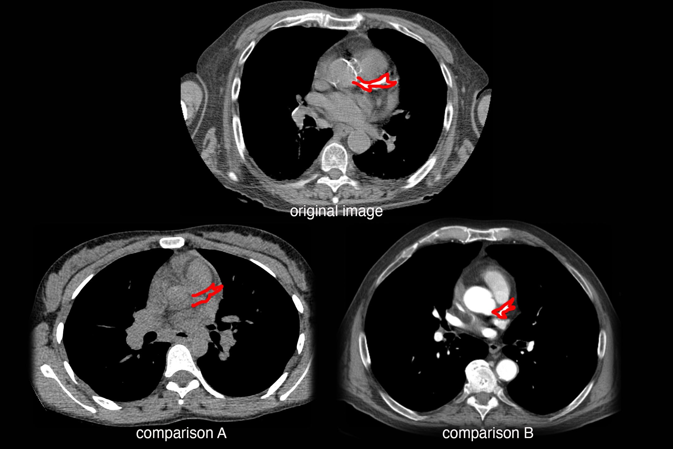
Here is a selected image from the prior CT series, along with two comparison studies from different patients.
Question 7:
What significant vessel appears most abnormal on this image? What are technical parameters for the studies?
In the original image, the left anterior descending coronary artery appears densely calcified, which is highly significant and indicates that the patient is at risk for myocardial ischemia. This study was done without IV contrast, and is displayed with soft tissue windows in the axial plane. For comparison A, no IV contrast was given and the vessels do not appear calcified. For comparison B, IV contrast was given and is filling the aorta and left coronary artery.
