


Case 2
One of the main imaging methods for the heart is cardiac echo. This is a challenging imaging study because the heart is encased in tissues that block ultrasound beams: bones of the chest wall and air-filled lungs on either side. There are only a few spots where a small ultrasound (US) beam can be effectively directed toward the heart.
Further Explanation:
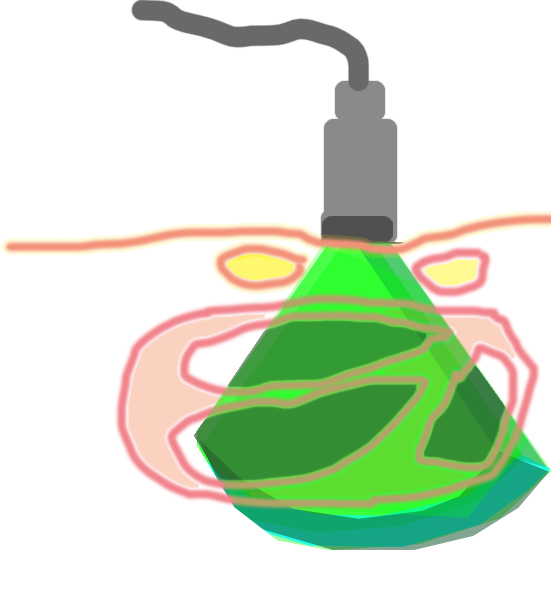
It is also important when looking at US images to realize that you are viewing a slice through the body, with the exact orientation depending on how the device is positioned and angled by the operator. Because the field of view is so small, labeling of images is crucial to show where the imaging is being done. For cardiac echo, special terms are often used for the standard views. The images are always shown with the transducer at the top of the image.

Further Explanation:
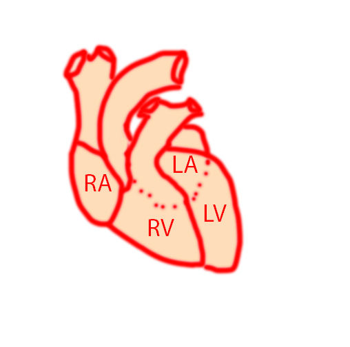
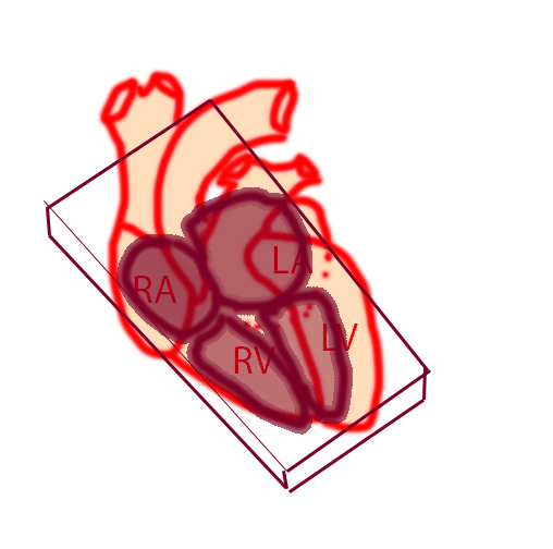
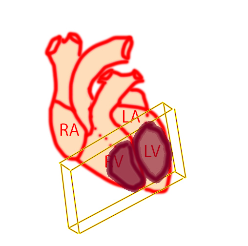
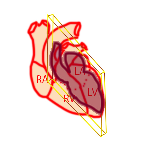
Some of the standard imaging planes for the heart are shown here. The apical four-chamber view is somewhat like a coronal plane, but angled for each individual patient to optimize the view of all four chambers. The 'apical' refers to the fact that the beam is coming from the apex toward the base of the heart, so the device (called a transducer) is placed on the skin either pointing up from under the xiphoid process, or from a left anterior intercostal space. If the transducer is turned 90 degrees from this position, this will give a short-axis view, which is somewhat like an axial slice. Again, the actual angle is adjusted by the operator to optimize the view for each individual patient. For the long-axis view, the transducer is rotated so that its long side is parallel to the edge of the sternum, and moved up and down to find an appropriate anterior interspace (between costal cartilages) to give a view that is similar to a sagittal slice.
Case 2
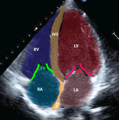
Normal four chamber view
Further Explanation:
Try to identify the chambers and valves in this view before clicking on the labels. This movie shows normal left ventricular contraction. Try to compare the appearance at diastole and systole on these images, which is used to calculate ejection fraction.
Case 2
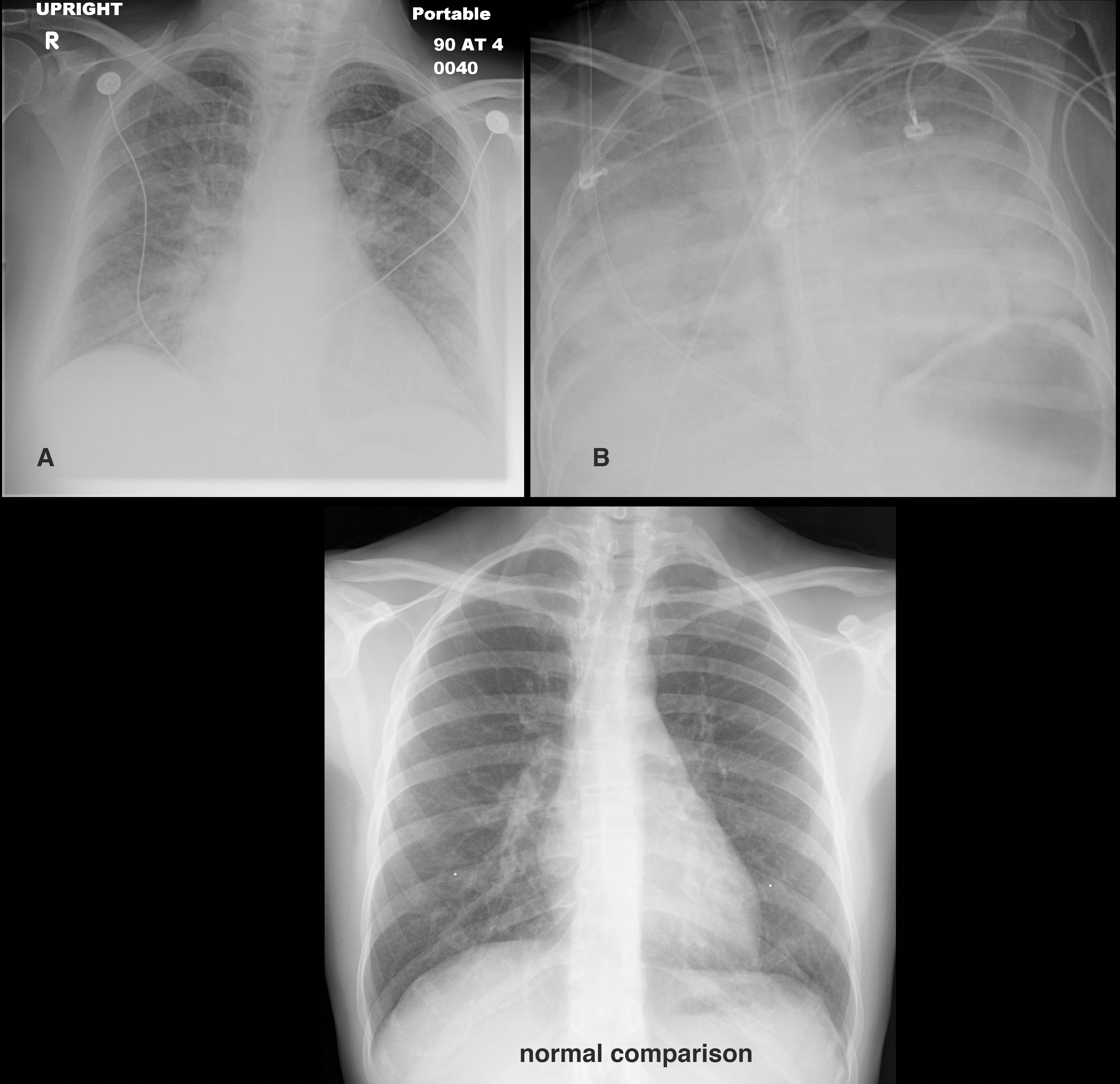
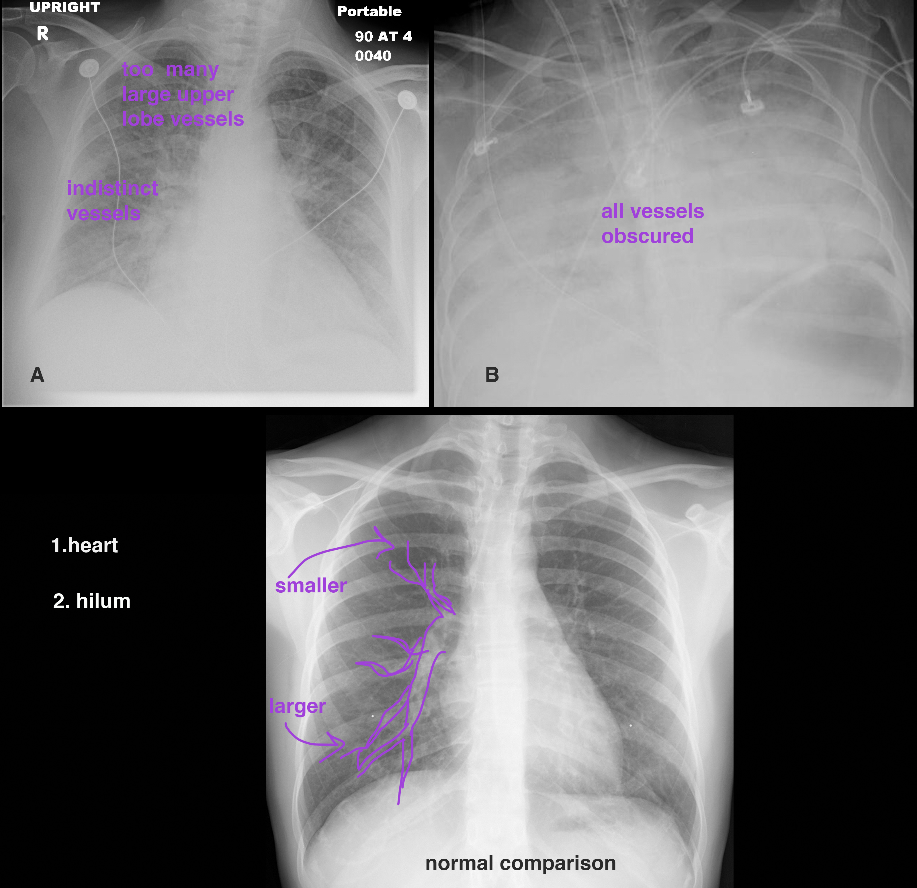
The chest radiograph (CXR) is a cost-effective and relatively sensitive way to assess for possible left ventricular dysfunction. You are shown two patients with symptoms of congestive heart failure (CHF), and a normal comparison radiograph. What areas should you focus on when assessing a CXR for possible CHF?
Further Explanation:

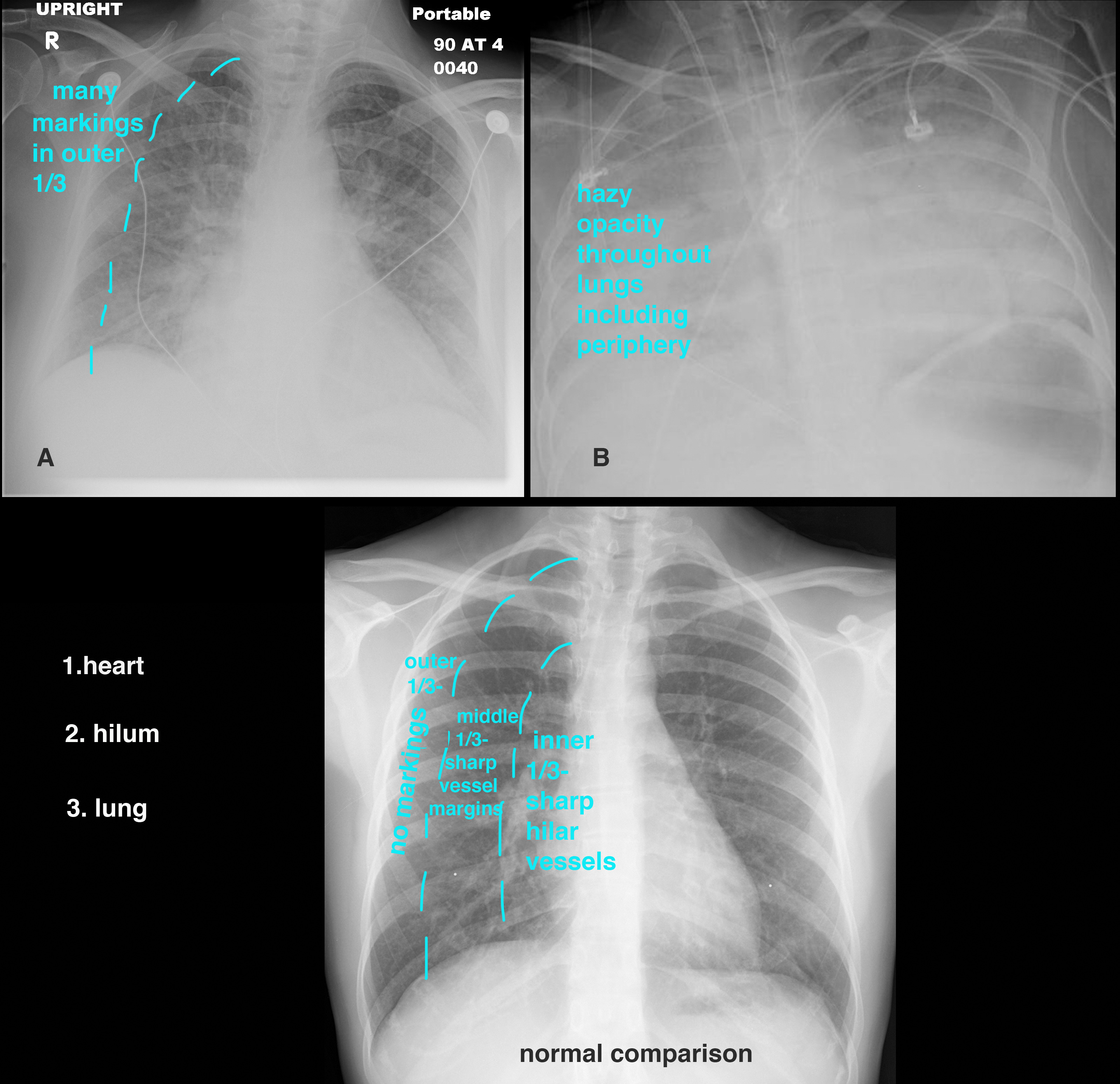
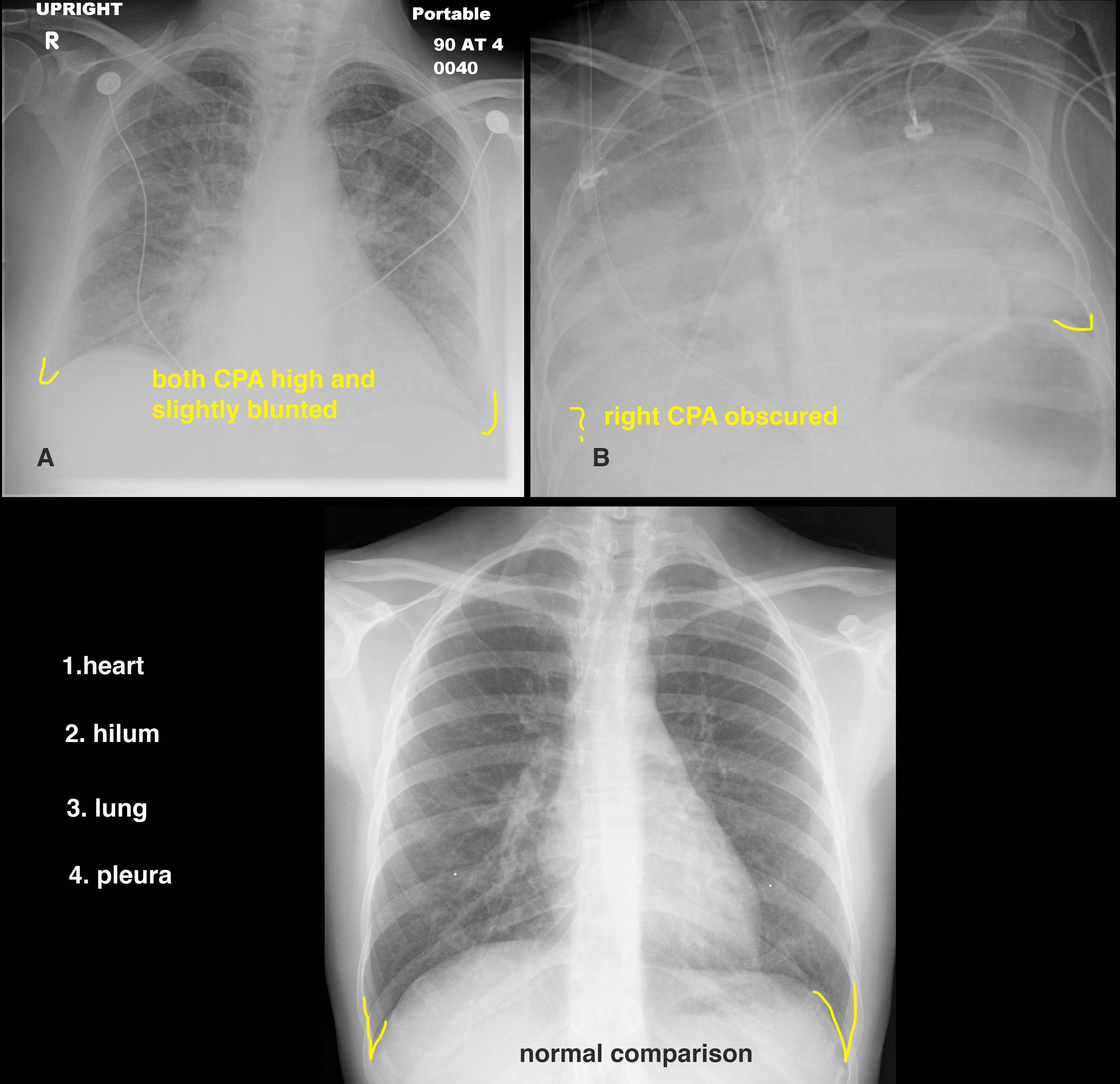
Case 2
This is another apical four-chamber cardiac echo.
Question 1:
How does this study compare to the previous normal case?
The left ventricular wall motion is minimal. In fact, when the ejection fraction was calculated, it was about 25%, which is indicative of left ventricular dysfunction.


