

Case 3
This patient has clinical symptoms suggestive of aortic stenosis.
Question 1:
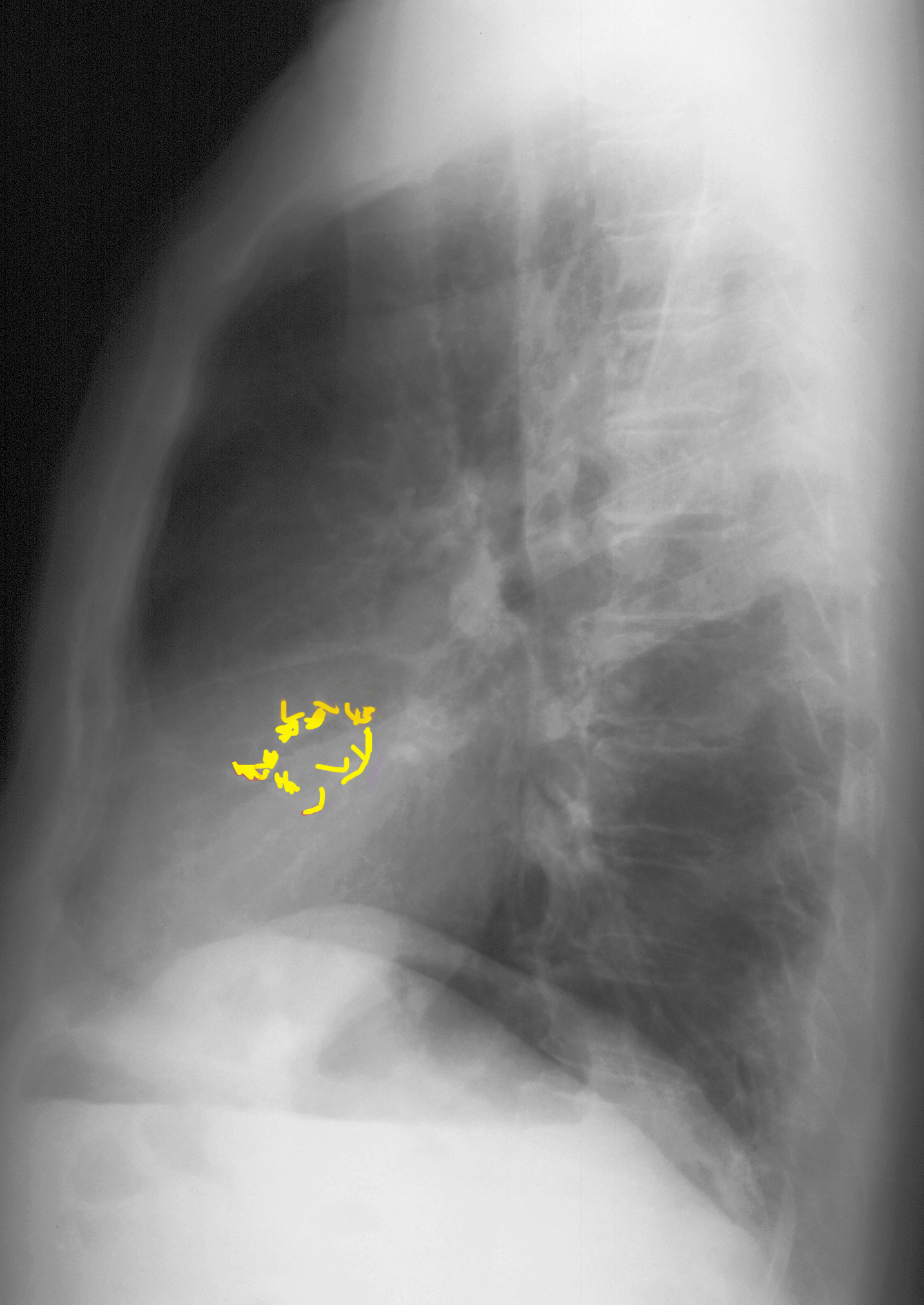
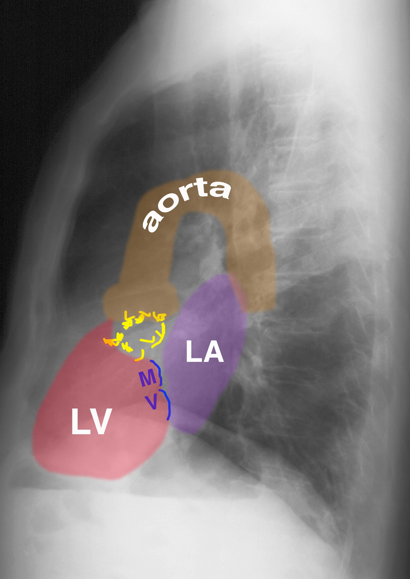
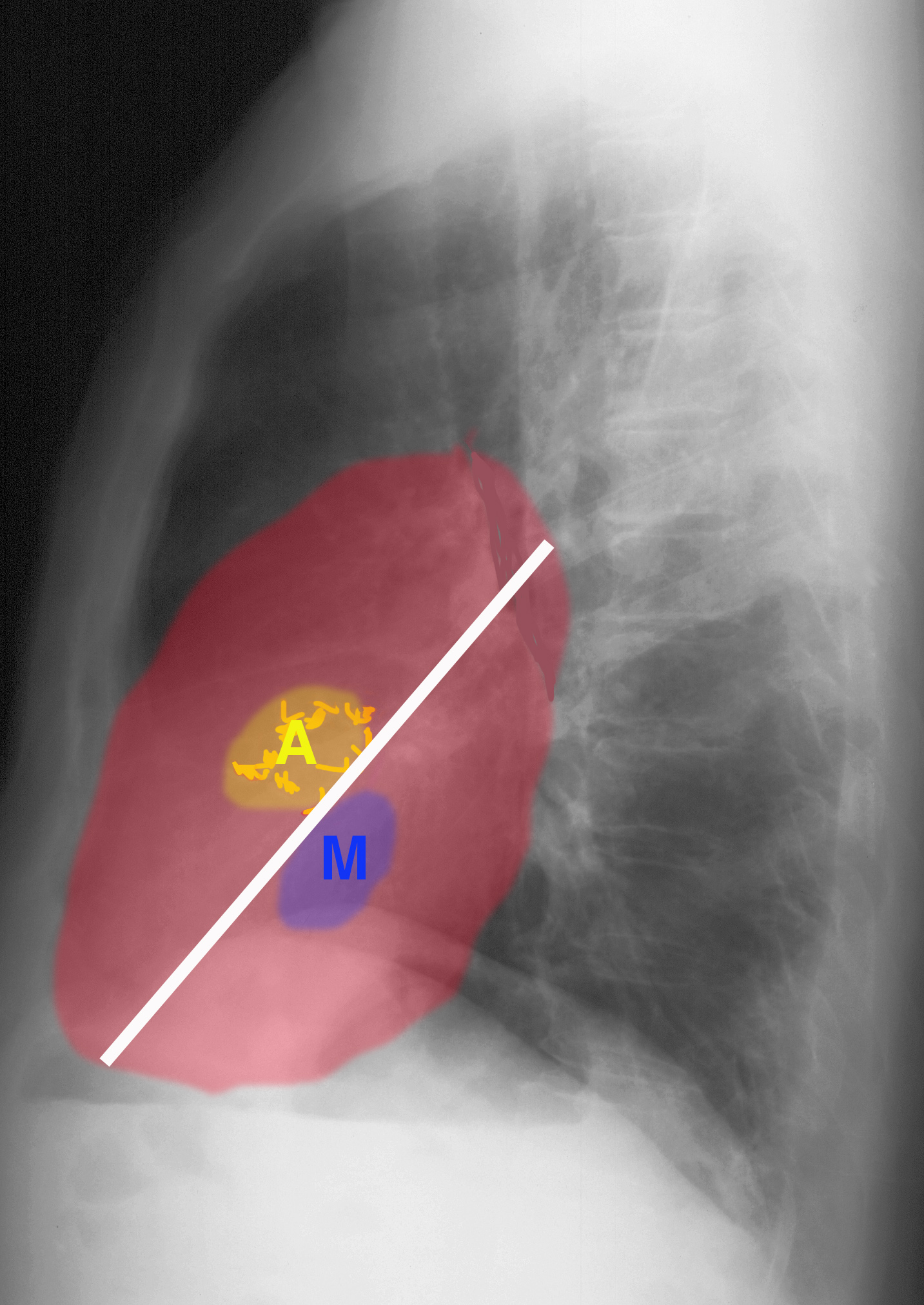
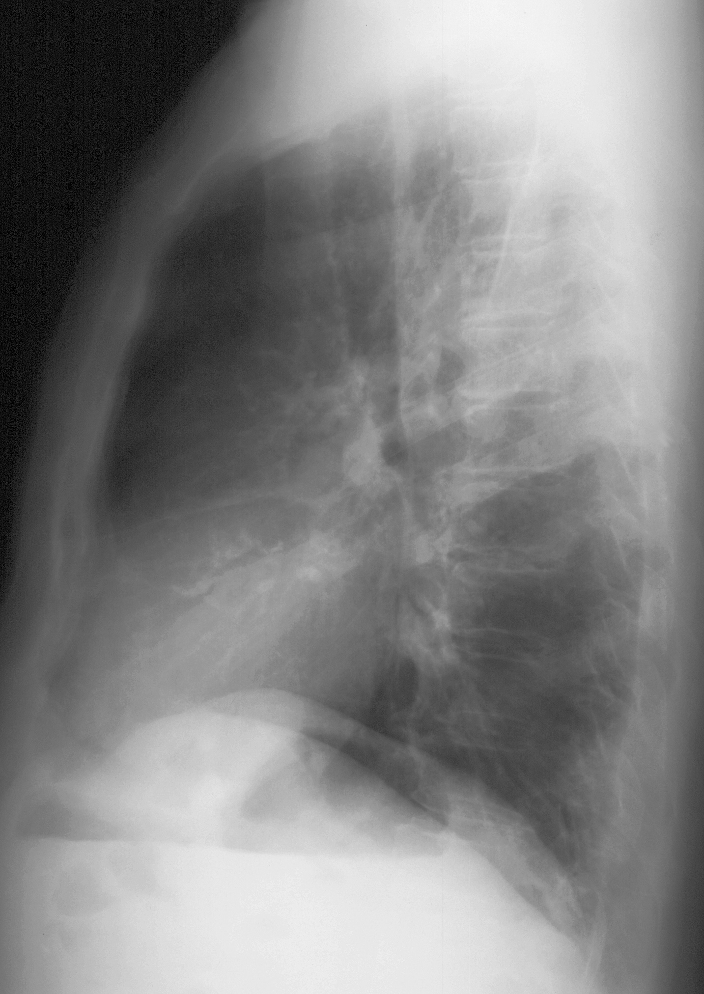
What do you think of this lateral chest radiograph? Where is the aortic valve located within the heart?
If you check the labels, you can see that there are irregular calcifications projecting right in the center of the heart, and they are not shaped like the typical tram-tracks of vascular calcifications. The aortic valve is located in the center of the heart.
Case 3
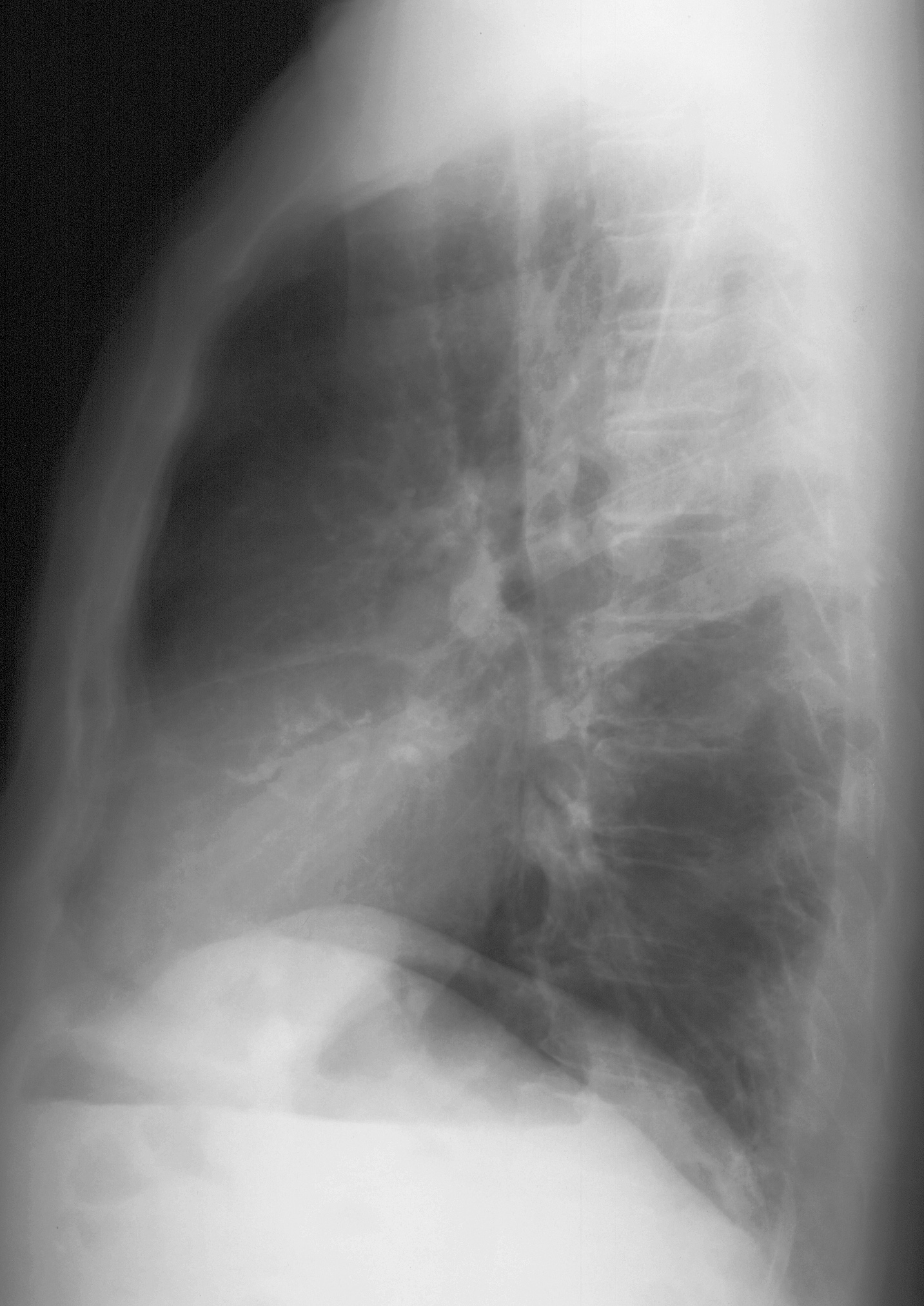
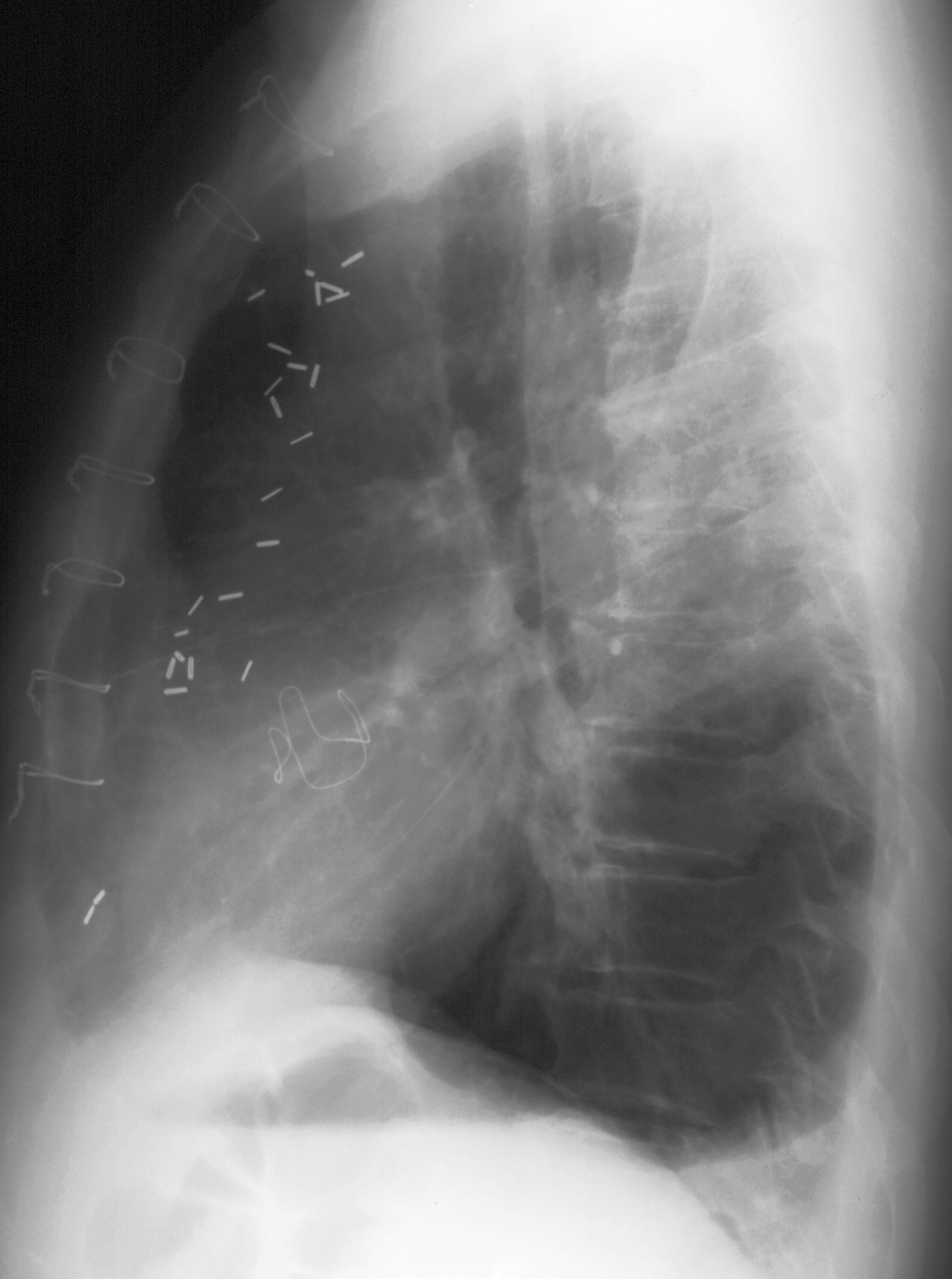
This patient underwent an aortic valve replacement for aortic stenosis.
Question 2:
What looks different on this post operative lateral compared to the pre operative image?
There are new sternal wires and mediastinal surgical clips, but the irregular calcifications are gone and an artificial valve is in their place.
Case 3
This is another and more sensitive way to detect aortic valve problems than a chest radiograph.
Question 3:
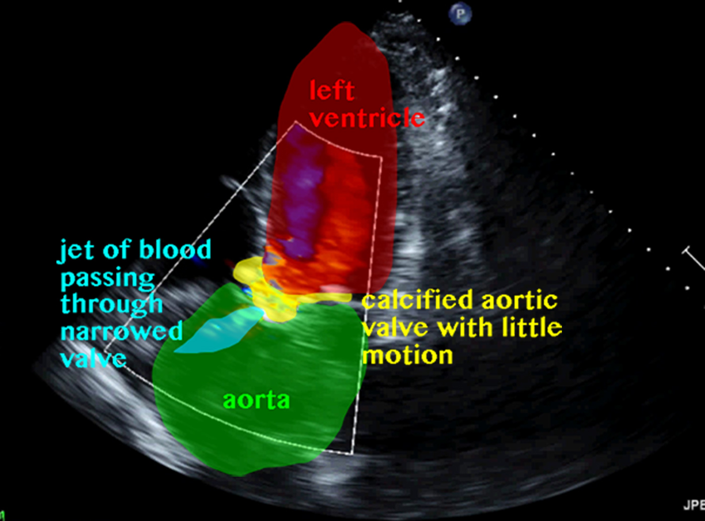
What is this study and what view is this?
This is a special kind of ultrasound called a Doppler. Within the box outlined in white on the image, flow is depicted with color. The colors do NOT reflect arteries and veins, but rather flow toward vs away from the transducer. This view is a variation on an apical four-chamber view where you slightly change the angle of your plane until the aortic valve comes into view in the center of the image. You can see that the flow across this valve is very turbulent, with a mix of red and blue. This is indicative of tight aortic stenosis.
Case 3
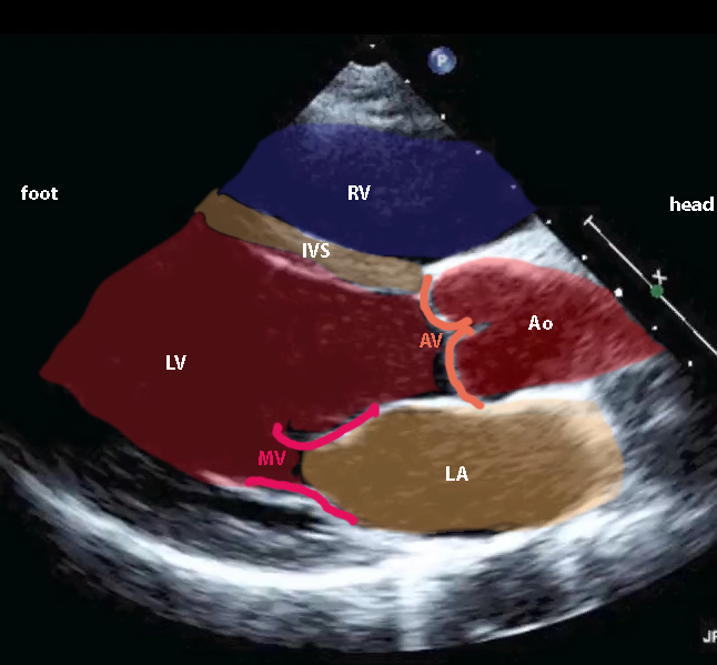
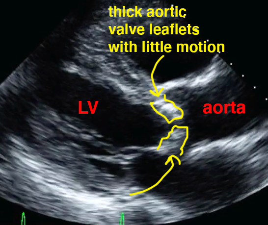
This is another cardiac echo view that gives a good look at the aortic valve. This is a normal example. You can click on the links to see a labeled view and an example of aortic stenosis.
Question 4:
What is this view?
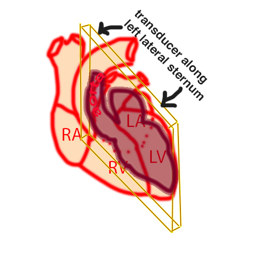
This is a longitudinal or long-axis view of the heart, often done to the left of midline to show the left chambers and valves. It is called a parasternal long axis view (PLAX, for short) because the transducer is along the left margin of the sternum. It produces a view that is somewhat like a sagittal slice. Unfortunately, it is typically displayed with the HEAD of the patient to the right of the image (not the way radiologists display sagittal images), so you need to flip the right and left to get this view.
