

Case 4
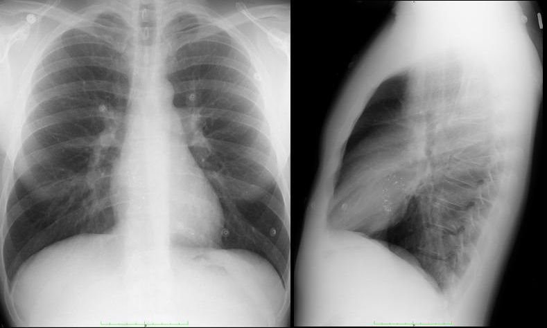
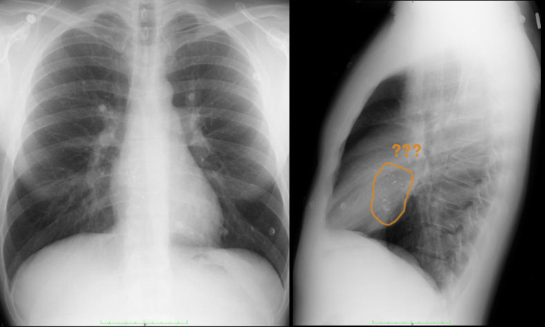
This is a chest radiograph of a patient with a murmur since childhood, and had some sort of treatment in late childhood but they are not sure exactly what was done.
Question 1:
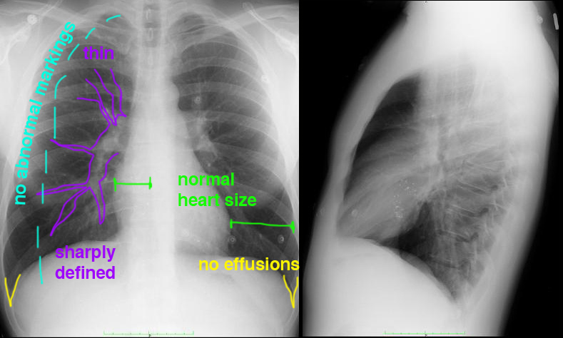
What do you think of the PA and lateral views?
The PA view looks essentially normal. Check out the labels to review what to look at: heart, hilum, lung and pleura. However on the lateral view there are opacities overlying the heart that are unusual looking.
Case 4
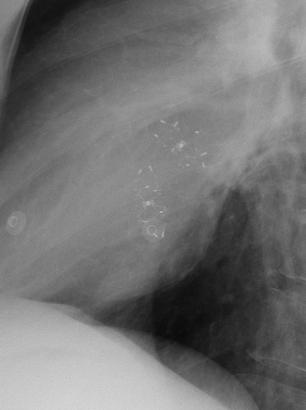
This is a closeup view of the heart from the previous lateral view.
Question 2:
Do these things look like calcified vessels or valves?
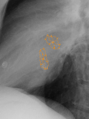
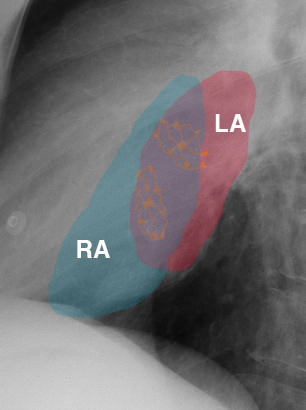
No! They look to regular in shape, and appear to be some sort of foreign object. Looking at the labels below for the chambers of the heart in the region, and knowing that the patient had a history of some sort of congenital heart problem that was corrected, what could they be?
Case 4
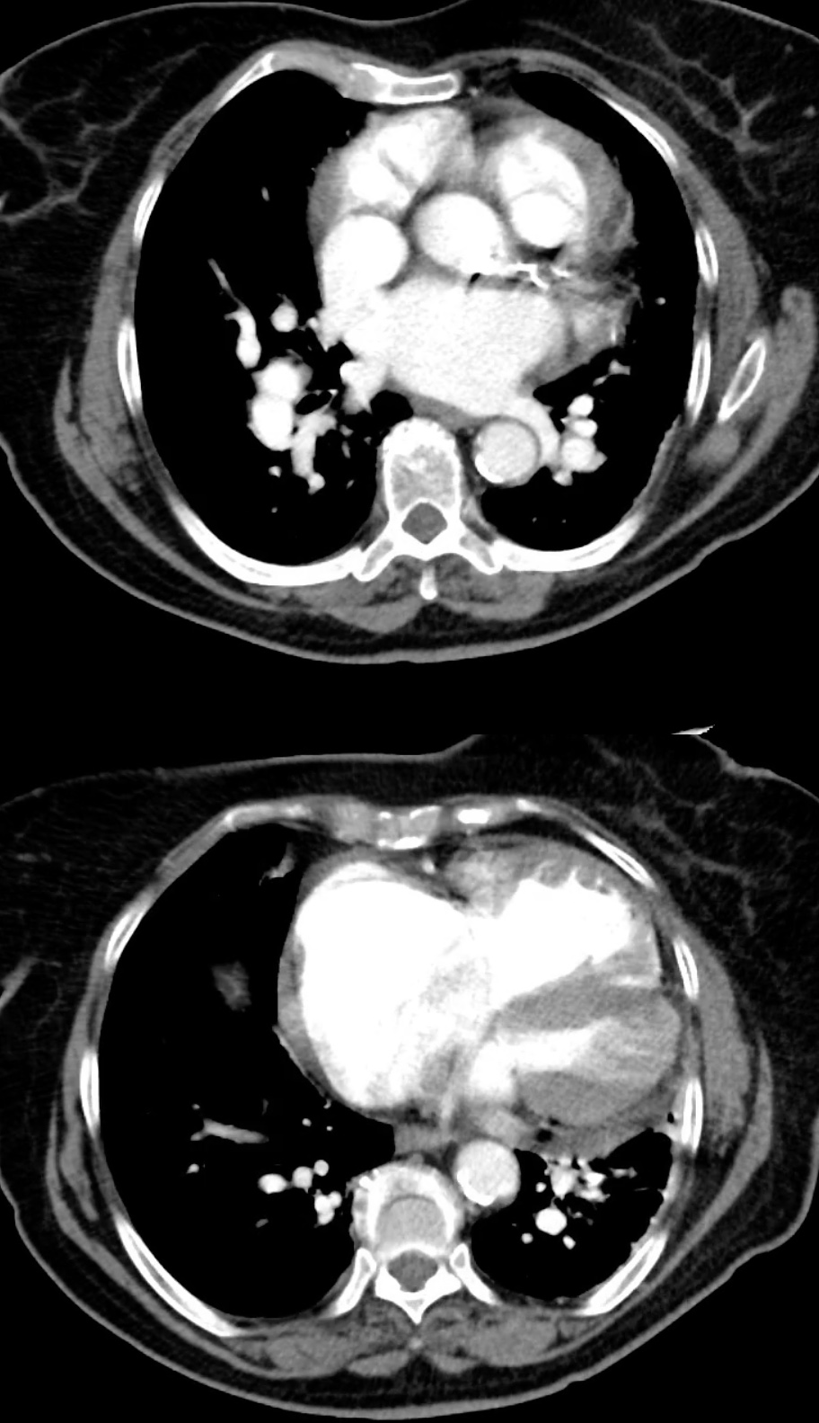
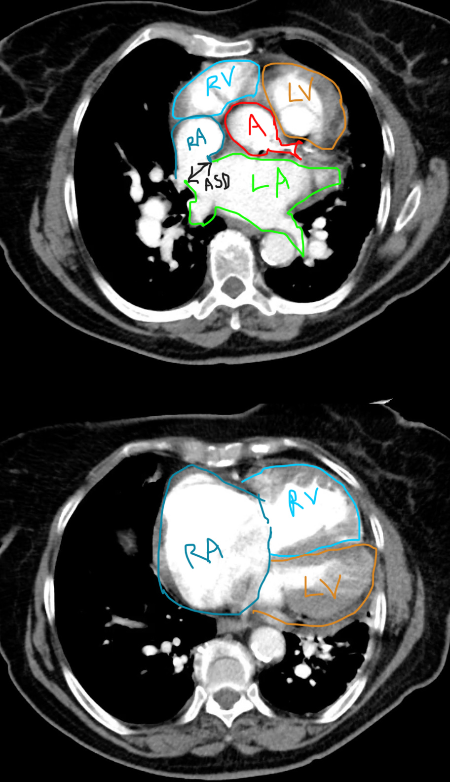
The previous patient had a minimally invasive closure of their ASD using intravascular devices that can be inserted via a catheter. This is a CT study on a different patient with a long-standing uncorrected ASD.
Question 3:
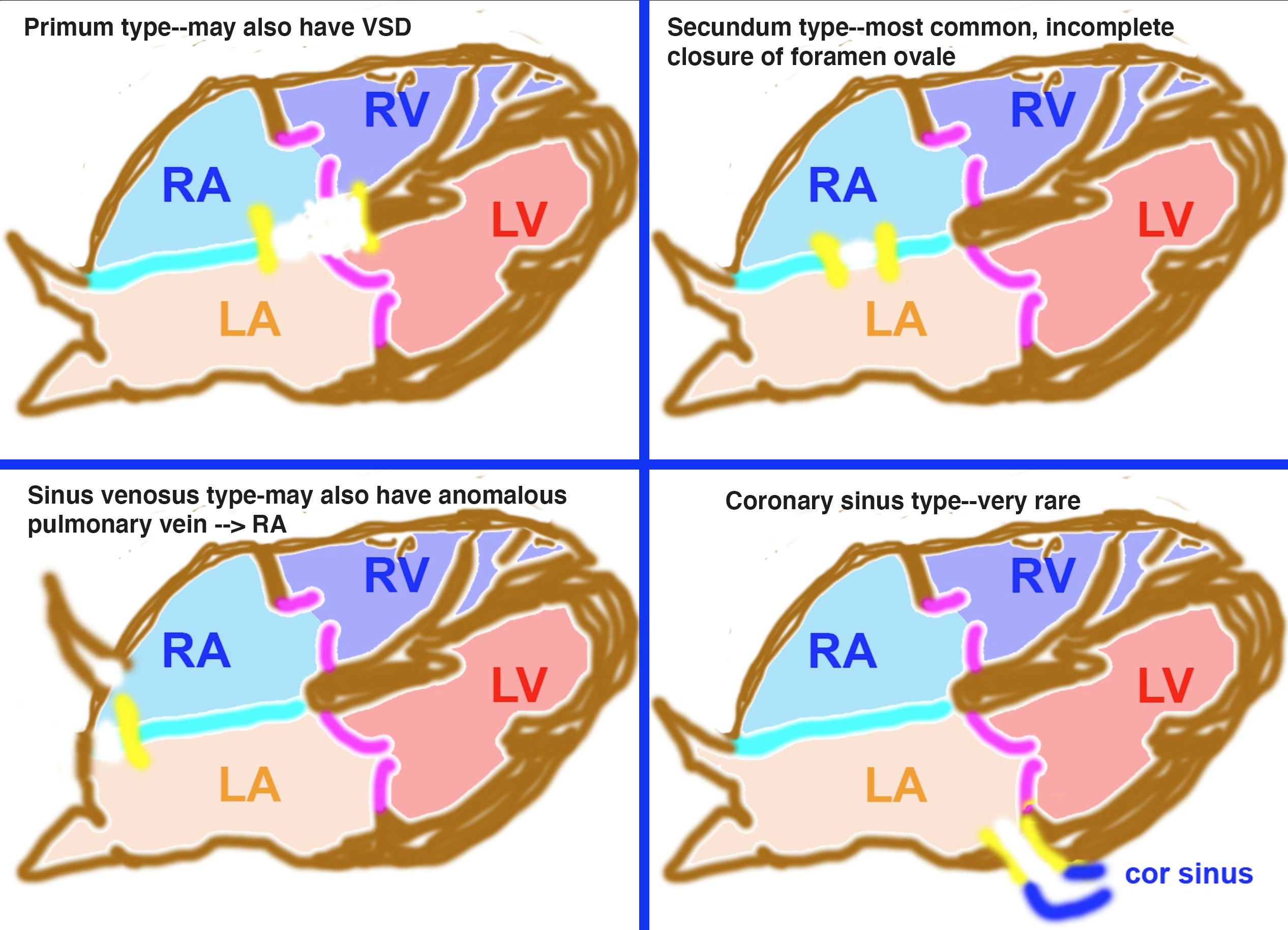
Try to identify the defect before checking the labels. What other chambers look abnormal?
The right atrium on the bottom image is very enlarged. The left atrium is also somewhat enlarged on the top image. And the muscular wall of the right ventricle appears thickened on the bottom image. All can be attributed to the overload of volume from this large ASD, probably sinus venosus type.
Case 4
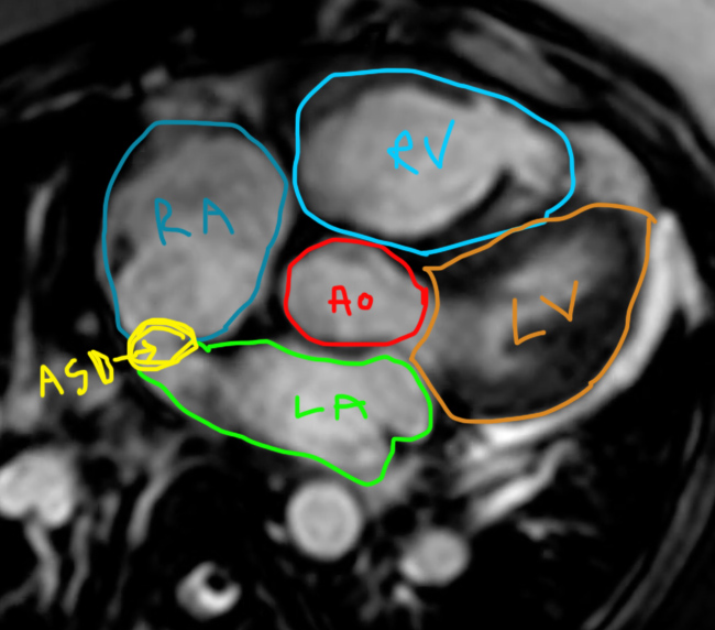
Other modalities are often used to evaluate congenital heart defects.
Question 4:
What is this study? What advantage would it have over CT?
This is an MR, and its main advantage is that there is no radiation involved. US also shares this advantage and is often used to evaluate congenital heart problems and to quantify the amount of shunt that is present.
