

Summary
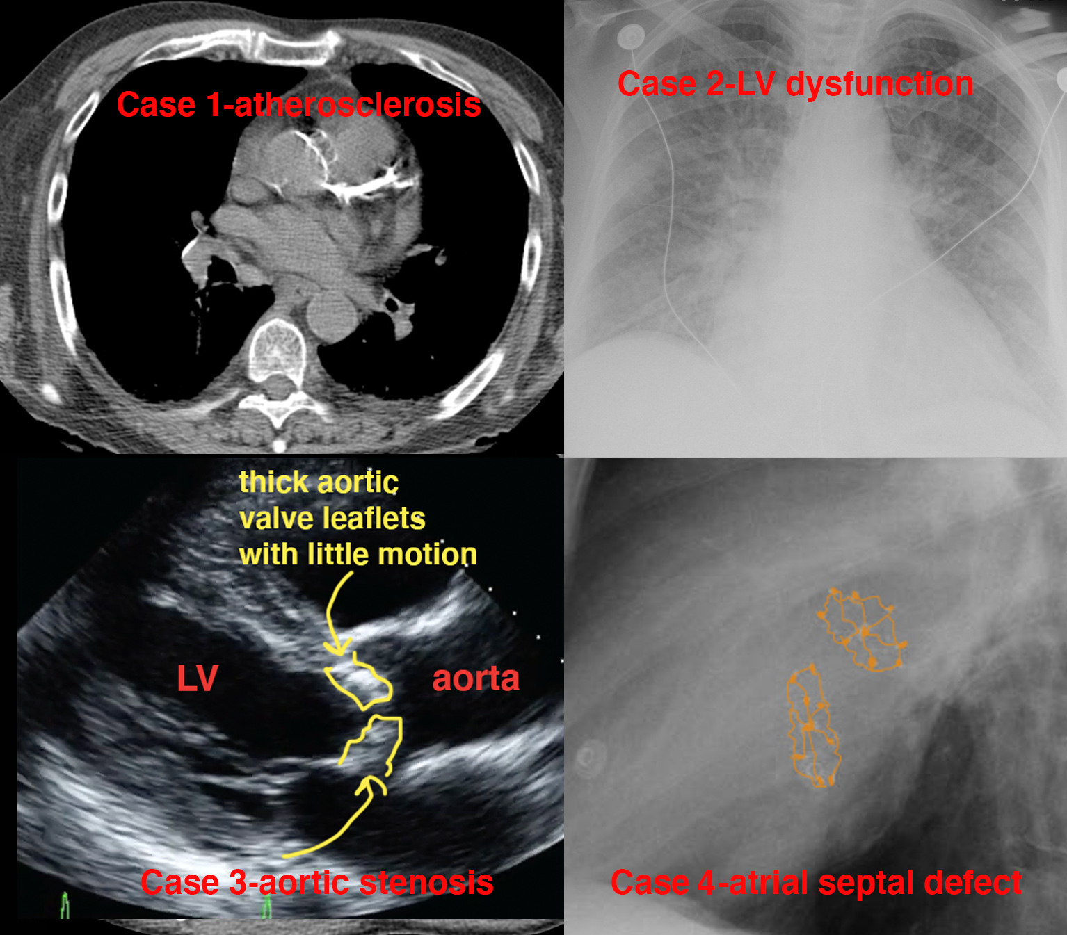
This is a summary of the imaging that was covered in this pre-class module.
Further Explanation:
1. Identify findings on imaging studies that indicate atherosclerosis--we reviewed typical tram-track appearance of abnormally calcified vessels on radiographs, mammograms, and CT, and showed one US image of normal carotids.
2. Recognize features of left ventricular failure on cardiac echo and CXR--we reviewed some standard cardiac echo views, including apial four-chamber, which allows evaluation of left ventricular wall motion, and compared normal to a patient with a very low ejection fraction. We discussed a systematic way to look at CXR for changes in heart size, hilar sharpness/vascular distribution, peripheral lung markings, and pleural effusions.
3. Describe typical CXR features of aortic stenosis, how to identify the aortic valve on radiographs and what aortic stenosis looks like on US--we saw that a calcified valve may be visible, particularly on a lateral view, and that the aortic valve is central in position in the heart, above a diagonal line across the heart on a the lateral view. We reviewed the appearance of aortic stenosis on Doppler US and on the PLAX view of cardiac ultrasound.
4. Demonstrate possible locations for ASD, and what types of imaging can be used to demonstrate these congenital defects--we reviewed the four basic types of ASD, and saw the appearance of closure devices on CXR, as well as how ASDs can look on MRI and CT, and discussed that ultrasound is also very useful for diagnosing and quantifying shunt fraction in ASD.

Further Explanation:
This is the end of the PrISM-CV pre-class cases for Imaging Correlation. Click here to return to the Case List.


