

Imaging Anatomy Pelvis Case 1
This is a 51 year old patient (pronouns: she, her, her) with pelvic pain
Question 1:
a) What are pertinent technical parameters for this study?
This is a CT study (cortical bone is white), in the axial plane, displayed with soft tissue windows, with oral and IV contrast.
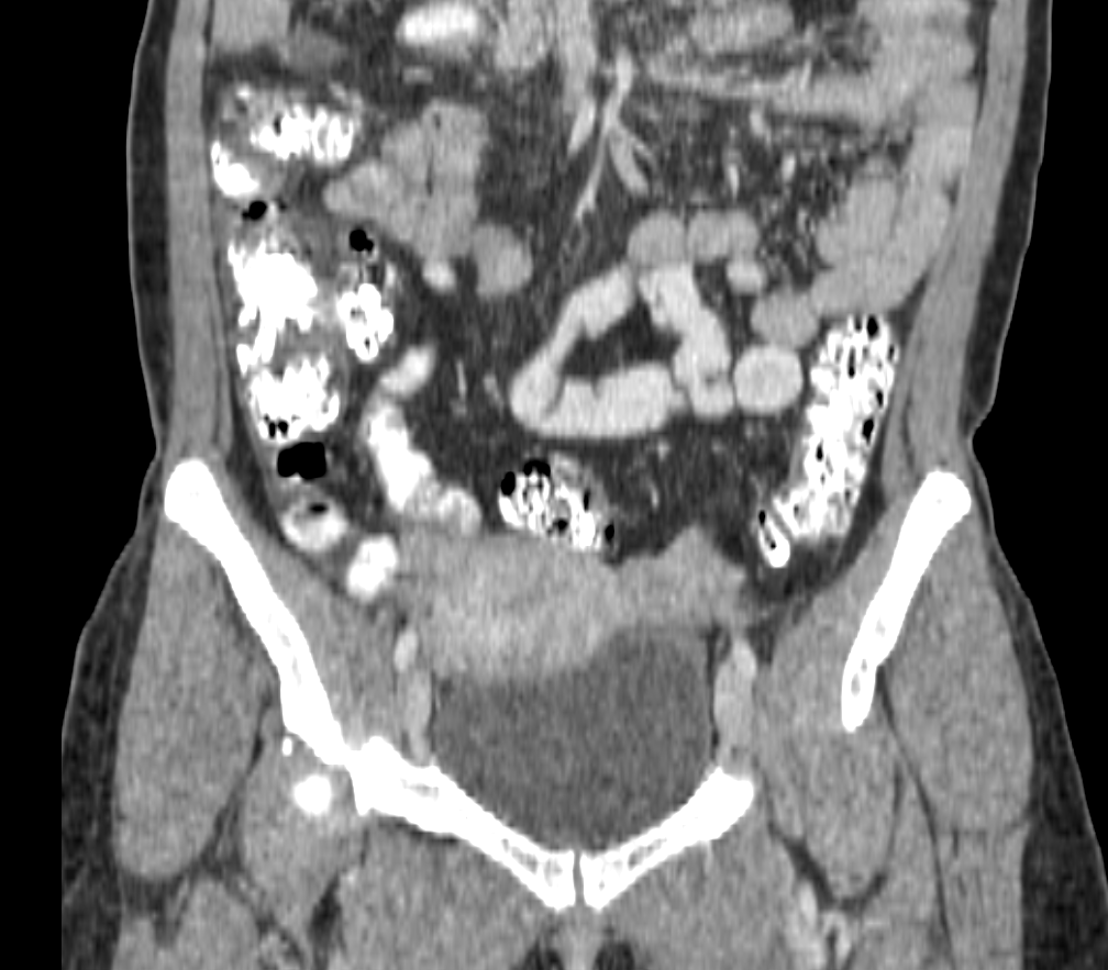
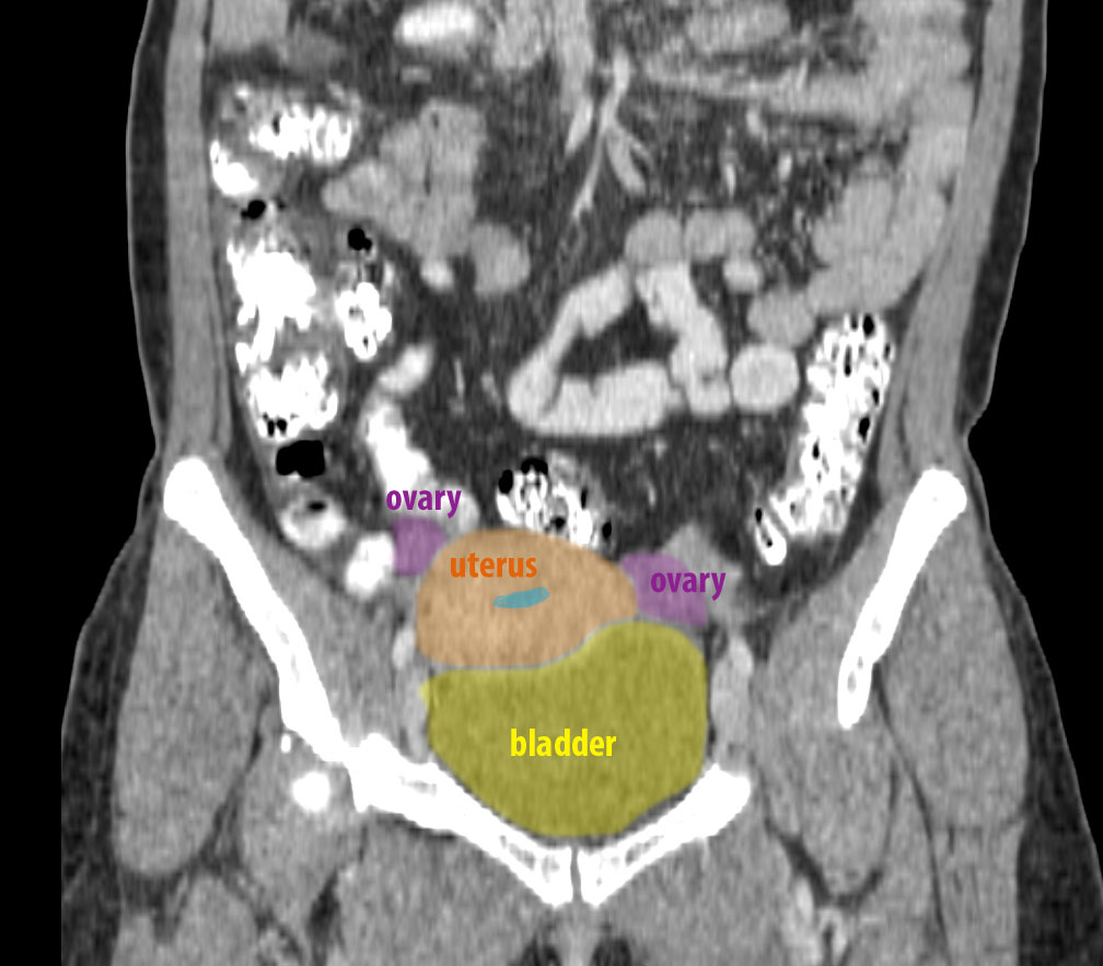
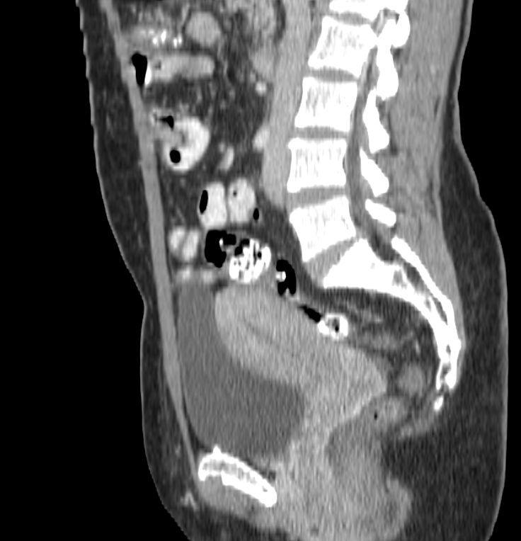
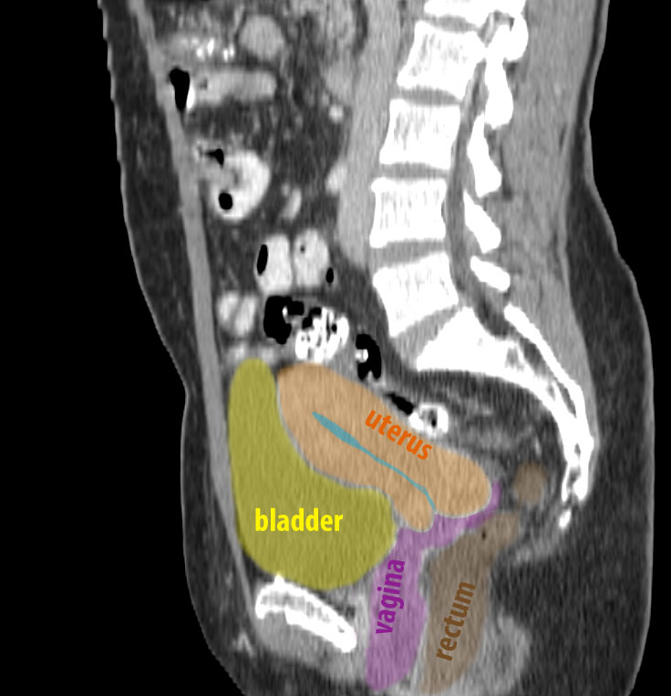
b) You are shown selected images below in other imaging planes. Is the uterus normal in position? What imaging plane is best for assessing uterine position?
It is easy to see on the sagittal image that the uterus is anteverted (meaning that the fundus is anteriorly positioned, sitting just superior to the bladder), which is normal. Uterine position can vary a lot from one patient to another, and the uterus can also move in one patient at different points in time. On the coronal series you can see that this uterus is slightly to the patient's right, which is also quite common and would be considered normal (unless there was a large mass pushing the uterus over, which is not present in this case).
Imaging Anatomy Pelvis Case 1
This is another study that can be done to evaluate the uterus.
Question 2:
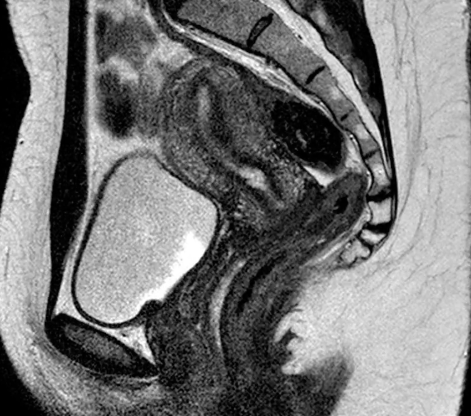
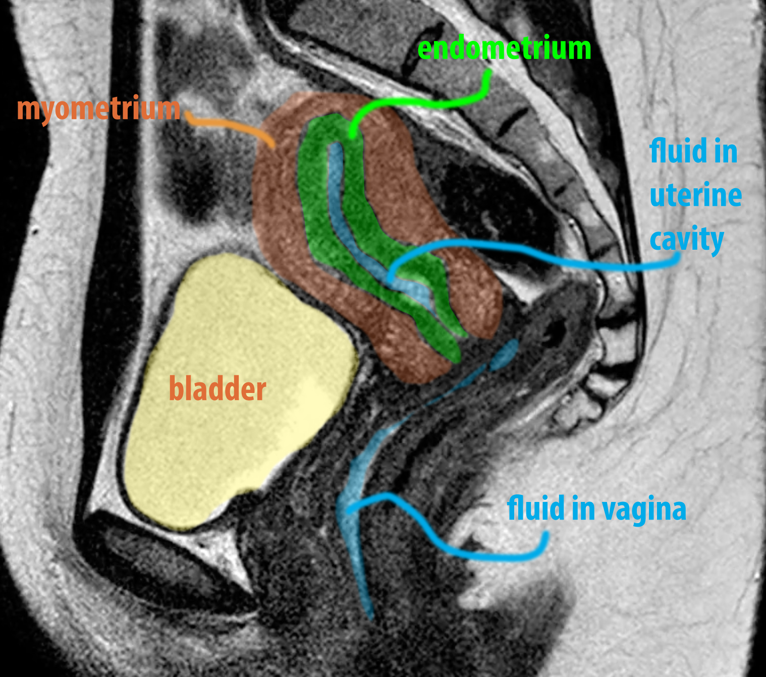
a) What are technical parameters for this study? How does the appearance of the uterus differ from what is seen on CT?
This is an MR (cortical bone is dark) in the sagittal plane, with T2-weighting (fluid in bladder and uterine cavity are bright) and no fat saturation (fat is bright). MR shows more internal detail of the structure of the various layers of the uterus than can be seen on CT. So while CT can show the overall shape, size and position of the uterus, it is not as sensitive as MR for identifying problems with the endometrium or myometrium as MR.
b) What other study is often done to evaluate the uterus (mystery study link below)? What specific advantages does this study offer compared to CT?
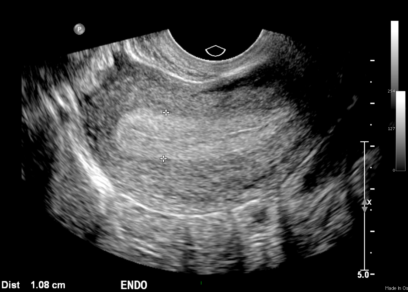
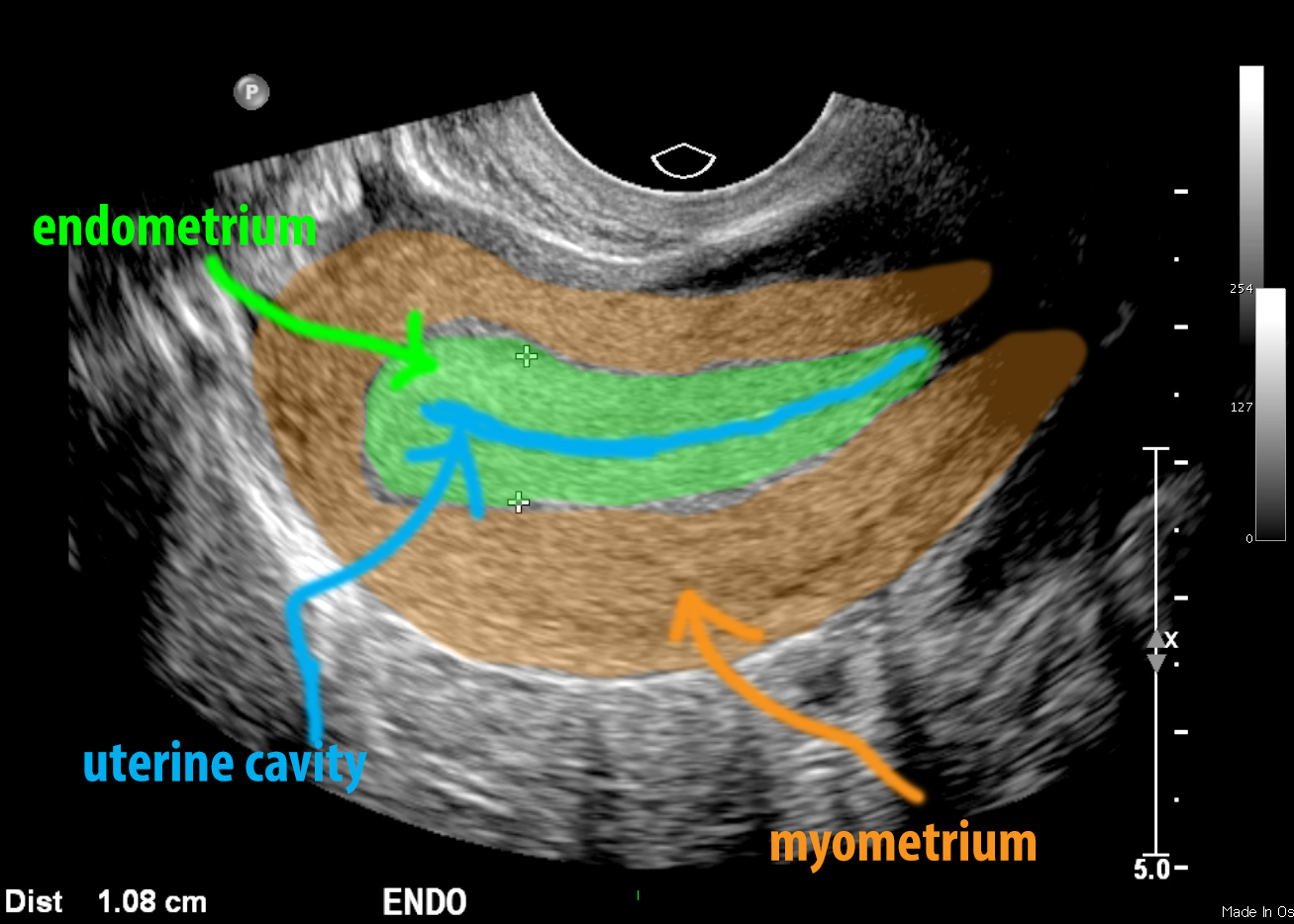
The mystery study below is a pelvic ultrasound. Ultrasound has the advantage of not using ionizing radiation, and it can be done on patients with claustrophobia, and less expensive than MRI. While the field of view can be a limitation if the uterus is enlarged, with careful imaging, the entire region can be examined (which is often done in pregnancy). Like MR, ultrasound shows more detail of the internal structures of the uterus than CT, particularly if a transvaginal probe is used. Imaging can be done either through the abdominal wall (requiring a probe that can penetrate deeper into tissue, but with sacrifice of spatial resolution), or using a probe placed in the vagina (about the size of a tampon, with high spatial resolution but limited penetration through tissue--because the vaginal placement brings it very close to the uterus, it can usually penetrate to give useful imaging of the uterus and ovaries). The movie below shows how the study looks while it is being done. The images are 'real-time', meaning that you can view them as you move the probe to different positions.
Imaging Anatomy Pelvis Case 1
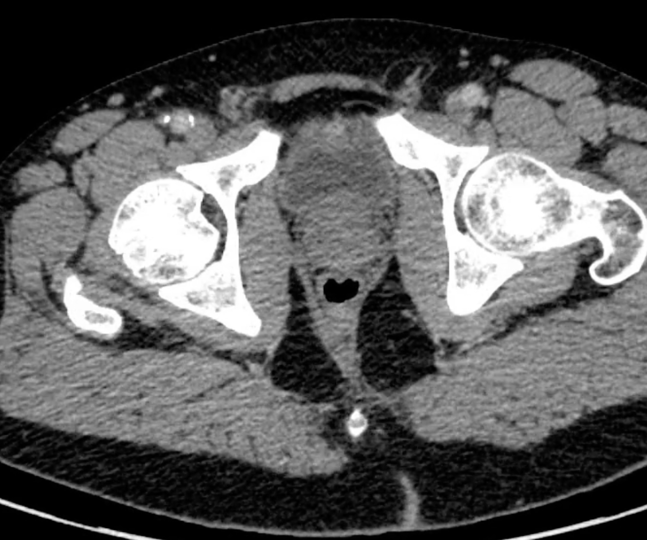
This study was done on a 65 year old male patient with difficulty urinating.
Question 3:
a) What are pertinent technical parameters for this study?
This is an axial CT scan displayed in soft tissue windows. There is very faint IV contrast present (the pelvic arteries are slightly more dense than muscle), so the patient likely received only a low dose of contrast, perhaps due to a malfunction of the IV line used for the injection.
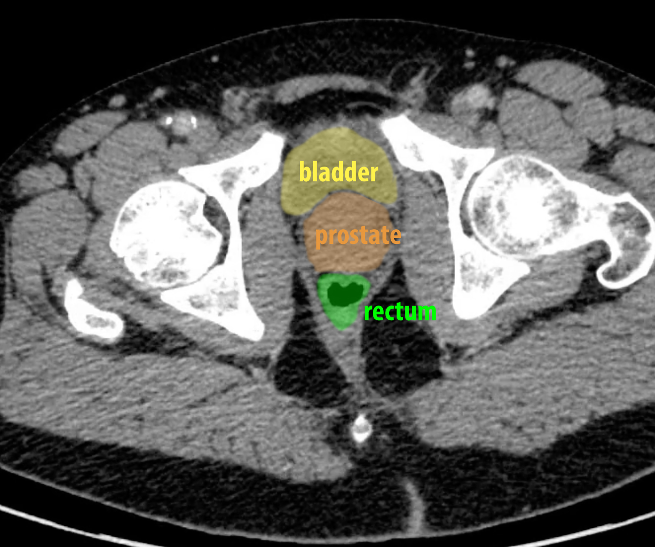
b) Try to identify the prostate on the image series before looking at the labeled image. Does the prostate show internal structures on this study?
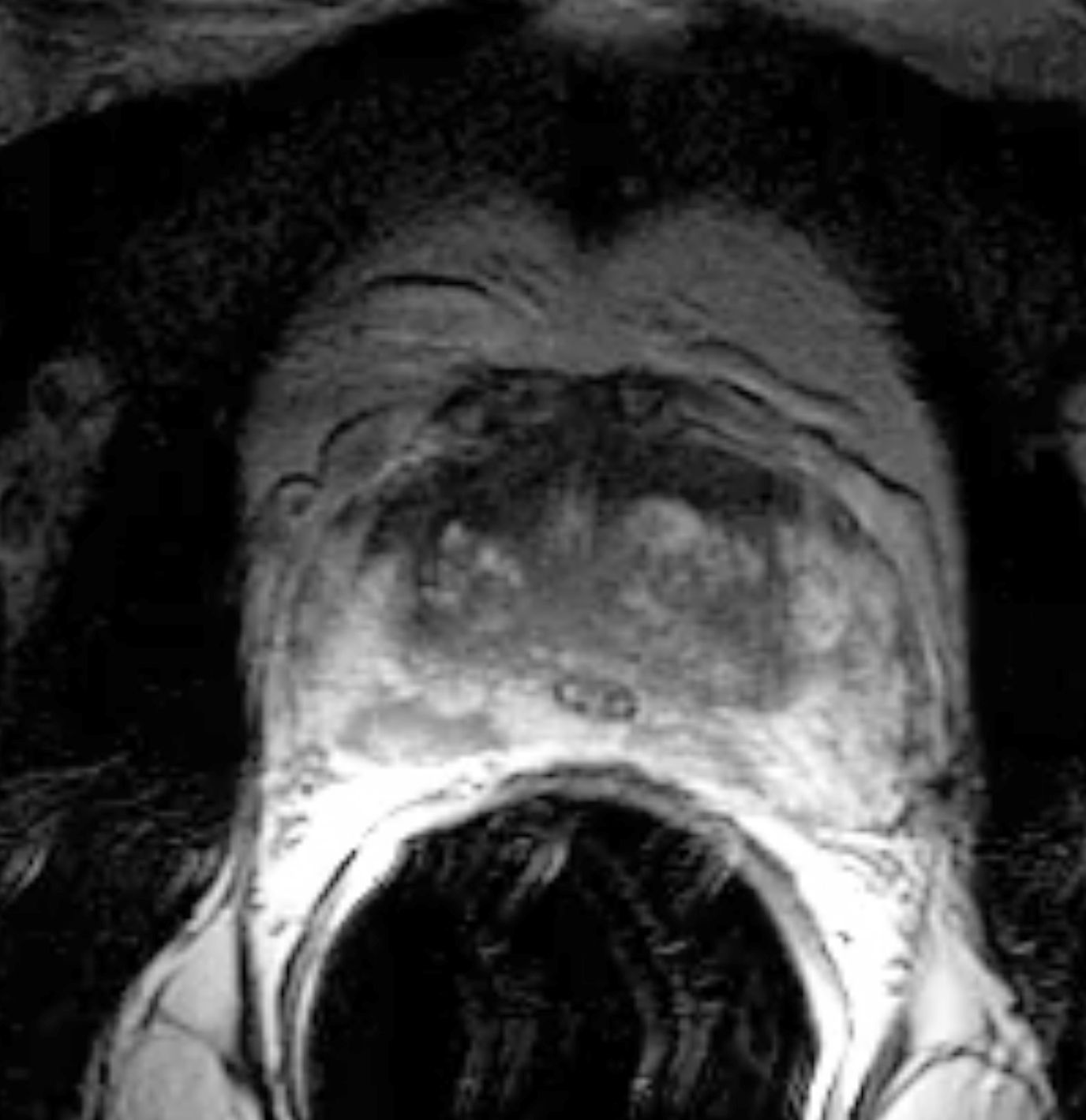
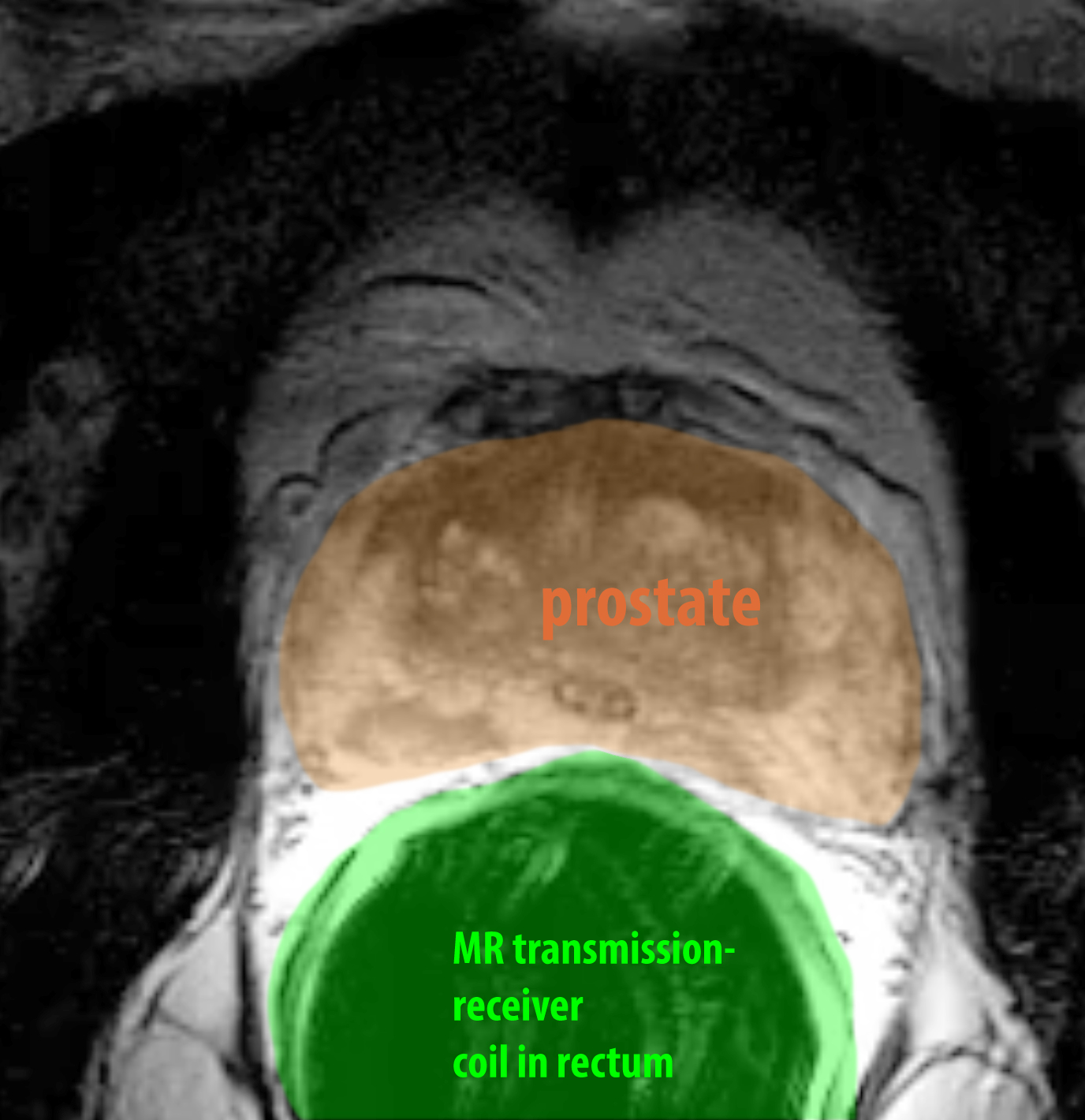
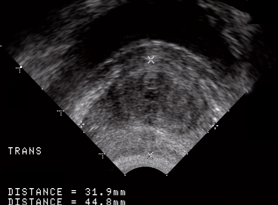
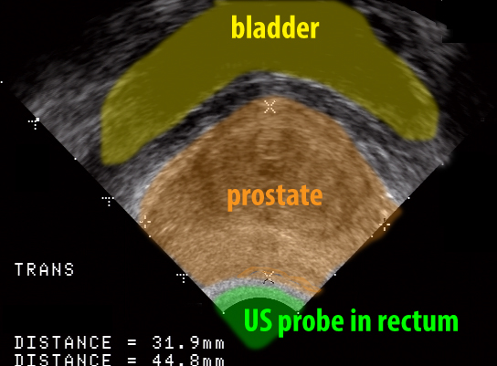
The prostate on CT scanning generally appears fairly uniform in density, similar to the uterus. So just like for the uterus, we often prefer to image the prostate with other modalities that can show more internal structure. You are shown examples of prostate MR and US below for comparison. For both of these studies, to increase the spatial resolution, a rectal probe (in the case of US) or a rectal coil (a source of radio-frequency pulses for MR) are used to get a clearer picture of the organ.
