

Imaging Anatomy Pelvis Case 2
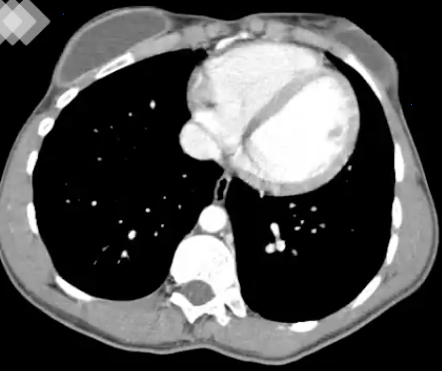
This 22 year old patient has vague left pelvic pain and is being evaluated for infertility. You are shown a series of CT images.
Question 1:
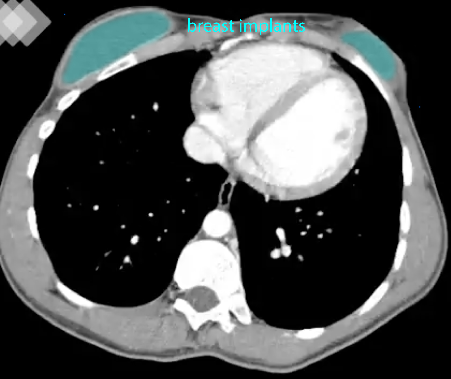
a) On the top few images, what do you notice when searching for fluid density? Are these images presented in the optimal window to detect fluid?
Click on the link below, which will outline very well-defined fluid collections just under her skin surface anteriorly, in the region of the breasts. The fluid is so well marginated that it should suggest that it might represent a foreign object. These are saline breast implants. This CT is shown in soft tissue windows, which are ideal for discriminating fluid from soft tissues. You can see that the fluid in the implants is less dense than muscle, and it would measure around 0 HU.
b) What do you notice about the kidneys in this patient as you scroll through the CT images. Was IV contrast given? What other imaging plane might be helpful for evaluation of this finding?
There is only a single kidney seen, on the left. IV contrast was given, as you can see by the density of the single kidney. Scanning was performed relatively soon after the contrast was administered, as the cortex is noticeably more dense than the medulla of the organ. If you waited a minute or two and rescanned, the cortex and medulla would appear similar in density, as contrast is extracted by the kidney and moves from the glomeruli in the cortex into the collecting tubules in the medulla. This single kidney is quite large, extending from the upper abdomen (at the level where the gallbladder is visible) to the upper pelvis (level of the iliac crests). This suggests that this is a congenital abnormality, rather than a result of surgical removal of the opposite kidney. When only one kidney is present from birth, it can hypertrophy (become larger) to allow relatively normal renal function. When a kidney is removed later, the remaining kidney does not generally enlarge very much.
Imaging Anatomy Pelvis Case 2
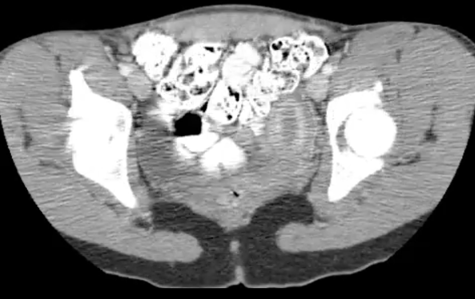
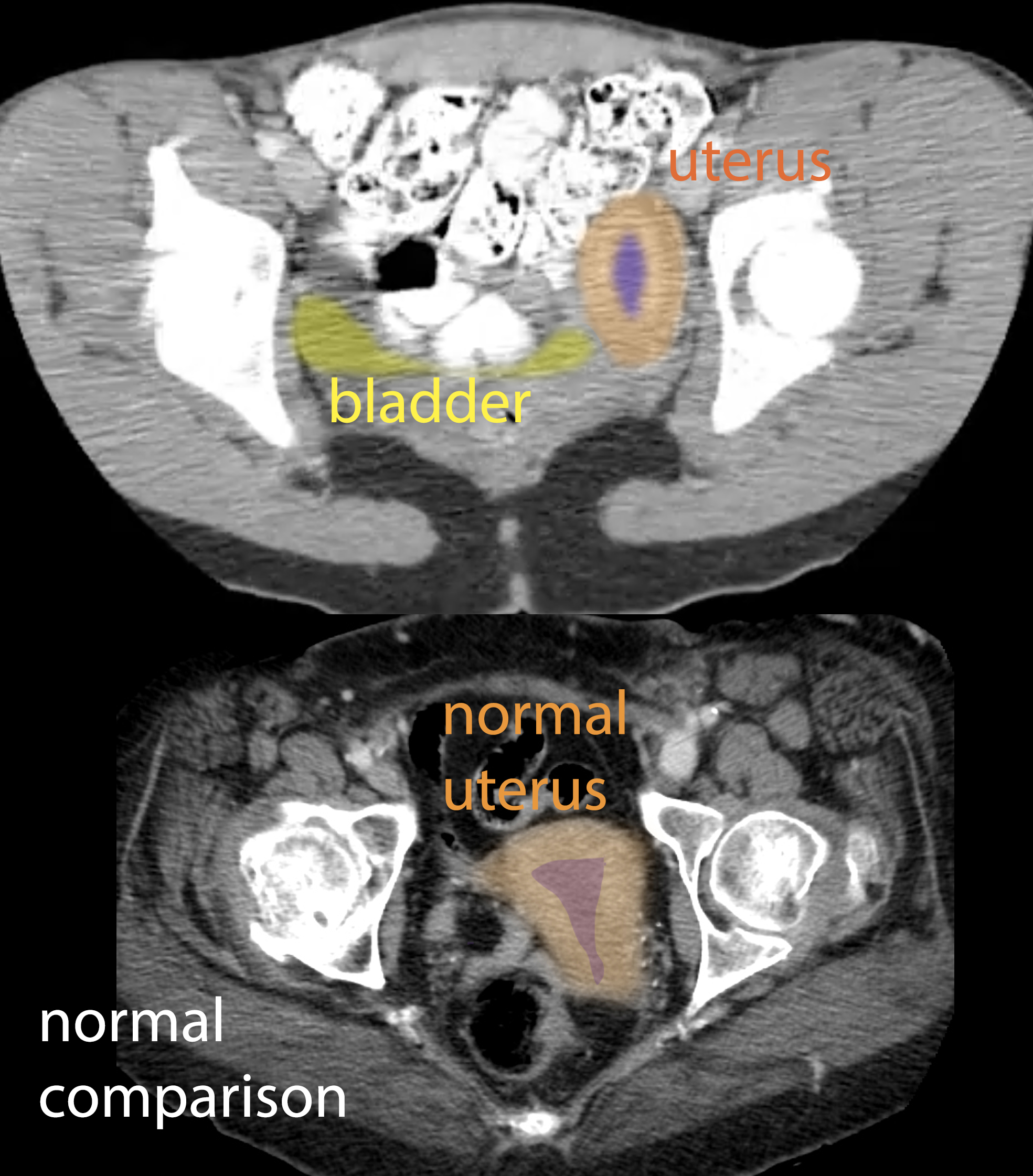
This is a selected image from the lower part of this CT scan in the same patient. Comparison images from a different patient are also shown.
Question 2:
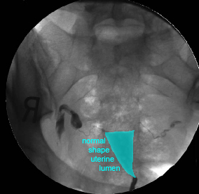
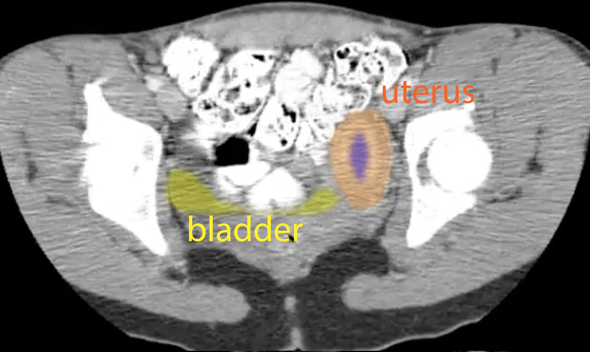
a) Can you identify the uterus on this image? Does it look normal in shape?
Click the labeled image below to see where the uterus is. On CT, the uterus sometimes has the appearance of many concentric layers of slightly differing density (like an onion), and may normally contain some fluid in the lumen. It is important when trying to figure out the shape of a complex three-dimensional structure like this to look at ALL of the images (scrolling through) to get an idea of the overall shape. A single slice may be deceptive, but all of the slices of this uterus look similar.
b) Was oral contrast given for this study?
Definitely. All of the GI structures on this image are as white as the bones on this soft tissue window. That means that the patient swallowed a liquid containing dense material (either a barium compound or an iodinated compound), and enough time elapsed to spread that material throughout the small and large intestine.
Imaging Anatomy Pelvis Case 2
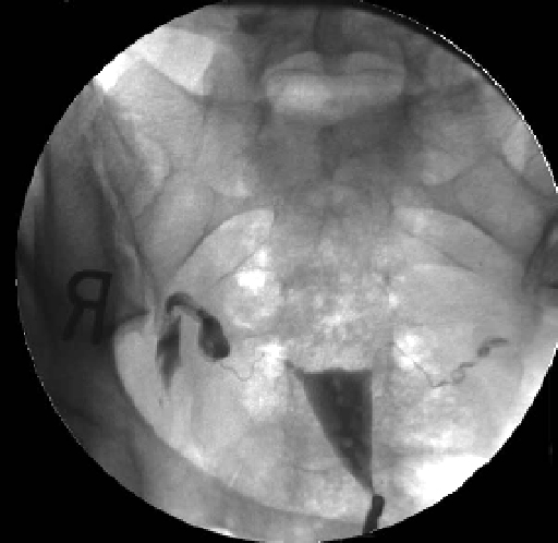
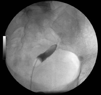
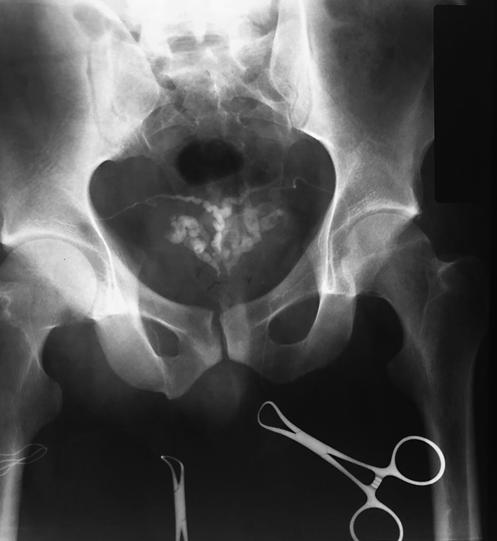
This is another way to look at the uterus that is more invasive. You are also shown interventional radiology images from a different patient showing a method to delineate the structures of the male genital tract.
Question 3:
a) How was this study done?
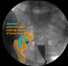
This is an example of an interventional radiologic study where contrast has been injected into the uterine lumen, to evaluate its shape and also to see if the uterine tubes are normal and patent. In a normal female, the uterine tubes (Fallopian tubes) are open to the peritoneal cavity, and if contrast is injected into them under pressure, it will travel from the uterus through the tubes and into the peritoneal cavity. The labeled image below shows contrast on the OUTSIDE of pelvic bowel loops, confirming normal connections.
b) What do you think of the study in our infertile patient? How does this relate to her renal finding?
Our patient's study (called a 'hysterosalpingogram') shows an abnormal uterine shape. The uterus normally forms from fusion of the tubular embryologic structures in the midline to form a somewhat triangular organ, wider at the top. In this patient, the uterus seems tubular and only on the patient's left side (patient left is on the RIGHT side of the image, as for a chest radiograph, axial CT, axial MR and transverse US). If only one kidney formed, then there was presumably a problem with the embryologic structures on the patient's right, and this might be predicted to also affect the formation of the uterus, as in this case. When a uterus is small and not normal in shape, this can result in infertility, as in this case.
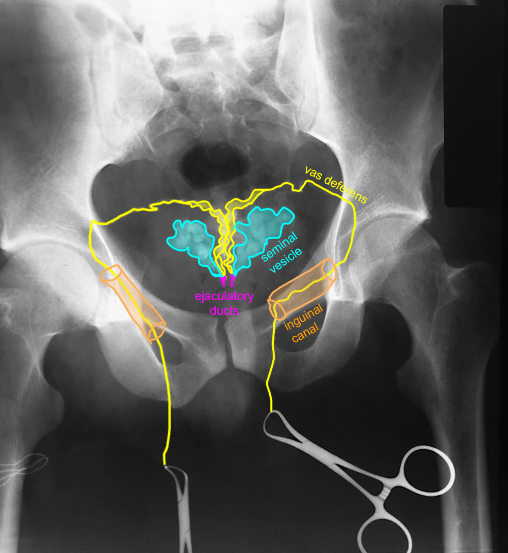
c) Below is another interventional study in a different patient. What are we seeing and how might this be done?
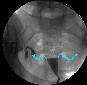
This is an example of a 'vasogram', or injection of contrast into the vas deferens, which can be part of a workup for infertility in genetic male patients. You can see the course of the vas from the scrotum through the inguinal canal to the seminal vesicles. A labeled study image is also provided. This study is sometimes done if a patient has a normal sperm count but is still infertile, to look for blockages or other abnormalities in the vas or ejactulatory ducts. Ultrasound is often done first to evaluate for blockage, but if no problem is seen on ultrasound, then this more invasive study can be done.
This is the last page of the mini-consolidation Pelvis Imaging Anatomy cases. To return to the main page for Foundations 2 Imaging Anatomy, click HERE. To return to the first page of the Pelvis cases for mini-consolidation, click HERE.
