

Case 2
This case deals with mediastinal structures and how they can appear on imaging
Question 1:
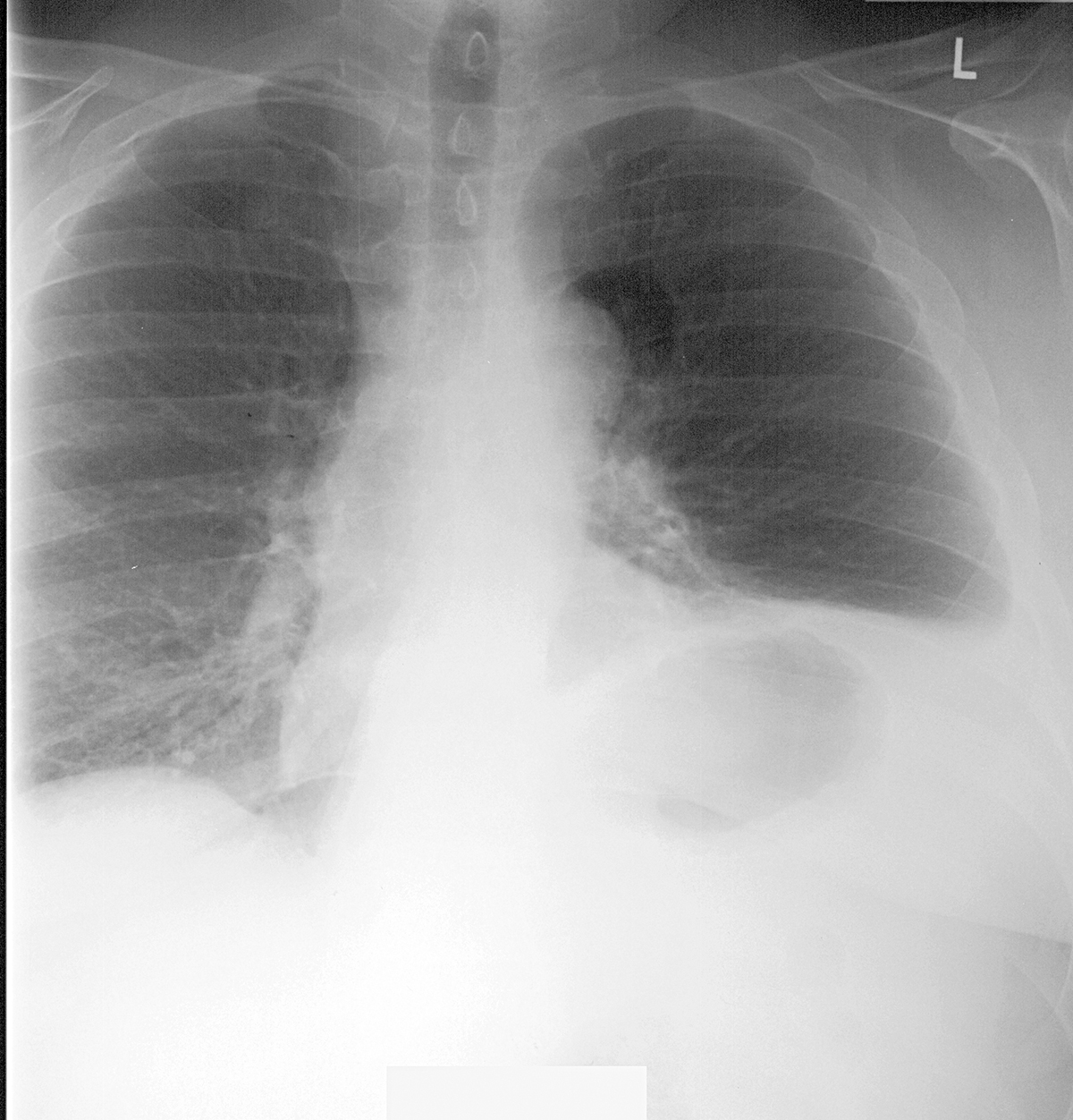
a) What things look abnormal on this chest radiograph? Consider lungs and diaphragms in particular.
There is an odd round lucency (too dark) on the patient's left at about the level of the aortic arch. The left hemidiaphragm is much higher in position than it should be.
b) This patient had a history of sampling of lymph nodes via anterior mediastinoscopy. What is that procedure and how is it done?
A mediastinoscope is a type of endoscopic equipment with a rigid tube (not a flexible one, like for GI endoscopy) that can be positioned within the mediastinum via two different types of skin incision. If the nodes to be sampled are near the trachea, the tube can be inserted via an incision in the jugular notch, and directed down along the tissues adjacent to the trachea to about the level of the carina (cervical mediastinioscopy). If the nodes to be sampled are in the anterior mediastinum, the tube can be inserted via an incision just lateral to the sternum, in between two adjacent anterior ribs (anterior mediastinoscopy).
c) If you click the "CT" button below, you will see selected images from this patient's study. What is the display window, and what is abnormal? How does this finding correlate to the abnormality see on the patient's chest radiograph?
The three CT images are in lung windows, and show herniation of the lung through the left anterior chest wall defect related to the surgical incision for the anterior mediastinoscopy procedure.


Case 2
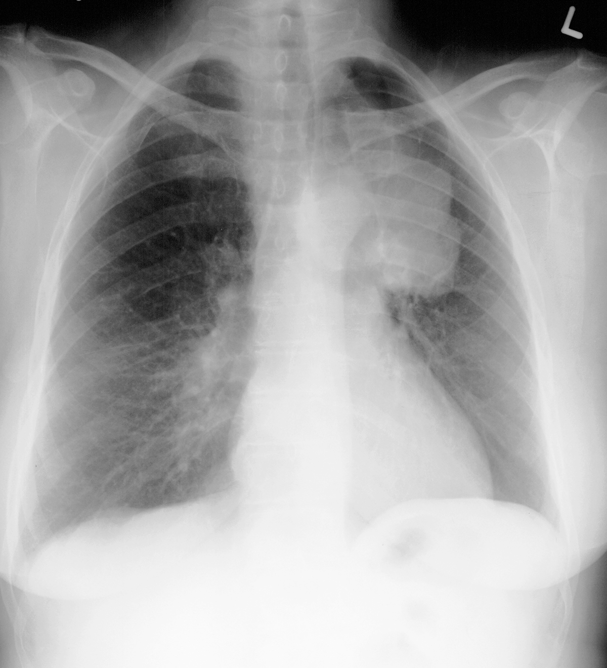
This is imaging of a different patient, who has a large left lung cancer and possible fullness of the right mediastinum.
Question 2:
a) If enlarged nodes were noted on this patient's chest CT near the right tracheal margin, what method could be used to sample them?
There are two ways to reach nodes that are adjacent to the trachea--with a flexible bronchoscope, you can biopsy nodes by directing a needle through the wall of the trachea and into the node. Or, you can do cervical mediastinoscopy with a rigid endoscope via an incision in the jugular notch region. This patient had cervical mediastinoscopy.
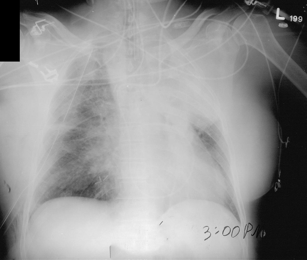
b) The 'next image' shows the appearance of the patient immediately after their procedure. What do you think of the image?
The patient looks acutely much worse with hazy lungs and many new support lines. This indicates that something went wrong during the cervical mediastinoscopy.
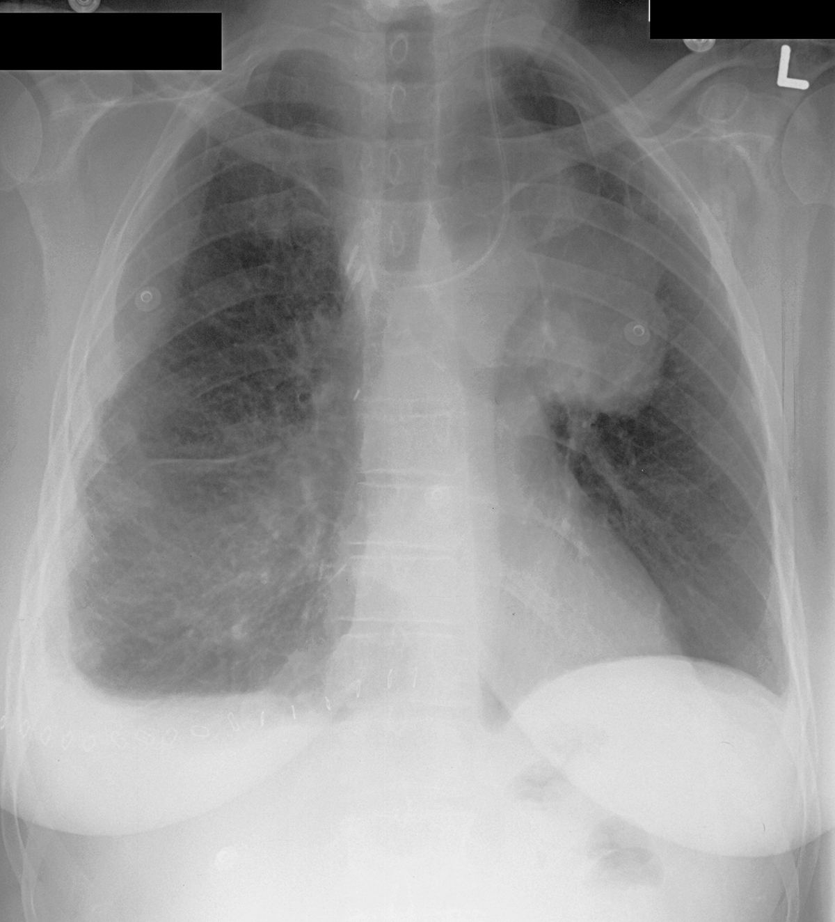
c) The 'last image' shows the appearance of the patient after another surgical procedure. There are new surgical clips present. Given their location, what anatomic structure might have been damaged during the cervical mediastinoscopy?
There is a cluster of clips along the lower right paratracheal region (the tissues just to the right of the trachea), at the level of the origin of the right main bronchus. This is the location of the azygos arch. This vessel was accidentally biopsied instead of the adjacent lymph nodes.
d) The last two links show another way to image the lymphatic system (a lymphangiogram), which shows the anatomy well but has been replaced by other methods. How might such images be obtained?
A lymphangiogram is performed by putting a very tiny cannula into a lymphatic channel, most often in the foot, and then VERY slowly injecting iodine-based contrast over time to fill the lymphatic channels including the thoracic duct. The nodes are also visualized. This procedure has been replaced by more functional imaging, such as PET, or PET-CT.


