

Case 4
This case focuses on lung and chest vascular anatomy
Question 1:
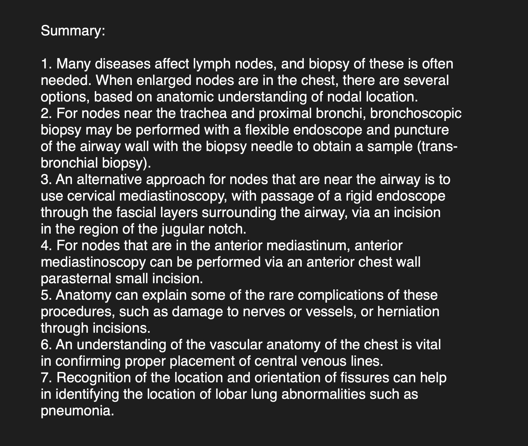
a) Where is the abnormality located on this image? How does the upper vs lower portion of the abnormality differ in terms of its margin with adjacent lung?
There is abnormal opacity in the right upper chest that is consistent with a right upper lobe pneumonia. The upper part of the pneumonia has a hazy margin with adjacent uninvolved apical segments of the upper lobe, but the lower part has a straight sharp margin, indicating a fissure. This is the horizontal, or minor fissure.
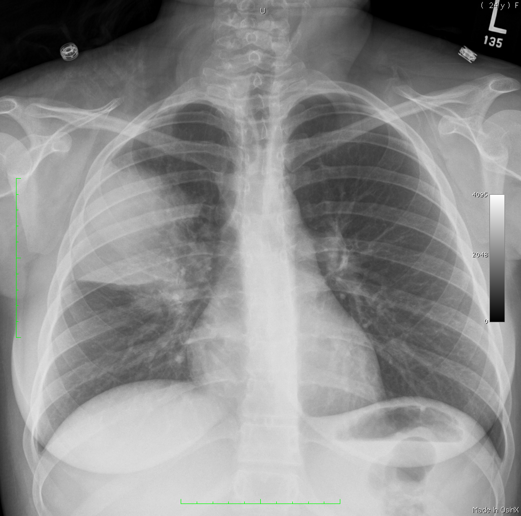
b) What do you call the image represented by 'another view of this patient'?
This is a lateral view, and shows the pneumonia abutting both the horizontal and oblique fissures (minor and major).
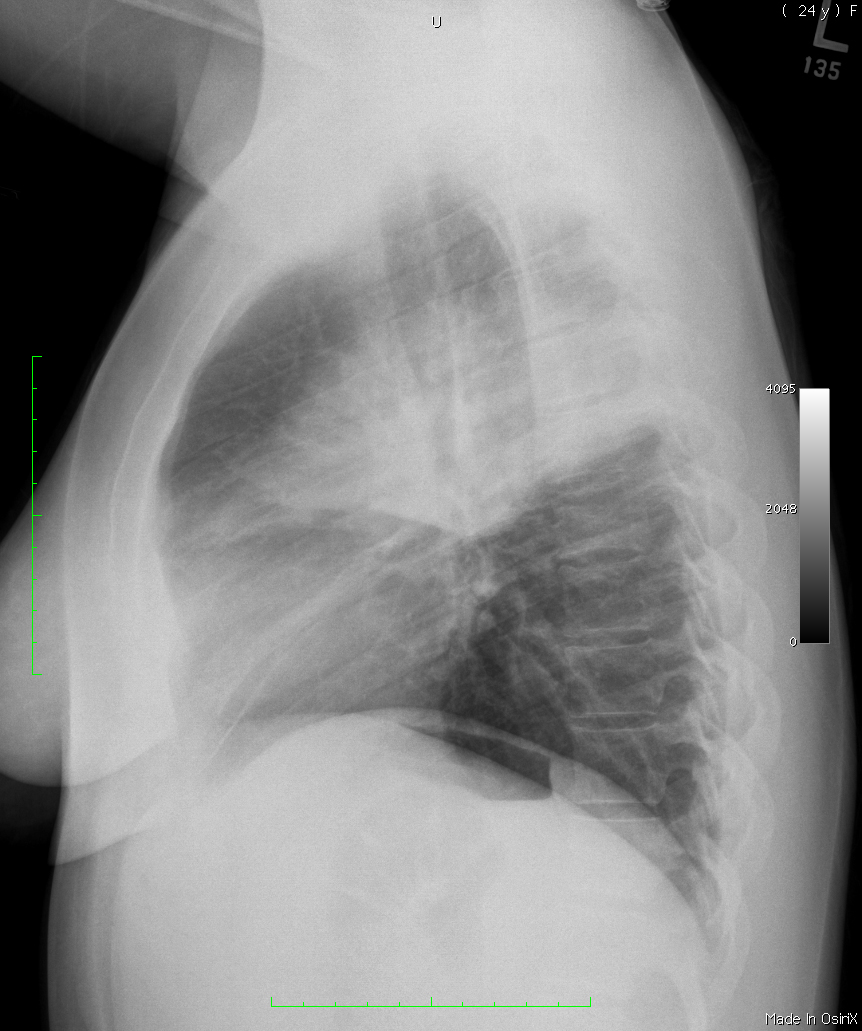
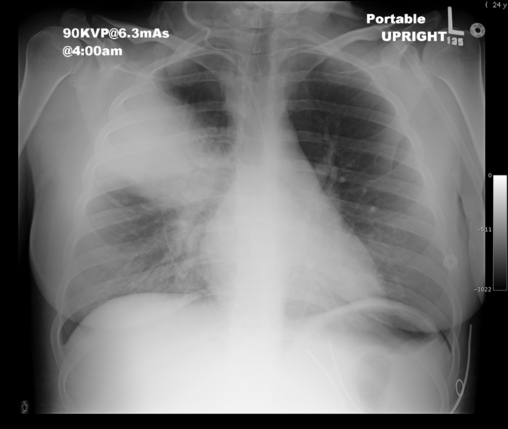
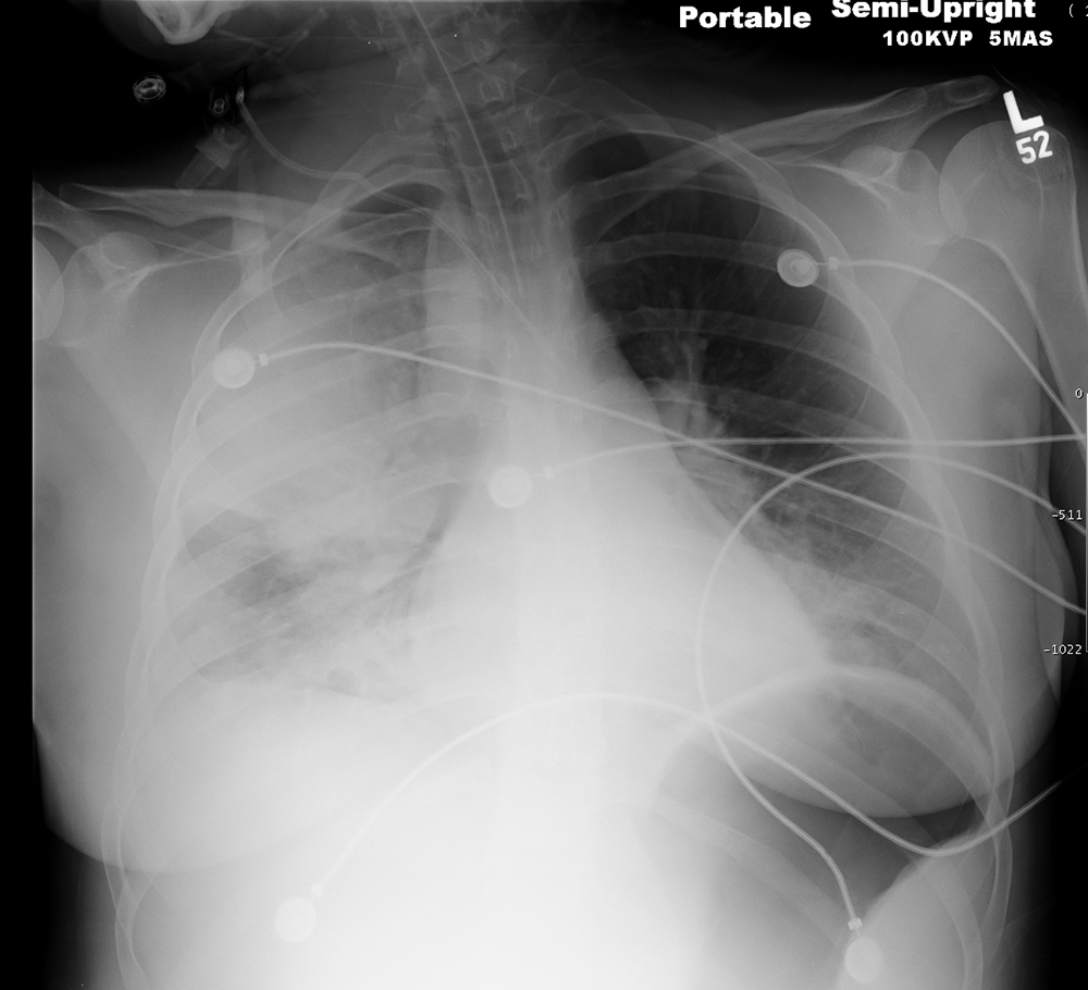
c) What has happened on the image listed as 'followup imaging'? And on 'another followup image'?
A central line has been inserted to administer antibiotics. It is not in good position. Decide what is wrong with its path through the chest, given that we want the tip to be in the superior vena cava. On 'another followup image', the patient is doing very poorly, and now has an endotracheal tube in their trachea.
Case 4
More patients with fever and pneumonia in different parts of the chest.
Question 2:
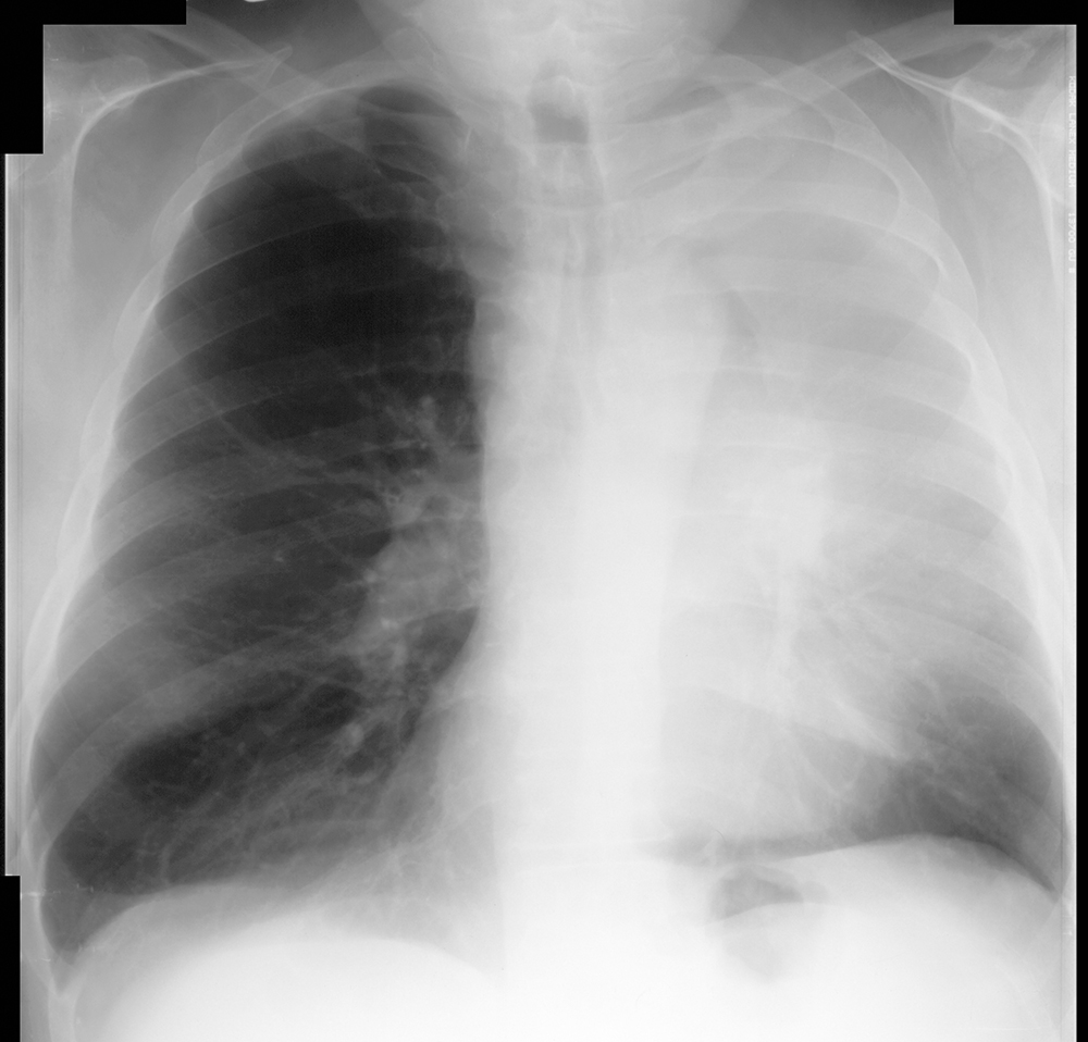
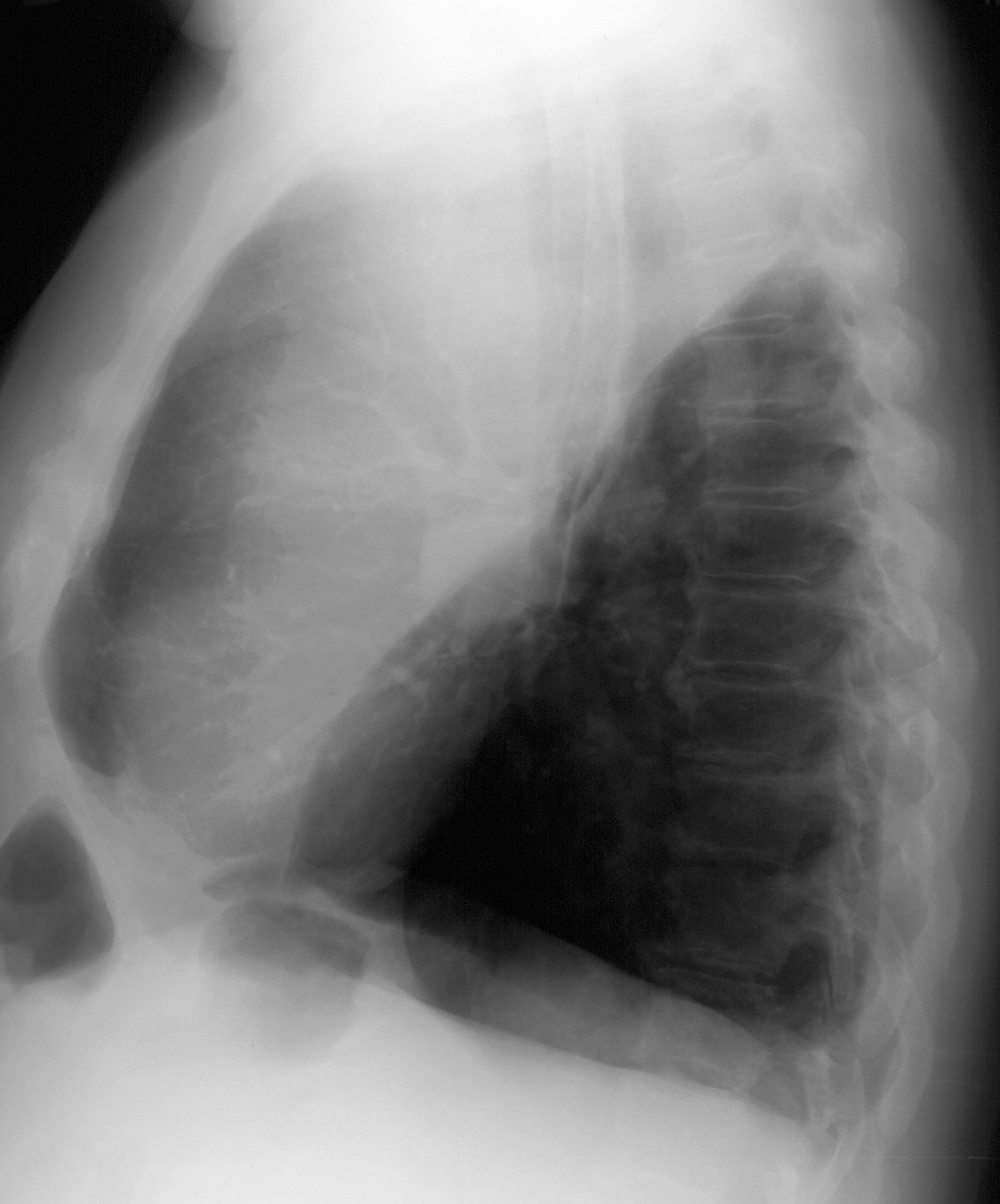
a) 'Another view of this patient' shows the lateral view for the first case. What lobe is involved, looking at both the frontal and lateral views?
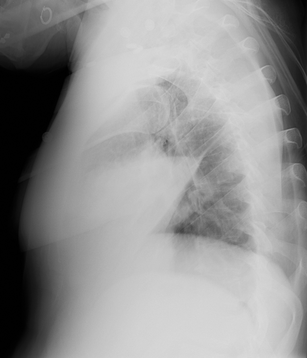
There is opacity in the left lung on the frontal view, with no sharply defined margins evident. But on the lateral view, there is a straight interface at the posterior/inferior margin of the pneumonia, which is the left oblique (major) fissure, indicating that the left upper lobe (including the lingual) is involved.
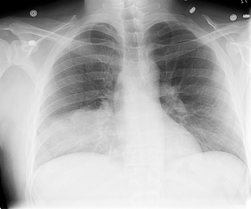
b) For the two views of the second patient, what lobe is involved?
On the second patient frontal view, the opacity obscures the right heart border but not the diaphragm, and has a slightly horizontal upper margin. On the lateral view, there is a triangular opacity overlying the heart, consistent with right middle lobe pneumonia.
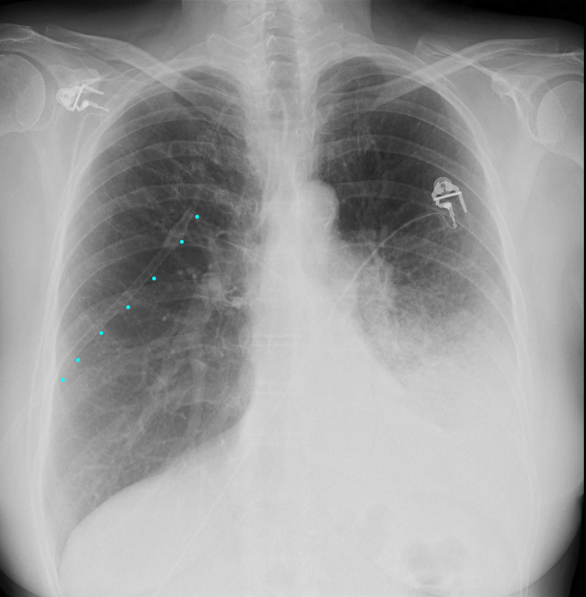
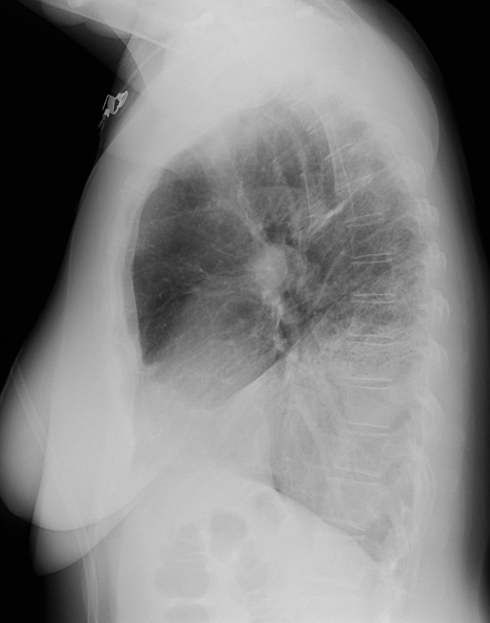
c) For the two views of the third patient, what lobe is involved? Ignore the tubing that is projecting over the right side of the chest (indicated by blue dots).
On the third patient frontal view, the opacity does not obscure the left heart border but does obscure the left hemidiaphragm. On the lateral view, there is a sharp straight oblique line through the chest, with opacity below it. This indicates a pneumonia in the left lower lobe.
Case 4
Summary of what was discussed relative to imaging anatomy of the chest.
Further Explanation:
