

Case 1
This case deals with findings related to the urinary system
Question 1:
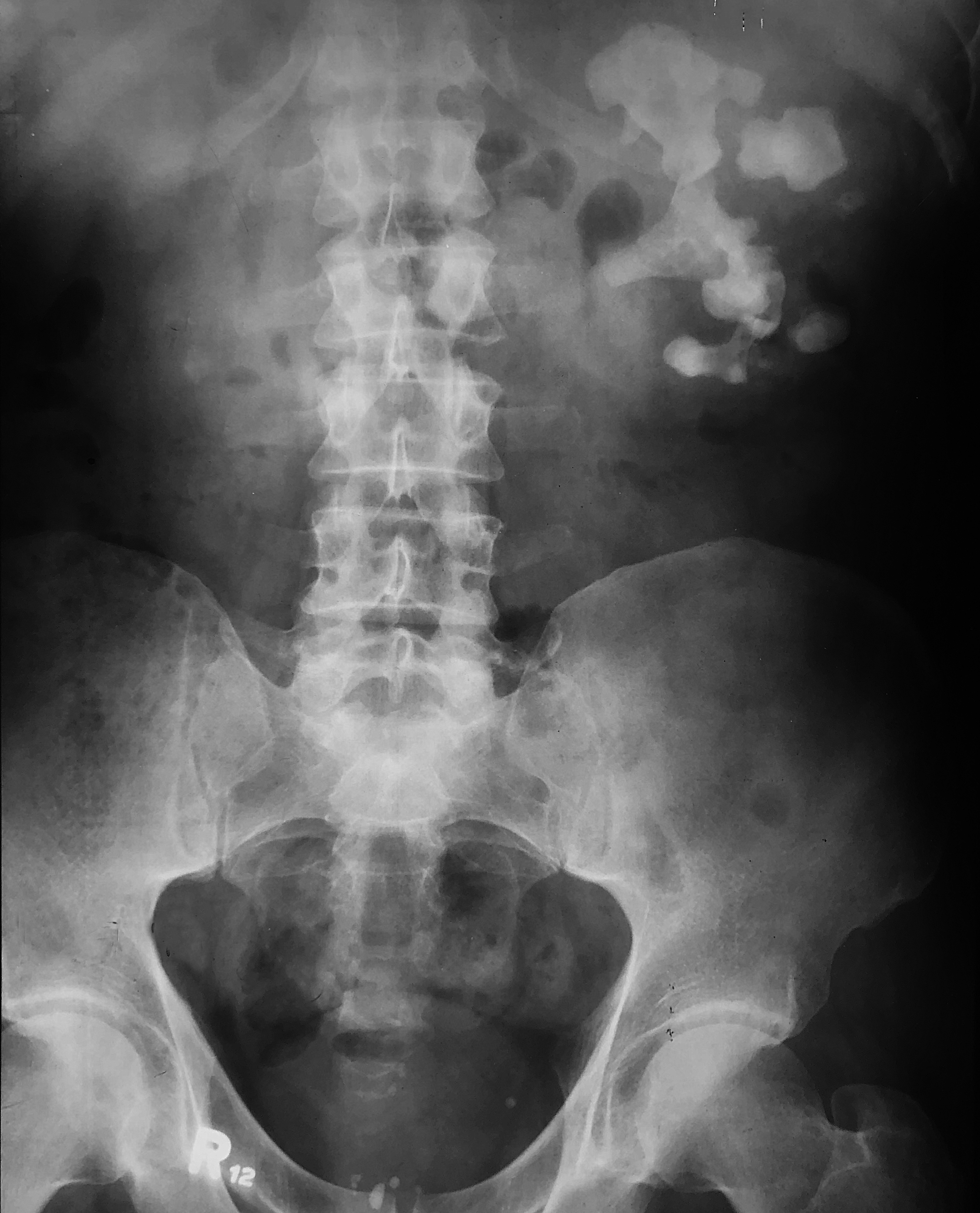
a) Calcium appears white (like bone) on radiographs, such as this abdominal image. In what three locations do you see abnormal calcifications, and what parts of the urinary system may be involved?
There is a large irregular calcification on the patient's left in the expected location of the kidney. This is likely in the collecting system of the kidney, based on its shape. There is a single tiny round calcification on the left in the pelvic region just medial to the left iliac spine. This could be the distal left ureter. There is a group of slightly irregular calcifications in the region of the pubic symphysis. These are likely in the prostate.
b) If you click the link 'another patient with a renal finding', you will see an axial CT scan displayed in soft tissue windows with no intravenous contrast. Try to find two tiny abnormal calcifications in this case.
There is one tiny calcification in the patient's prostate, and another calcification in the distal right ureter at the ureterovesical junction. It is easy to miss. It is producing very slight increase in size of the right renal pelvis suggesting mild hydronephrosis.
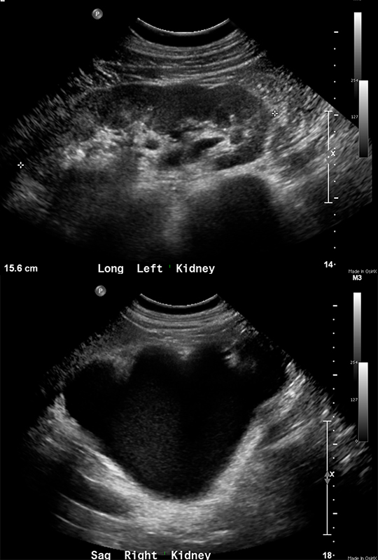
c) If you click 'another way to view the kidney', you are presented two renal ultrasound images. Ultrasound is a cross-sectional modality, just like CT and MR, but the imaging planes are much more variable, depending on how the transducer is held. What appears abnormal in this case?
The upper image shows a normal appearing kidney, with a label indicating that it is a longitudinal (long) view of the left kidney. On the right, the view is labeled 'sag' for sagittal, and shows a severely dilated renal pelvis and calyces with very little visualization of renal parenchyma. Fluid (both blood and water, as well as bile) appear black on ultrasound while calcifications are focally white with a dense black shadow behind (quite different from their appearance on x-ray based imaging).
Return to case list for abdomen


Case 1
Another case of a renal abnormality
Question 2:
a) What is the imaging plane for this CT sequence? Do you see abnormal calcifications in this case? What do you think of the right kidney?
This is a coronal CT sequence shown in soft tissue windows with IV contrast present. There are a few calcifications in the left kidney, with a relatively large one in the renal pelvis. There is no normal right kidney, just a large dilated collecting system. This indicates a high degree of obstruction over a long time period, which has produced atrophy of the renal parenchyma.
b) What do you think of the imaging on the patient with flank pain after hysterectomy?
This is an axial CT shown in soft tissue windows with IV contrast present. There is marked dilatation of the right collecting system and of the right ureter, down to the last image, where there is a surgical clip. This was accidental ligation of the ureter during hysterectomy.
c) What is abnormal in the third patient, with flank pain and hematuria?
This time, the marked dilatation of the collecting system and ureter is on the left, and extends down to the region of the bladder, where there is a large bladder carcinoma obstructing the distal left ureter.
Return to case list for abdomen


