

Case 2
These cases deal with hernias
Question 1:
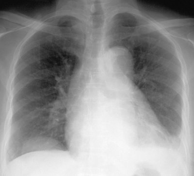
a) What is abnormal on this chest radiograph? Where is the likely location of the finding, anatomically? What other view would be helpful?
There is a rounded opacity in the lower right chest that seems to be centered on the diaphragm. A lateral view would be helpful in figuring out where this is, and showed that the rounded mass was projecting posteriorly, up from the abdomen. This suggests a hernia. If you click 'another way to image this problem', you will see selected CT images showing the posterior hernia, containing the kidney.
b) What is abnormal one the second patient, and what are the three views that are given to evaluate this case?
The first set of images are axial CT images, showing a very large posterior diaphragmatic hernia containing the entire kidney. Hernias in this location are often congenital and are called Bochdalek hernias. Most are quite small and only contain retroperitoneal fat. As is seen on the sagittal and coronal reformatted CT images shown, this patient has a very large hernia.
c) What feature of the embryology of the diaphragm might predispose toward hernias?
The 'embryology' link shows the various normal holes present for passage of the aorta, inferior vena cava, and esophagus. Hernias through the esophageal hiatus are quite common. The movie also shows the various complex components that must fuse properly for the diaphragm to be normal (pleuroperitoneal membranes, body wall components, esophageal mesentery, septum transverum). Weaknesses can lead to hernias either posterolaterally (Bochdalek) or anteromedially (Morgagni).
Return to case list for abdomen

Case 2
A patient with abdominal pain
Question 2:
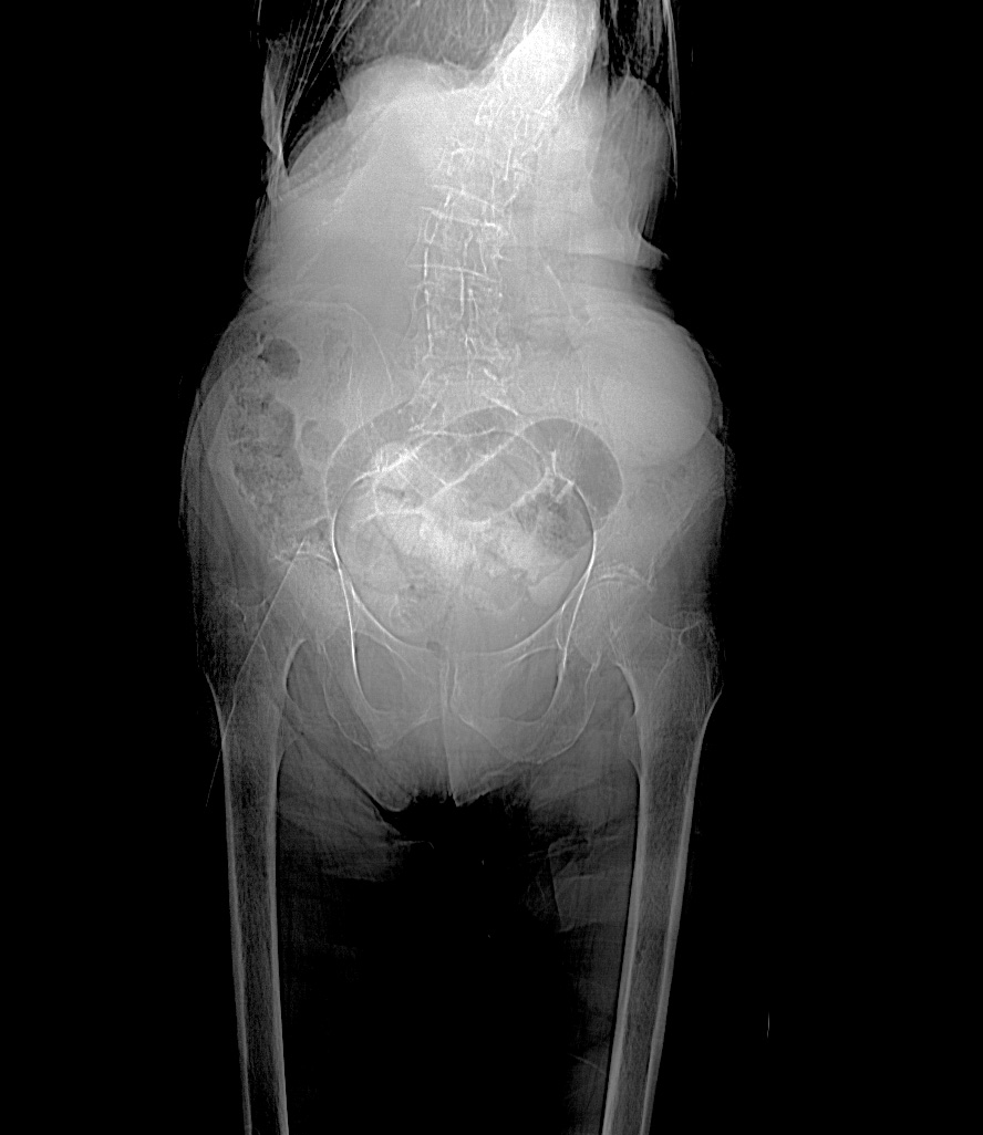
a) What is this image and what is abnormal?
This looks like a radiograph of the abdomen, but the field of view is too large. No x-ray receptors can show the body from mid-chest almost to the knees. This is a special type of image obtained at the start of a CT scan to allow the technologist to determine where to start and stop the scan. It is called a topogram or scanogram. There are several dilated loops of small intestine overlying the pelvis, and the spine is markedly curved (scoliosis).
b) The CT (shown by clicking 'more imaging of this patient'), is in the axial plane, soft tissue windows, with oral and intravenous contrast, and shows an unusual shape of the chest wall on uppermost cuts. What do you think is the cause of this?
The shape of the entire torso is affected by the scoliosis that was shown on the topogram. As you scan through the series, you can see the spine first to the left of midline and then to the right.
c) Focusing in the lower left portion of the CT, at the level of the pubic symphysis and below, can you identify the patient's hernia?
There is a loop of small intestine that passes through the left obturator foramen--a much rarer site for hernia than the inguinal canal. These hernias are actually passing through the tiny opening in the obturator membrane that allows the obturator vessels and nerve to exit, as the rest of the obturator foramen is covered by the dense fascia of the obturator membrane. So this is a particularly serious type of hernia and the bowel is often ischemic or necrotic shortly after passing through this very tight space. In this case, there were several feet of small intestine that needed to be resected at surgery because of ischemia.
d) The patient shown in 'a different type of hernia' has two hernias--one very common and one very rare. Look at the level of the diaphragm and at the lowermost cuts on the patient's right to identify them.
In the upper cuts of this axial CT displayed with soft tissue windows, and with no oral or IV contrast, there is a large hiatal hernia. In the lower cuts on the right, there is a tubular high attenuation structure that is in the inguinal canal. This is the appendix, and it contains contrast from prior oral contrast, as do several diverticula in the sigmoid. These locations (diverticula and appendix) are blind-ended, so when oral contrast enters, it can stay around for a long time afterwards.
Return to case list for abdomen

Case 2
Summary of Abdomen topics covered
Further Explanation:


