

Case 1
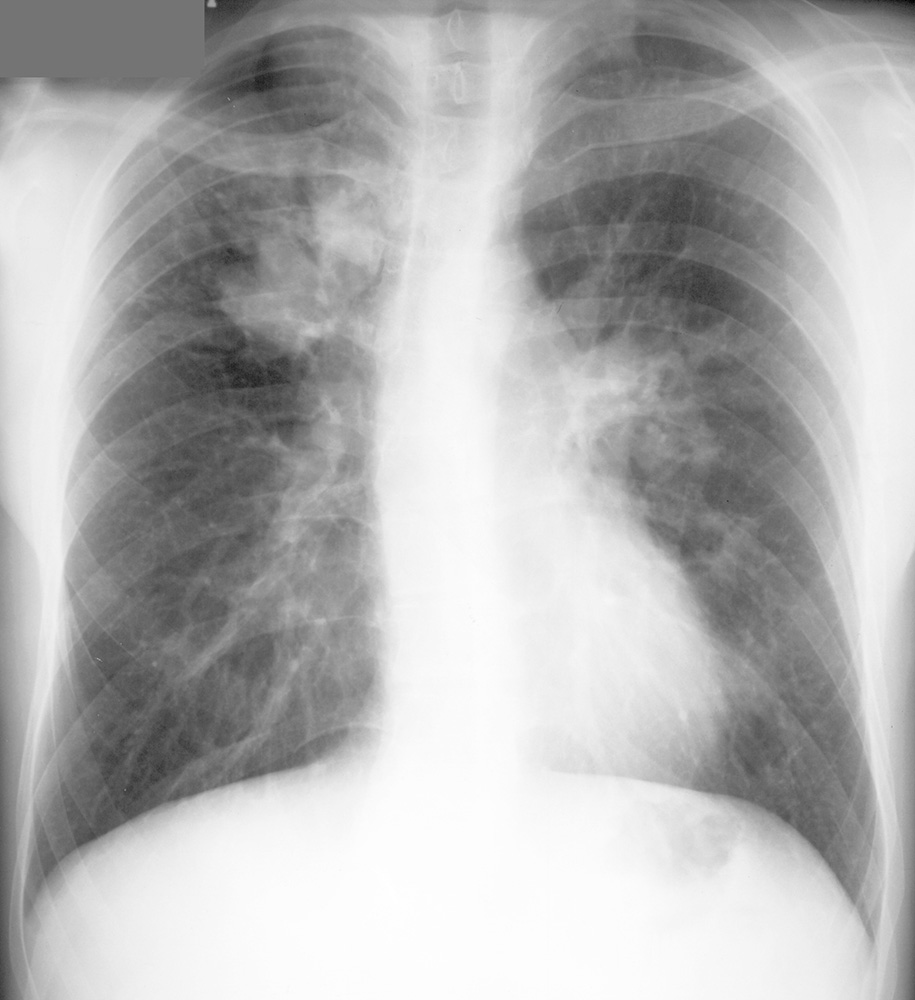
This young patient is short of breath after exercise. Try to figure out the answers to questions before clicking the links.
Further Explanation:
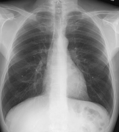
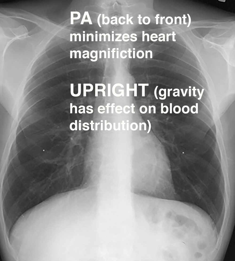
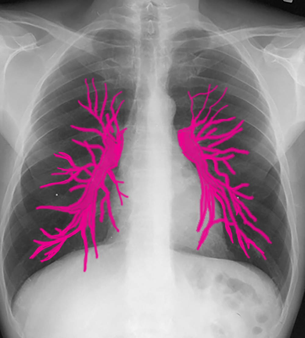
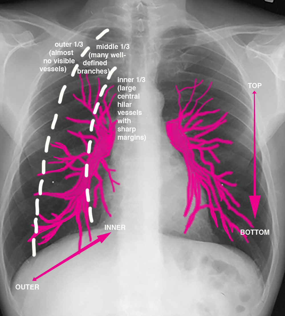
What is the normal distribution of pulmonary blood flow seen on a PA chest radiograph? Try to identify pulmonary vessels on this image, starting from the hilum and moving more peripherally. Consider how the vessels to the upper lungs look different from those to the lower lungs. How would this image look if it was performed on the International Space Station? Recall how the patient is positioned for a routine chest radiograph, and how the beam enters and leaves the body. The tiny white dots are metallic markers indicating the location of the nipples.
Further Explanation:
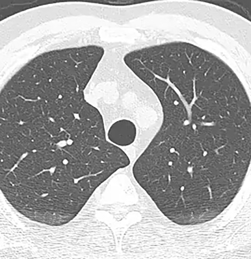
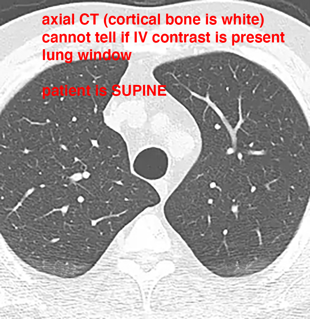
What is this study? What technical parameters can you identify? How was the patient positioned for this exam? Try again to identify and follow pulmonary vessels and consider how they are distributed in this normal study. Think about distribution front to back as well as central to peripheral. What does the lung parenchyma look like surrounding the vessels?
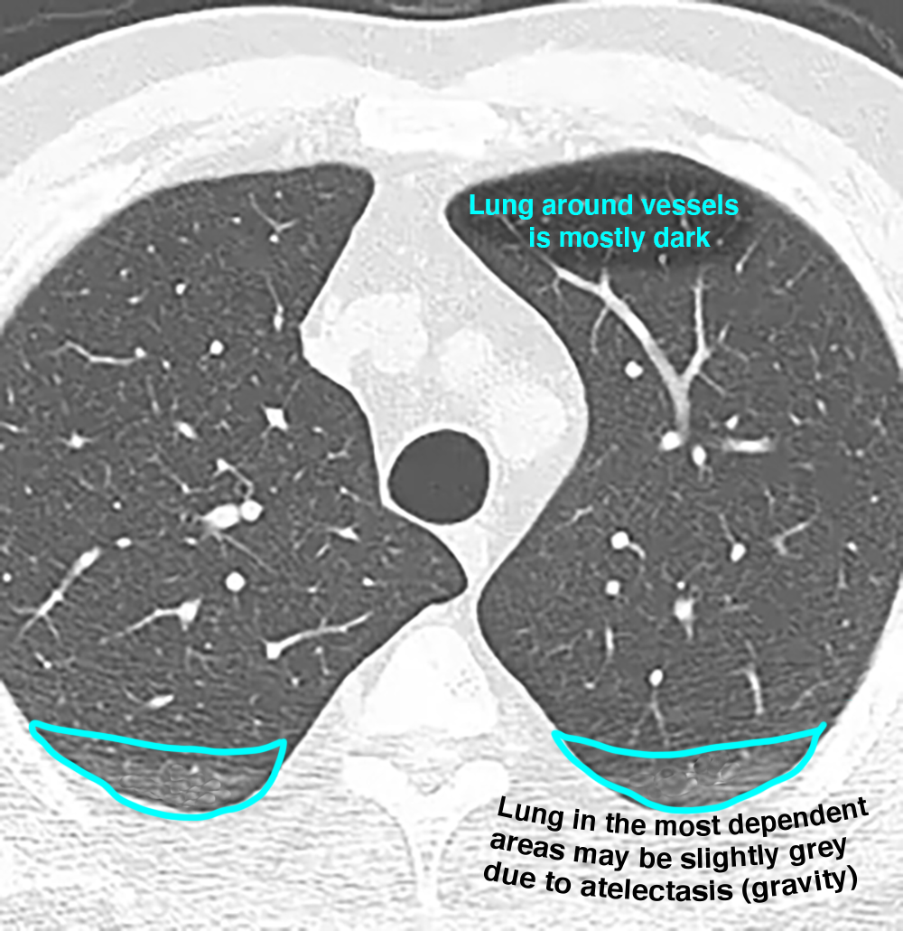
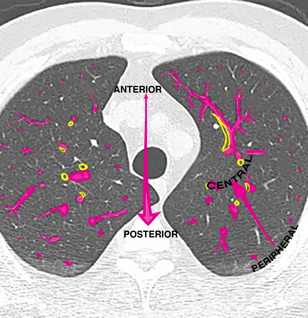
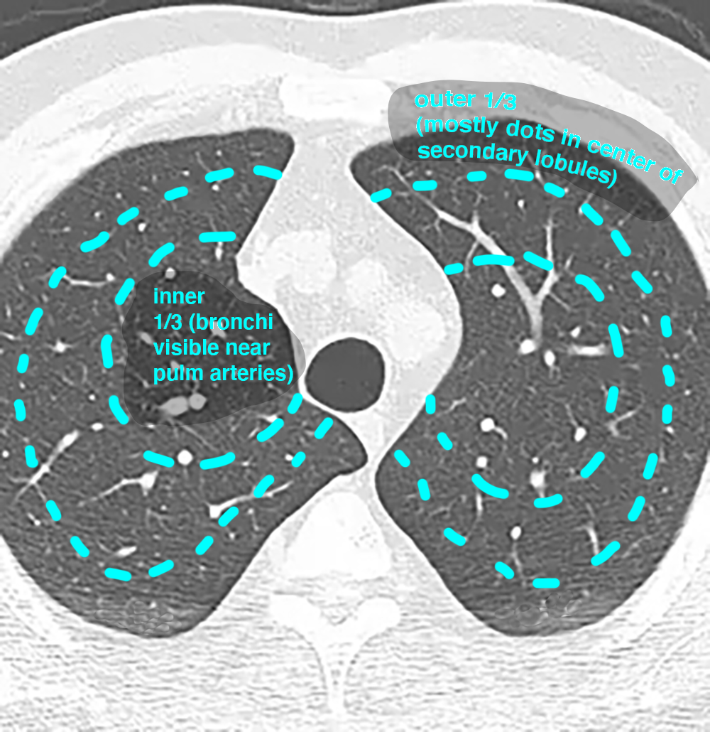
Case 1
Case 1, continued
Further Explanation:
What is this view called and what aspect of lung volume is best assessed on this view?
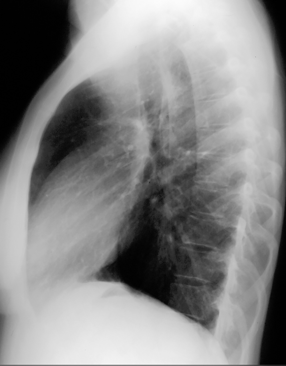
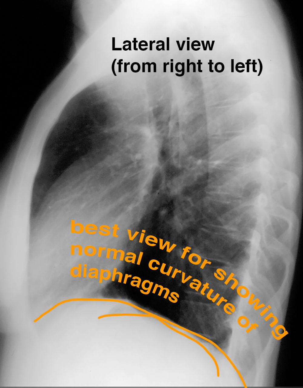
Case 1
Case 1, continued
Further Explanation:
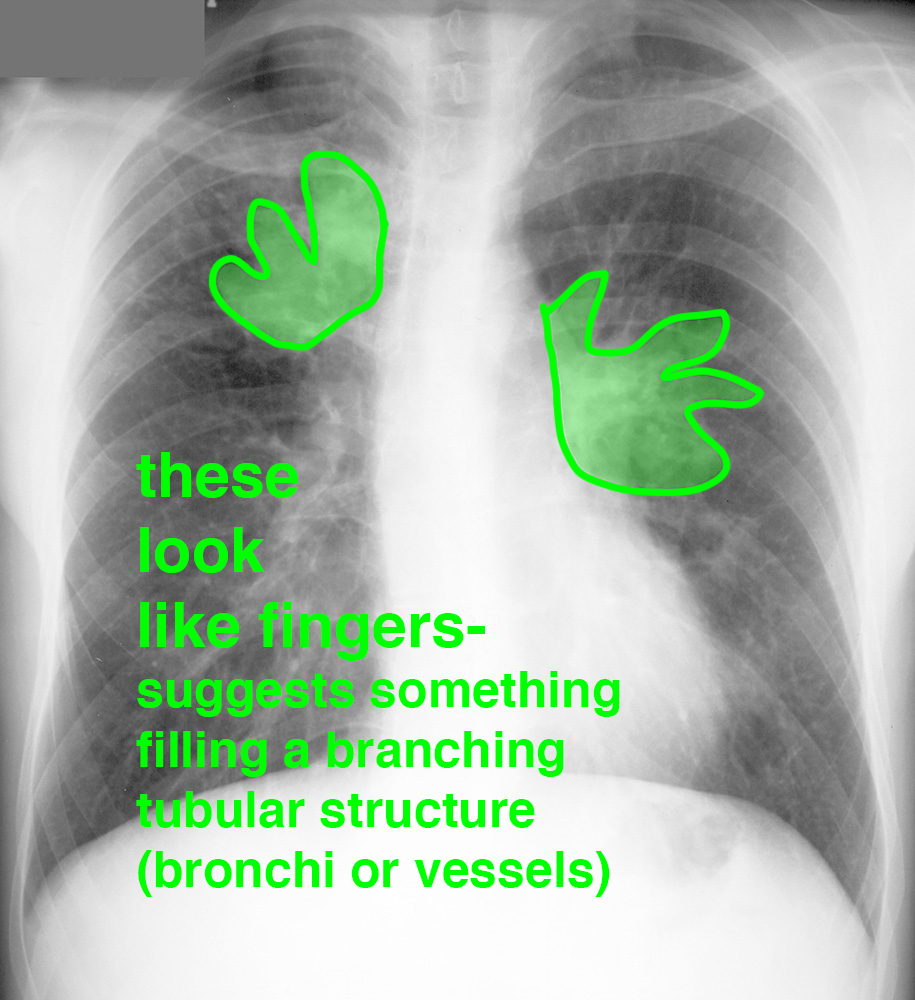
What looks abnormal on this patient, who presents with chronic cough and shortness of breath? How would you describe the SHAPE of the lung abnormalities, particularly what is seen in the right upper lung?
