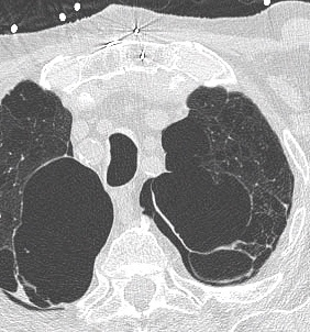


Case 2
This middle-aged patient has been chronically short of breath, with gradual progression of symptoms.
Further Explanation:
What is this image? How was the patient positioned? Do the lung volumes look normal? How do you assess lung volumes on this type of image?
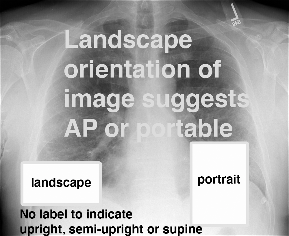
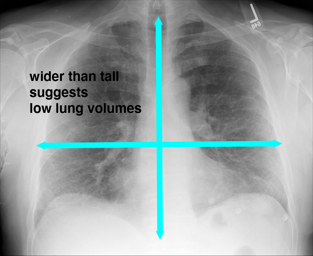
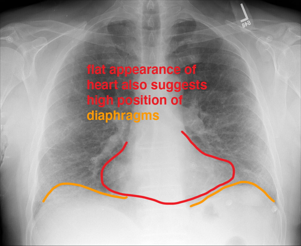
Case 2
Case 2, continued
Further Explanation:
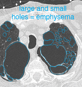
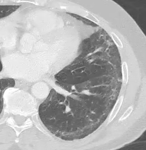
This is a closeup view of one image from the patient's CT scan. How does this look different from the normal CT image shown in the previous case? What descriptors would you use to indicate the findings in the lungs? Where are the abnormalities located?
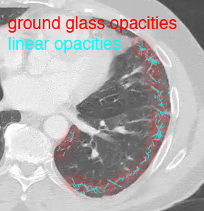
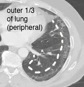
Case 2
Case 2, continued
Further Explanation:
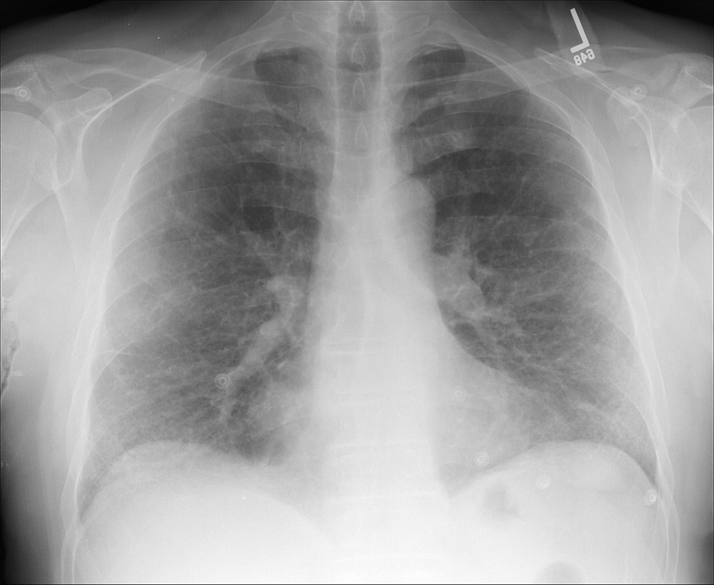
This is a different patient with shortness of breath. How does the appearance of this patient's chest radiograph differ from the previous patient?
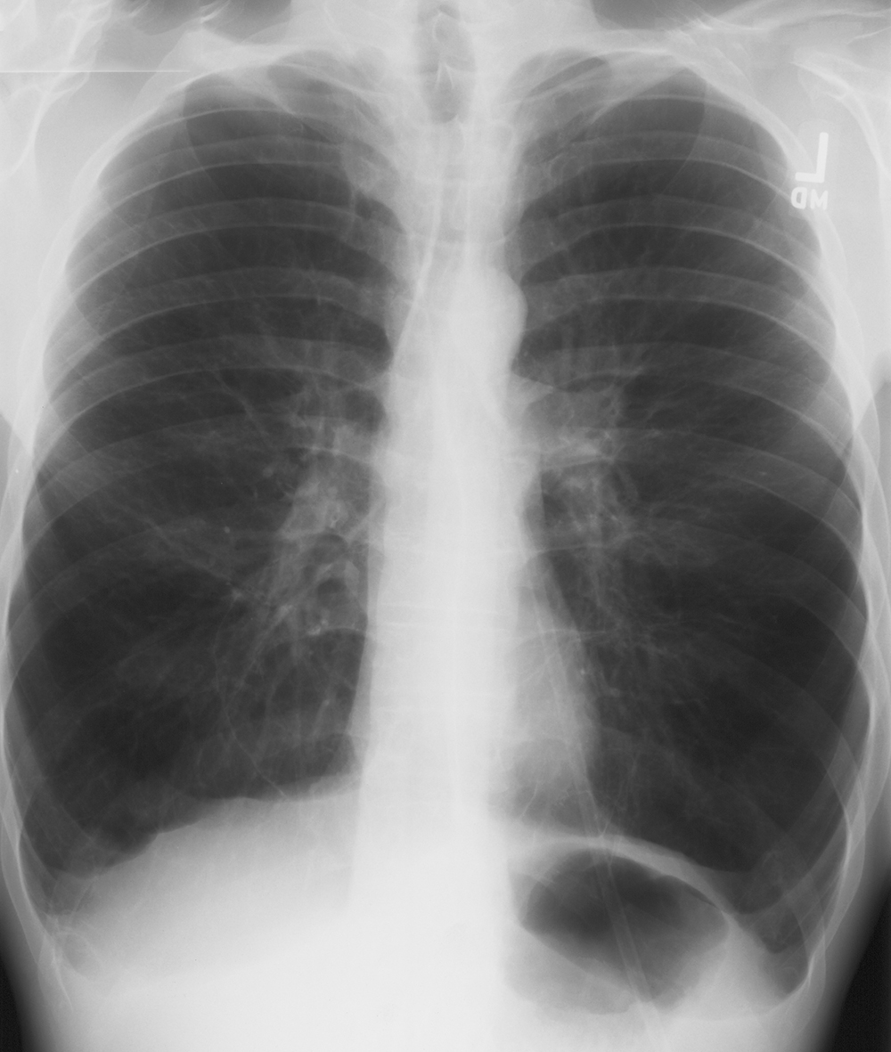
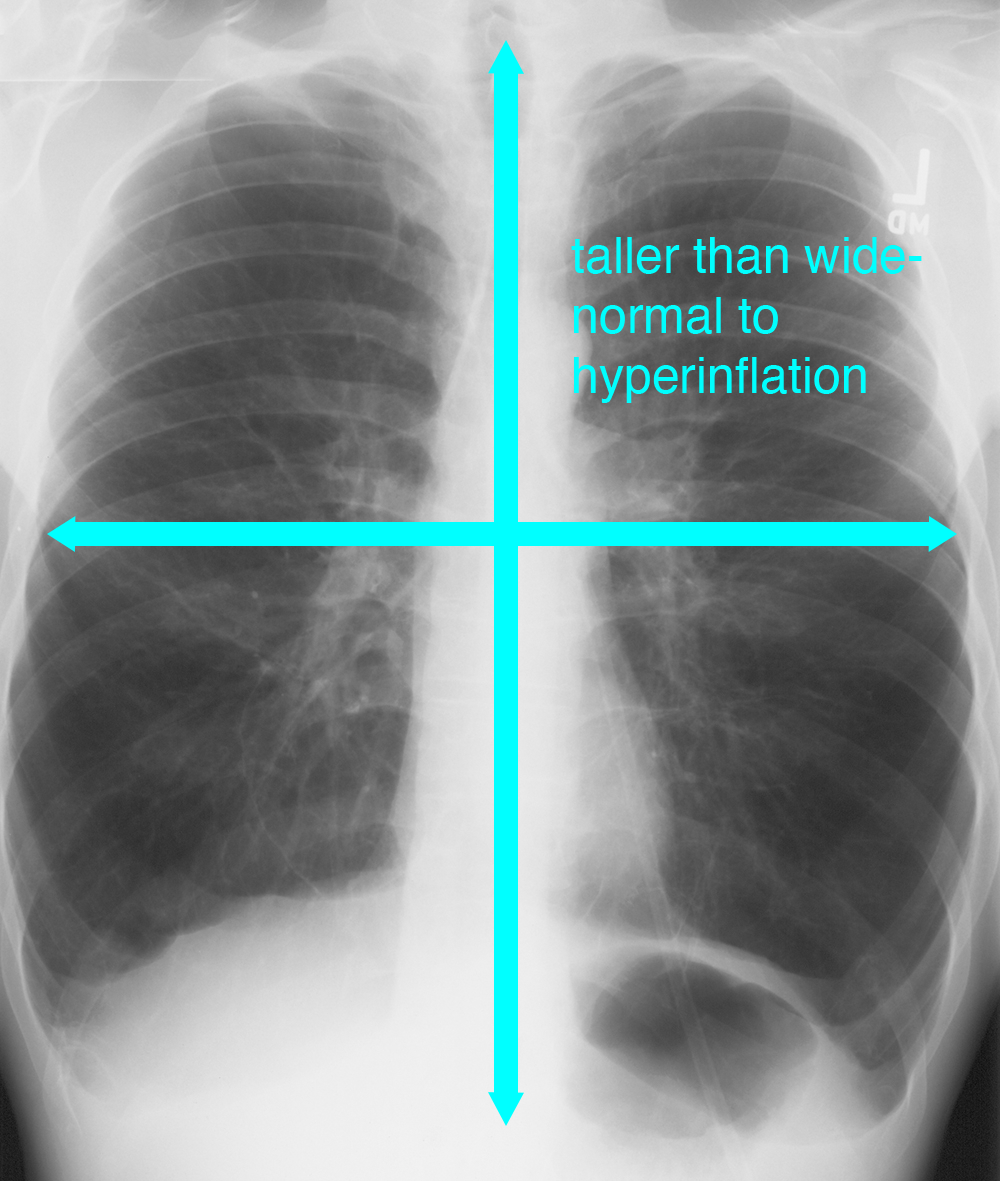
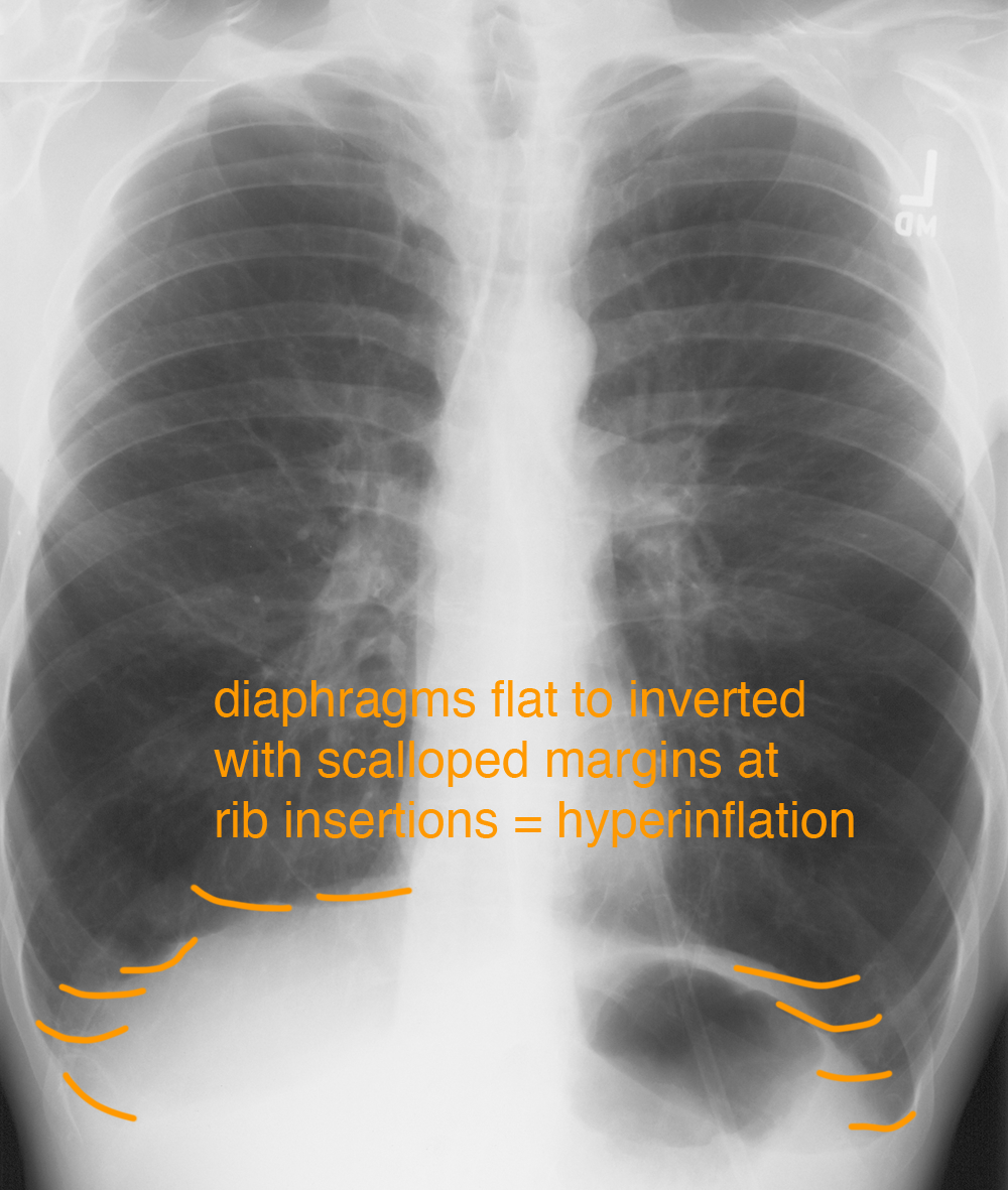
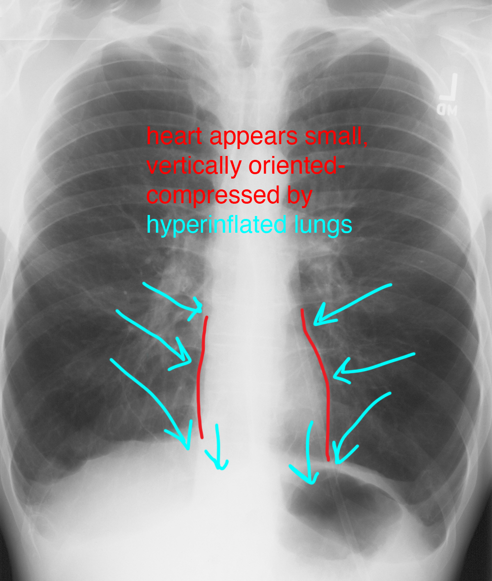
Case 2
Case 2, continued
Further Explanation:
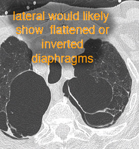
What do you think of the lung markings on this single CT image in another patient with shortness of breath? What might you expect to see on a lateral radiograph in this patient?
