

Bonus Case
This case will be discussed at the end of the in-class session. This patient was admitted with recent onset of fever and respiratory symptoms.
Question 1:
a) What specific questions would you ask her if you were concerned about COVID-19?
Loss of sense of smell, GI symptoms, whether she has been wearing a mask regularly when outside her home, recent exposures to large groups of people
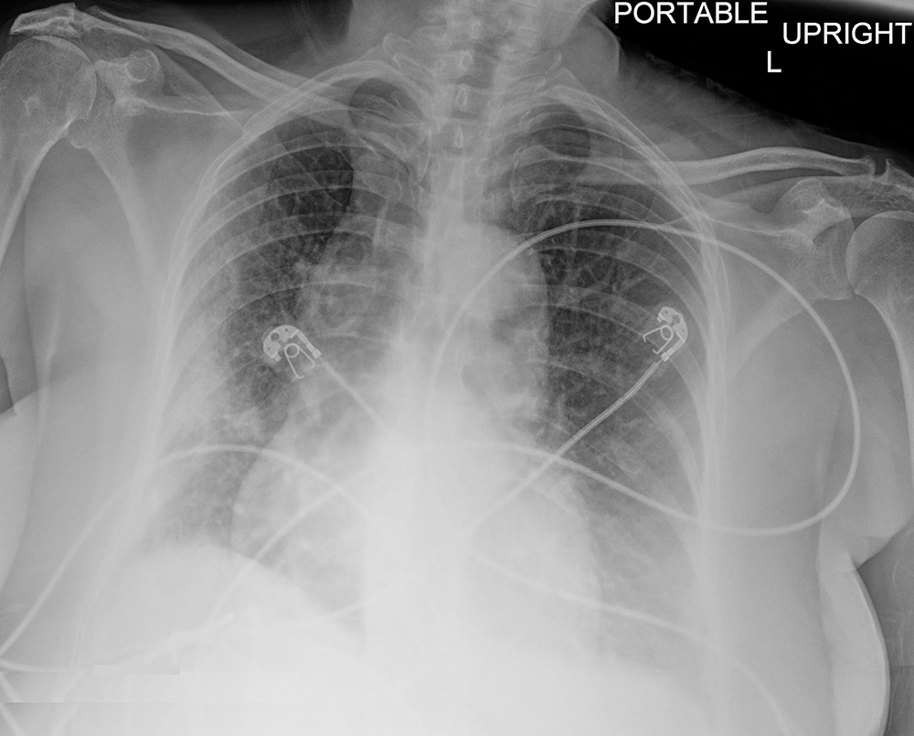
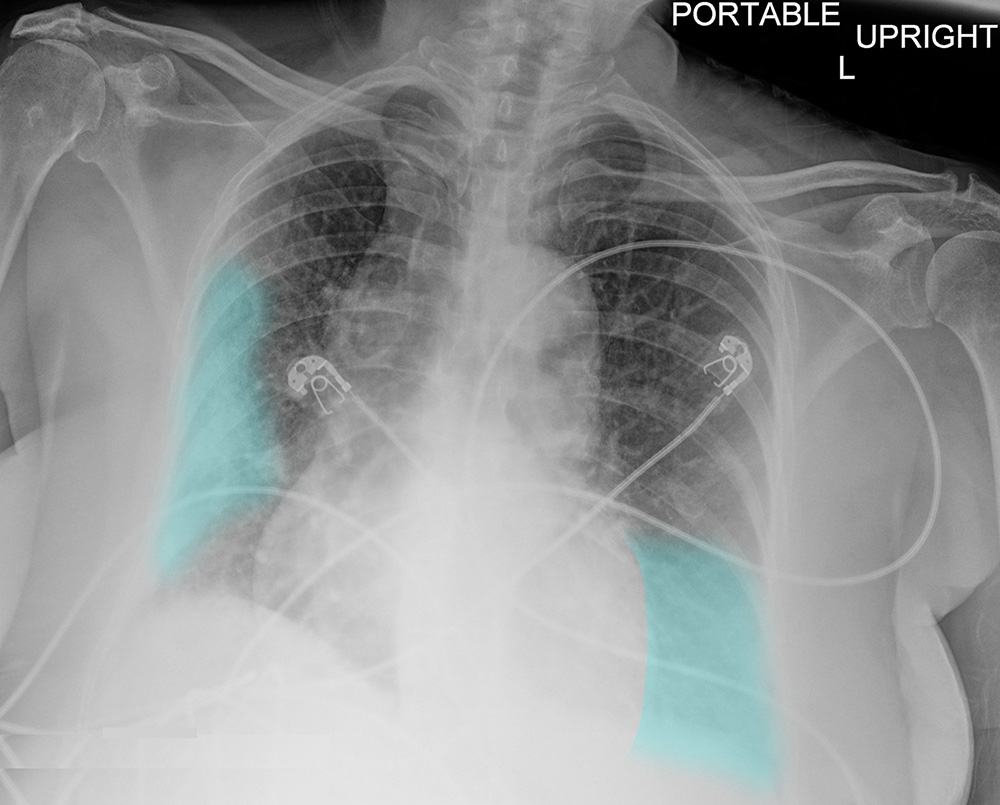
b) What is abnormal on this chest radiograph? Is this finding specific for a particular diagnosis?
There is hazy opacity in the lateral part of the right lung. There is also vague haziness at the left lung base. These are very nonspecific findings and could indicate COVID-19 infection, but could also be many other infections, hemorrhage, tumor, or infarct related to PE. Given the acuity of her symptoms, fibrosis is the only condition we discussed in the other cases of this module which does not seem likely. Click the link below to see the region of lung abnormalities
Bonus Case
The patient's respiratory status rapidly deteriorated. You are shown an image from a later time during her ICU course.
Question 2:
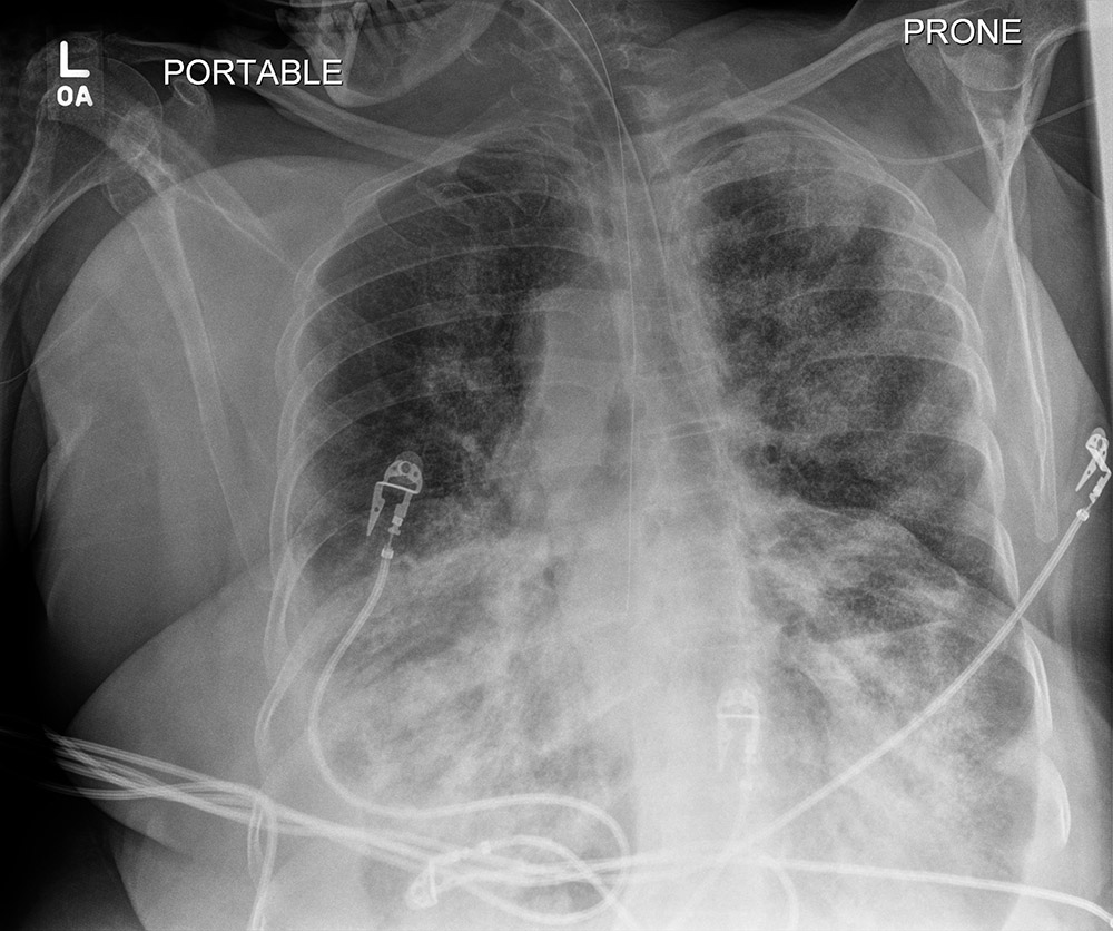
What are technical parameters of this image?
This is a portable chest radiograph obtained while the patient was in prone position, so this is actually a portable PA study, since the beam entered from the posterior side of the patient's body. As will be discussed in class, there are reasons in severe respiratory disease that patients may be more adequately ventilated when in the prone position.
Bonus Case
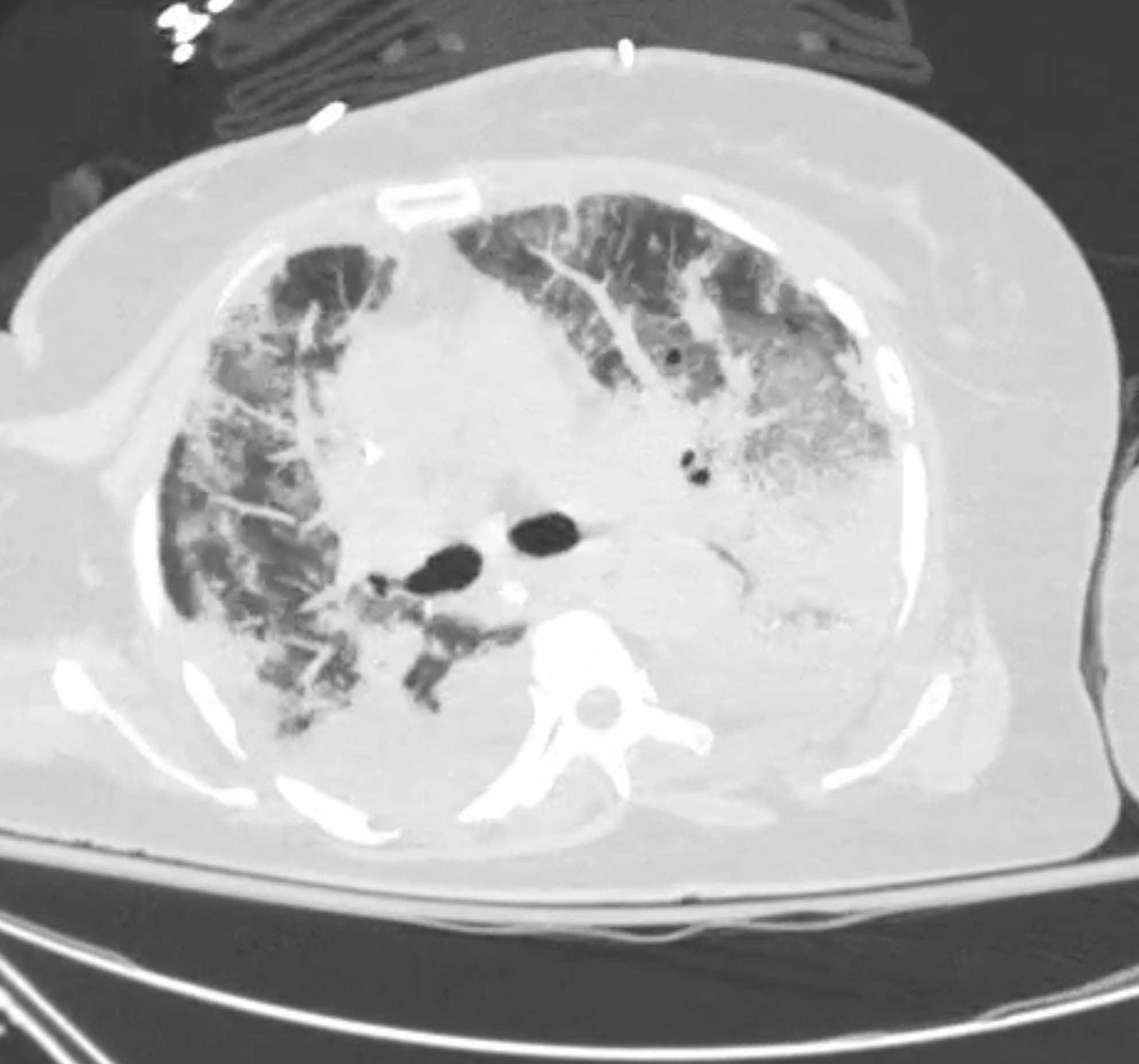
This is a study done in the mid portion of this patient's hospital course.
Question 3:
a) What are technical parameters for this image?
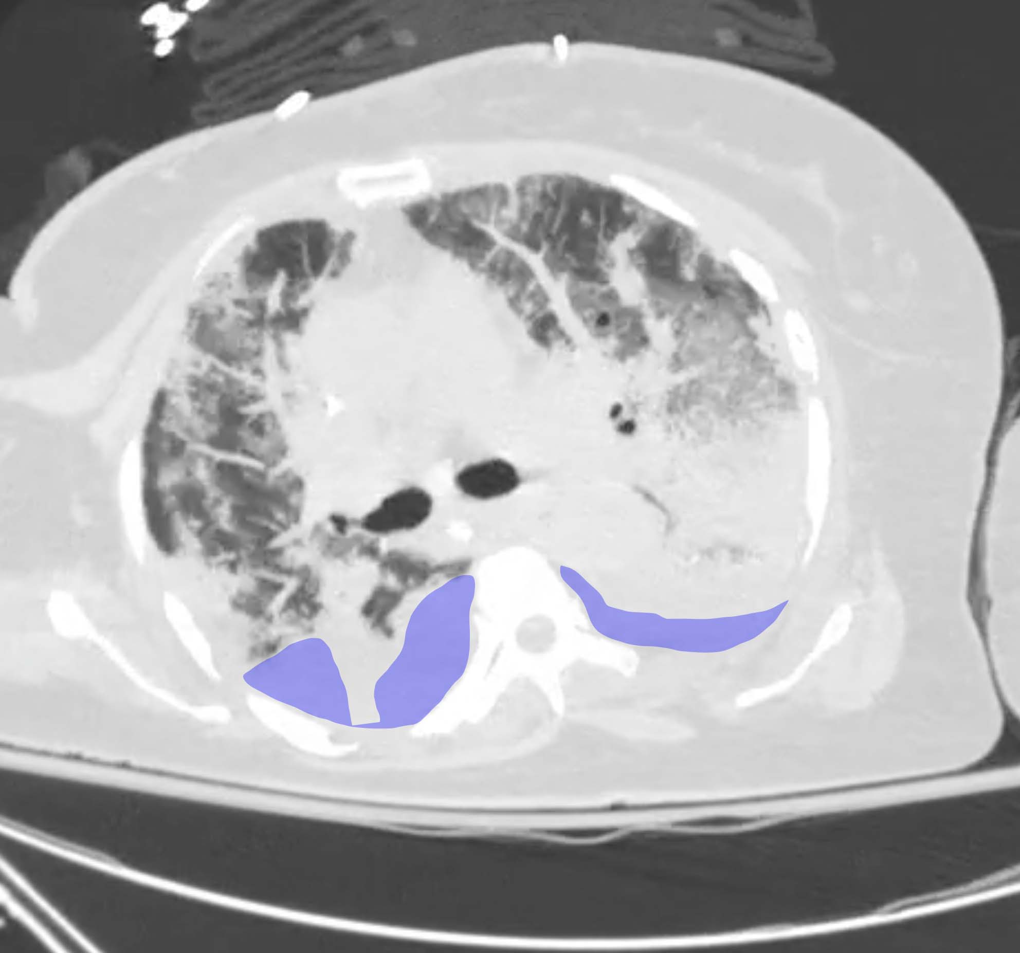
This is an axial CT scan displayed with lung windows. The patient was in supine position for this study. You can tell that she is no longer prone because you can see the movable support of the scan bed lying adjacent to the posterior part of her body. It is possible to scan patients in the prone position, if necessary. There are small bilateral pleural effusions present but they are hard to see with these lung windows. They are labeled below based on their appearance on soft tissue windows (not shown).
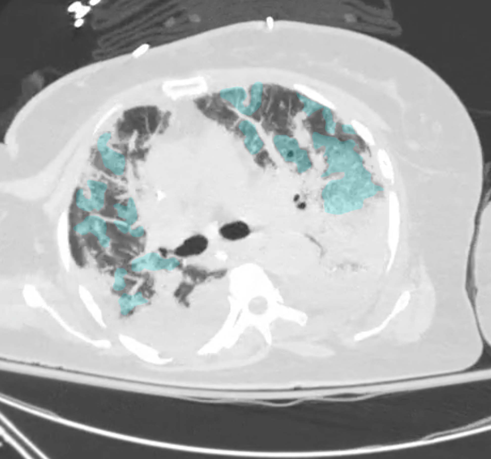
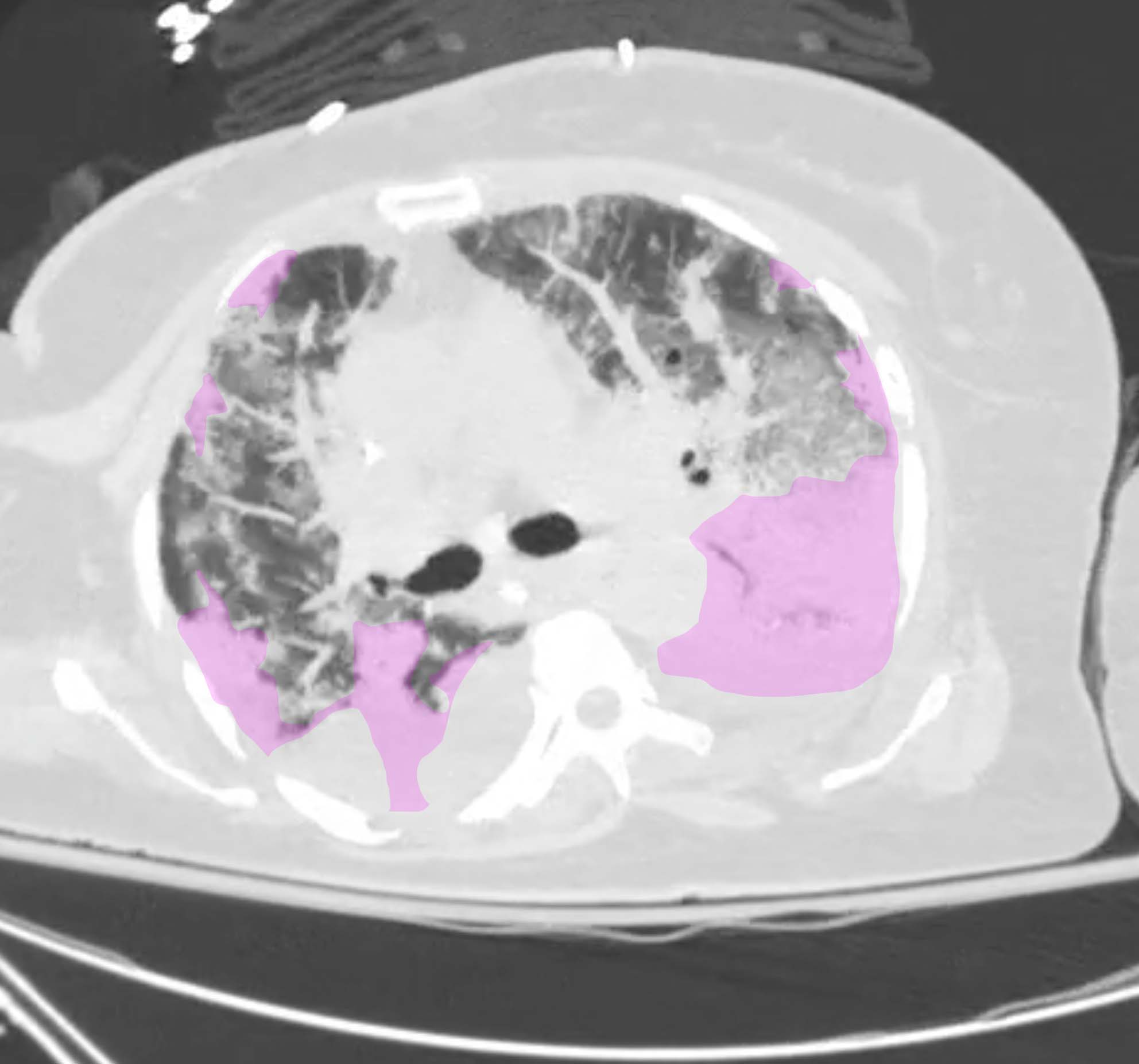
b) What two descriptors would you use for the findings in her lungs on this study? Are they specific for a particular diagnosis?
Labels below show lung abnormality 1 to represent ground glass opacity (GGO), which can be seen in many parts of her lungs on this scan. Labels below show lung abnormality 2 to represent denser abnormalities (obscuring adjacent pulmonary vessels), consistent with consolidation. A mixture of GGO and consolidation is commonly seen in COVID-19 but it not specific for this diagnosis. Many other infections or inflammatory processes can also produce these findings.
