

Summary
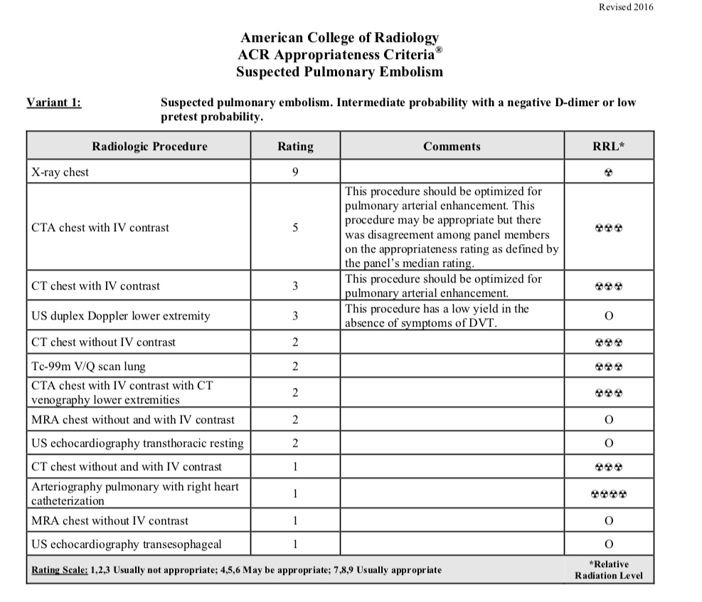
Case 1 and 2
Further Explanation:
Case 1 is an example of reversible airways disease (asthma), with normal lung imaging, with a comparison case showing non-reversible airspace disease. This case offers the opportunity to review the normal appearance of lungs on CXR and CT. Case 2 shows lung parenchymal fibrosis as a cause of shortness of breath, and displays comparison studies of obstructive disease (emphysema) to contrast with restrictive disease. In fibrosis, lung volumes are low and interstitial markings are increased, with a reticular pattern early and later honeycombing, often worst in the lung periphery. In obstructive disease, lung volumes are high and CT shows cysts or air-filled spaces in the lung where tissue has been destroyed. Pulmonary function tests in fibrosis show low volumes and loss of oxygen diffusion capacity. In emphysema, lung volumes are high with obstruction to air flow.
Summary
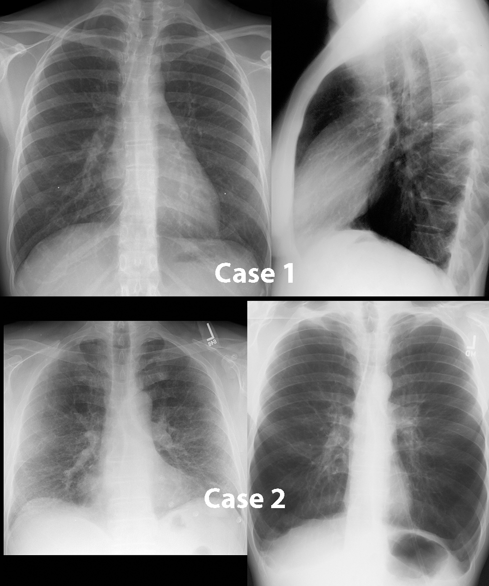
Case 3 and 4
Further Explanation:
Case 3 is an example of vascular disease as a cause of shortness of breath due to embolism, with imaging of both lungs and lower extremity. This case offers the opportunity to review the various imaging modalities that can show disease in the vascular system, in the pulmonary circuit as well as in the lower extremity veins. A filling defect in the pulmonary artery, seen only with properly timed IV contrast present, is the best diagnostic indication of pulmonary embolism. Case 4 is an example of diffuse lung disease due to capillary leak, with examples of complications that can result from mechanical ventilation as well as other examples of causes of fluid accumulation in the lung parenchyma. Due to use of high ventilatory pressures, air can leak from ruptured alveoli into the adjacent connective tissues, resulting in pneumomediastinum. Air can also enter the pleural space from rupture of peripheral air spaces, resulting in pneumothorax.
Summary
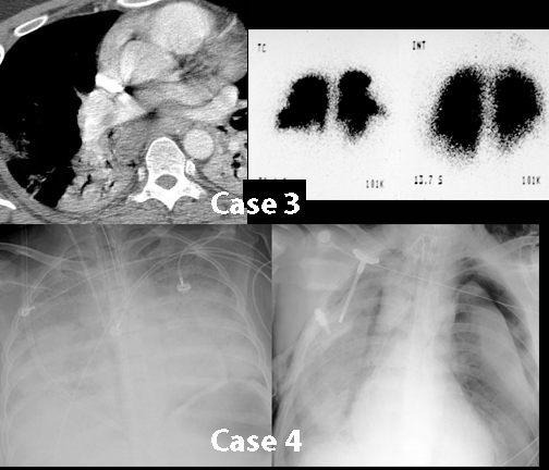
final grid of cases
Further Explanation:
Summary
Summary: What to Order
Further Explanation:
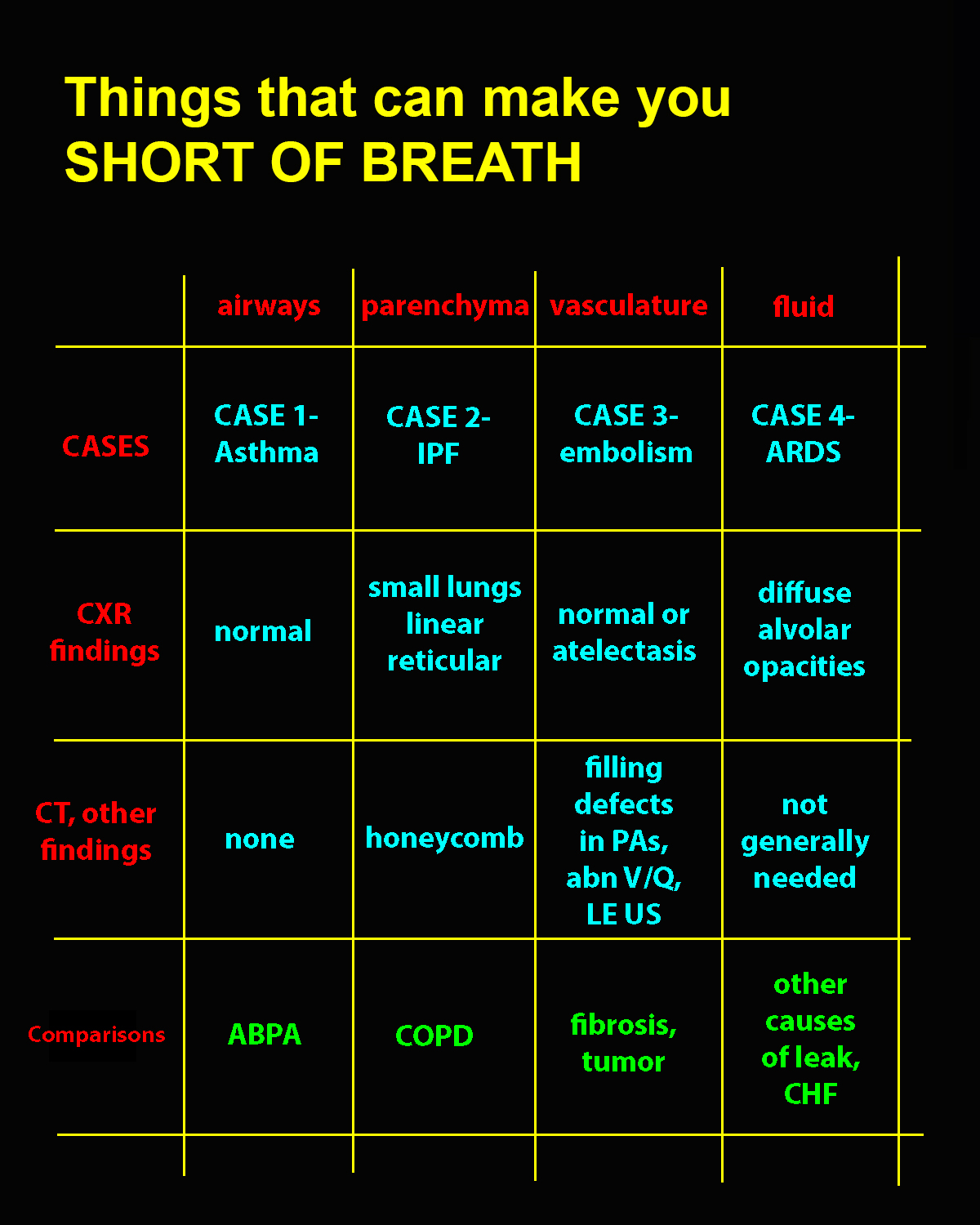
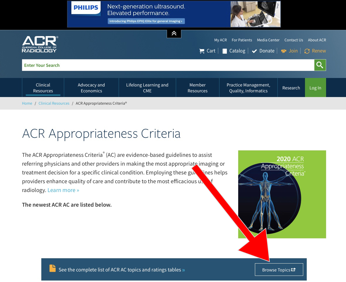
Use the ACR Appropriateness Criteria website to help you decide what the most appropriate imaging is to order in common clinical scenarios. The website is free, requires no login, and is organized by broad subspecialty topic areas organized by clinical presentation. If you search for 'ACR Appropriateness', you will likely find it to be the top choice.

Further Explanation:
Once you enter the site, choose 'Browse Topics' to bring up a list of all available topic areas, organized by body system. There are separate lists for Diagnostic (including Breast, Cardiac, GI, Gyn, Trauma, MSK, Neuro, Pediatric, Thoracic, Urologic and Vascular) and interventional Radiology (including many specific vascular and non-vascular topics). Topics are updated frequently.
Further Explanation:
For Thoracic imaging, there are 16 different categories and each includes a list of appropriate types of imaging as well as a review of the available literature on the topic. This is an excellent resource for evidence-based ordering of imaging studies for most common clinical complaints. You can search through the topics to find the one that best matches your patient's clinical setting, then click on the appropriate link to bring up a list of potential studies you might consider ordering.

Further Explanation:
Each topic area is organized by presenting clinical scenario. This shows one of several clinical scenarios for patients with suspected pulmonary embolism, with a listing of all possible imaging studies, a rating of how appropriate each is in this setting, and indication of relative radiation doses for each. Each section also includes a detailed bibliography and explanation of the references that were used for these recommendations at the end of the section.
