

Case 4
This young patient has become severely short of breath after a complicated but acute hospital course for presumed infection.
Further Explanation:
What type of study is this and how would you describe the findings? What part of the respiratory system seems to be most involved? Consider bronchi, blood vessels, pleural space, lung alveoli or lung interstitium. What support lines are present?
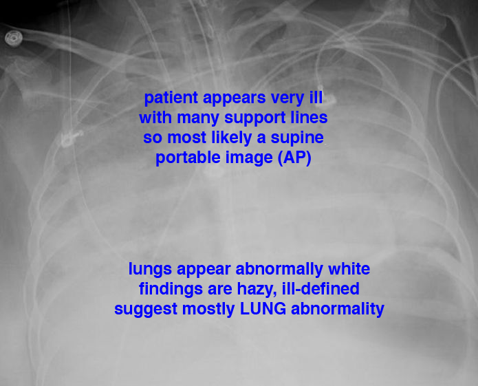
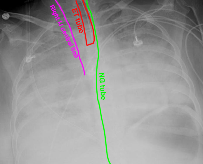
Case 4
Case 4, continued
Further Explanation:
This is a different patient with a similar history, who was intubated and placed on positive pressure ventilation. What is abnormal on this CT image and how might it relate to this history? in what space is the abnormal air located?
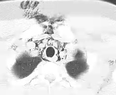
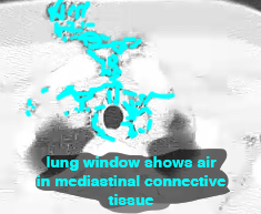
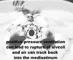
Case 4
Case 4, continued
Further Explanation:
This portable chest radiograph is from a different patient with a similar history. This patient was also on positive pressure ventilation. What complication do you see on this image?
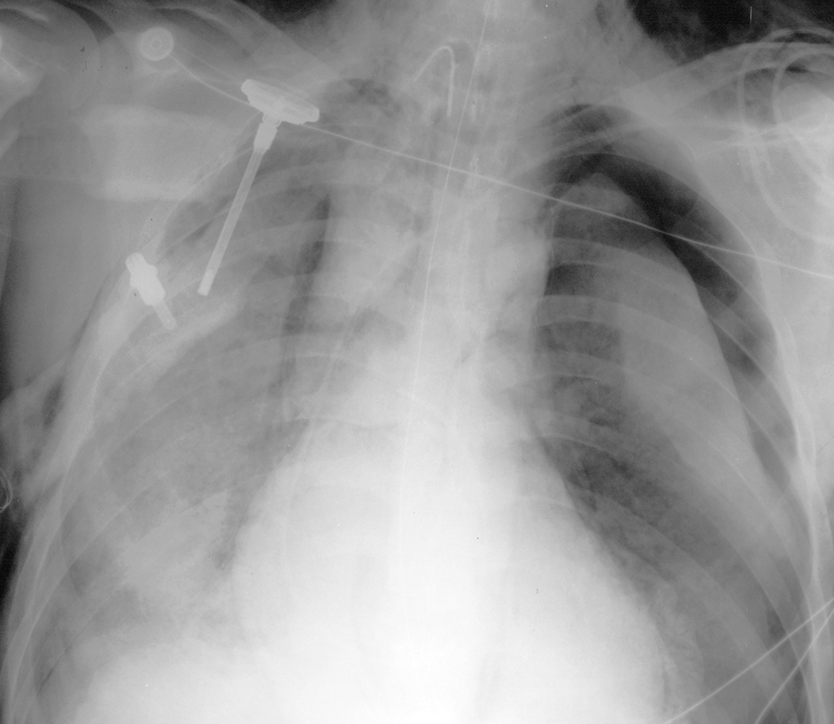
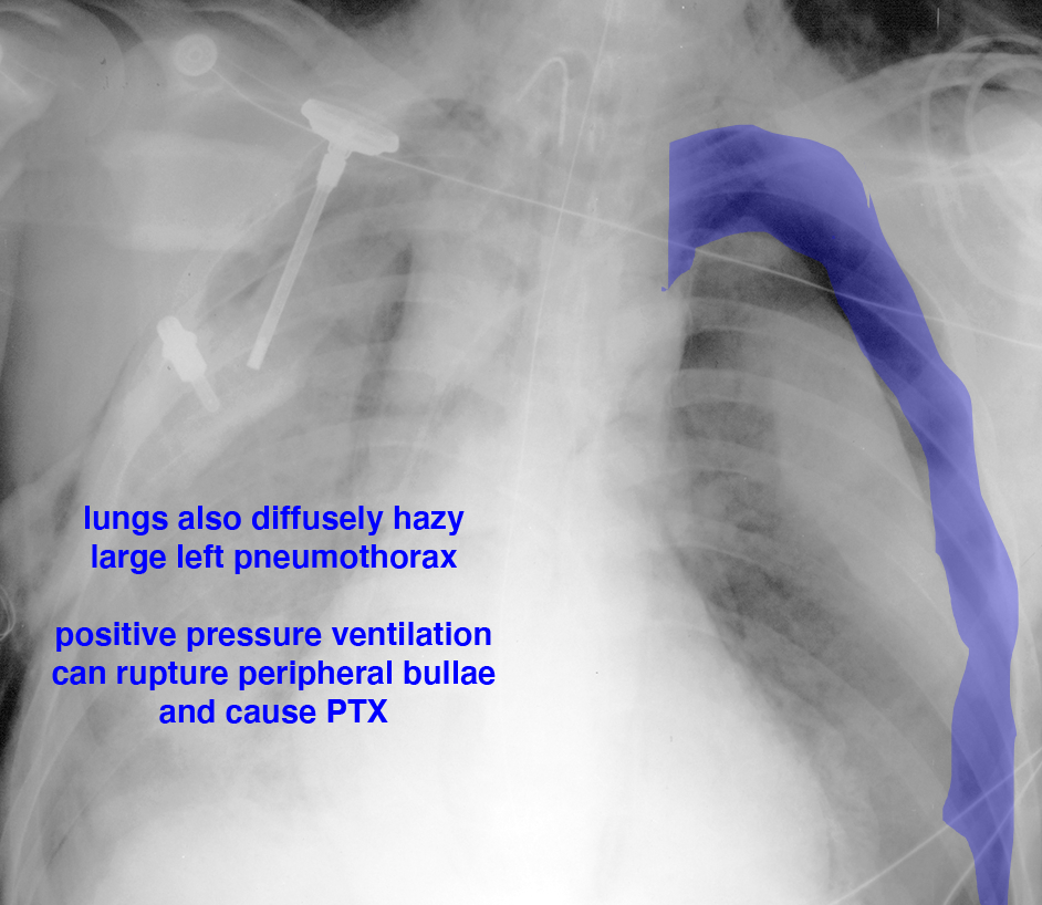
Case 4
Case 4, continued
Further Explanation:
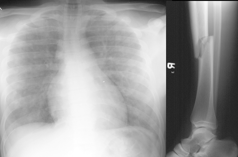
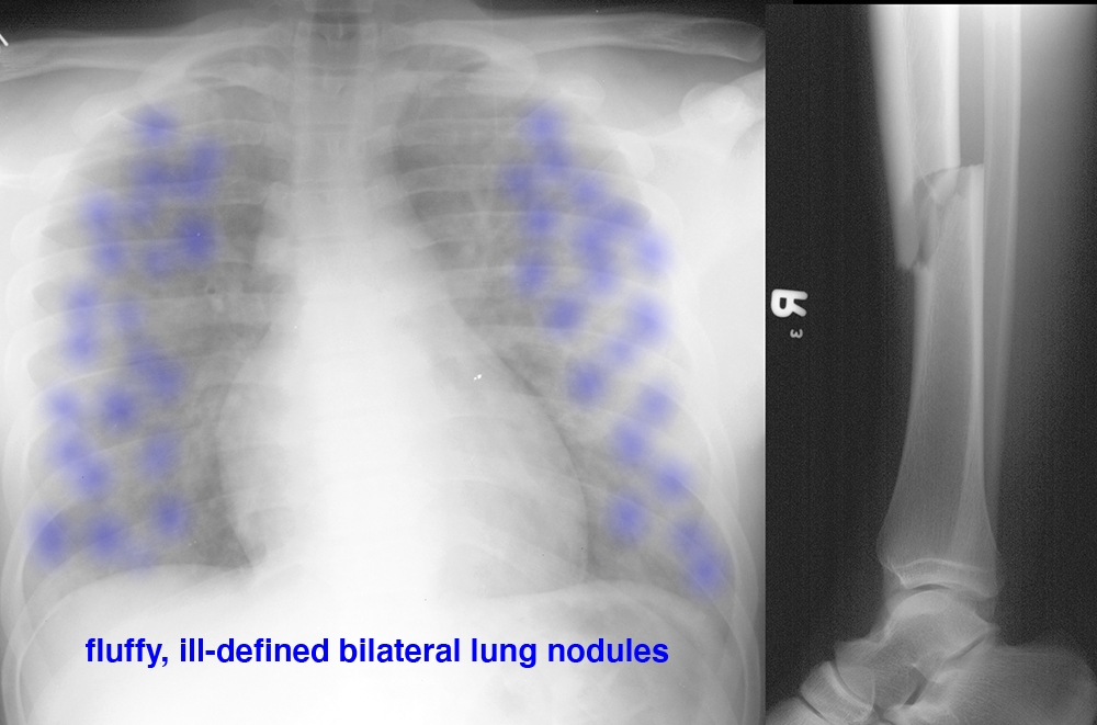
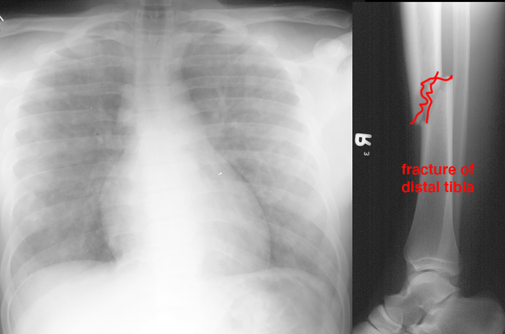
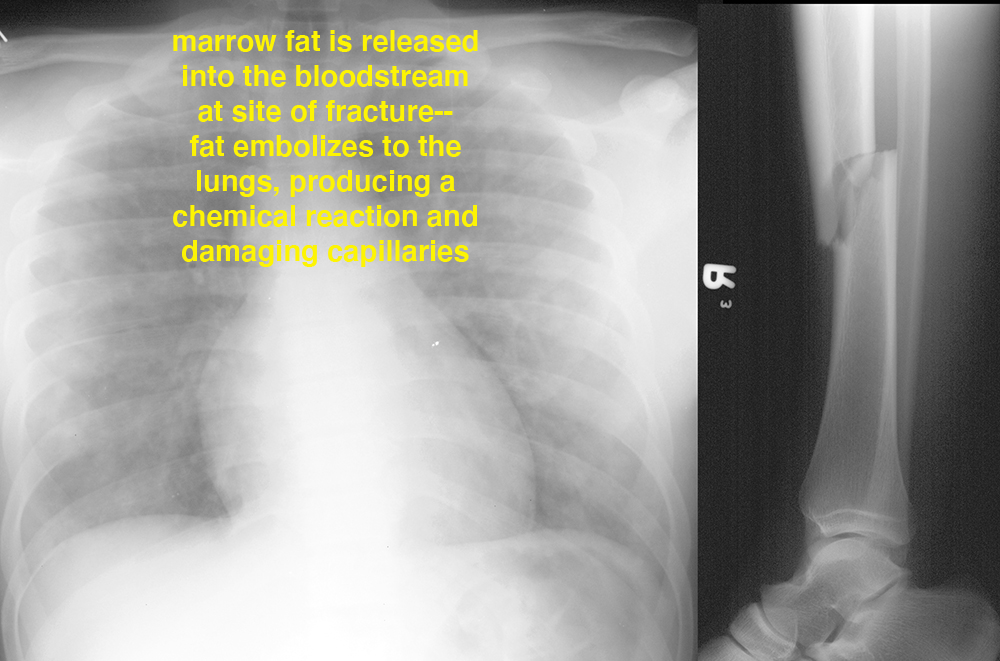
These images show studies done on a patient after a motor vehicle collision. The patient was acutely more agitated and had new petechiae (dark spots on the skin due to bleeding). How can you tie the two images together given these clinical findings?
