

Case 1
44 year old woman with breast mass
Question 1:
a) Is the listed history sufficient for ordering a breast ultrasound?
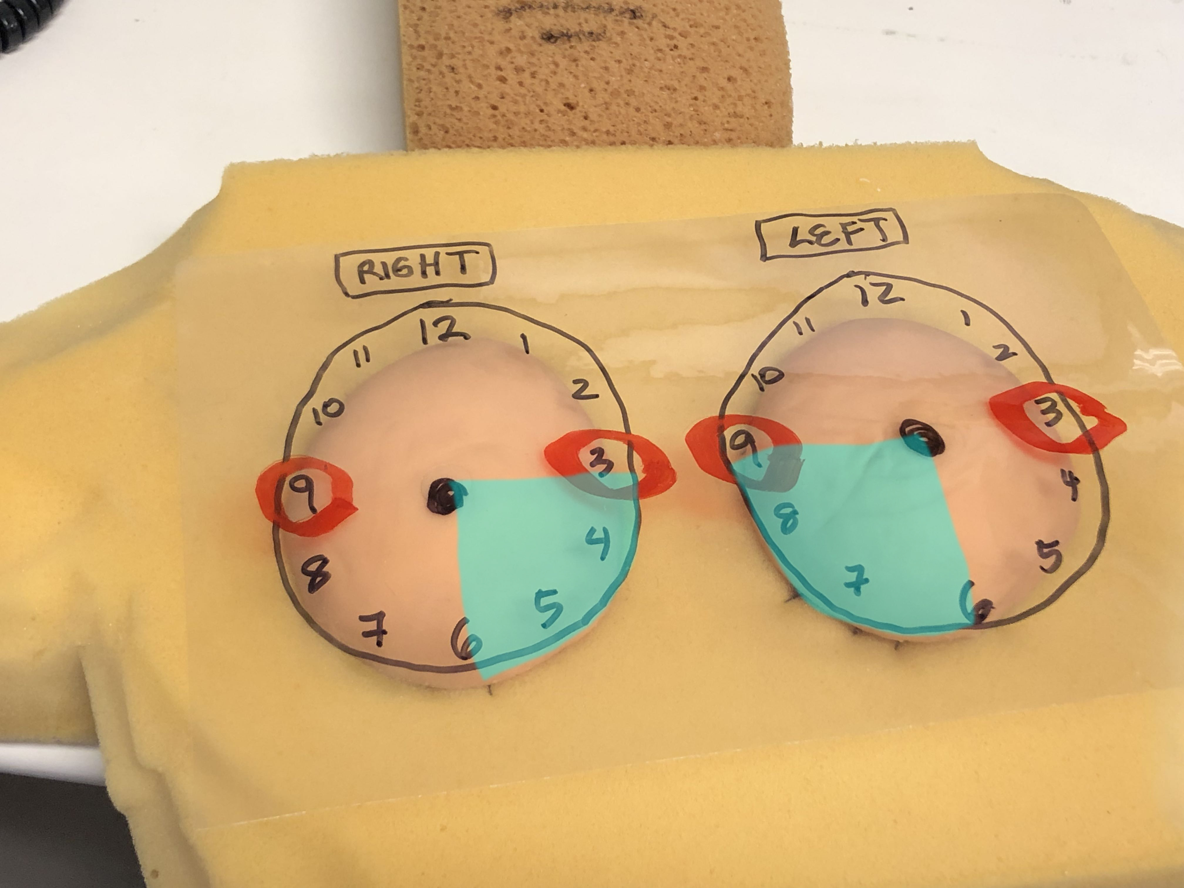
NO!! Breast ultrasound is usually a focused exam, meaning that the radiologist needs a very specific LOCATION to examine. You must indicate the SIDE, as well as the LOCATION, usually using a radial coordinate system centered on the nipple. So a better history would be '44 year old woman with left breast mass, 3:00 position at a distance of 6 cm from the nipple.'
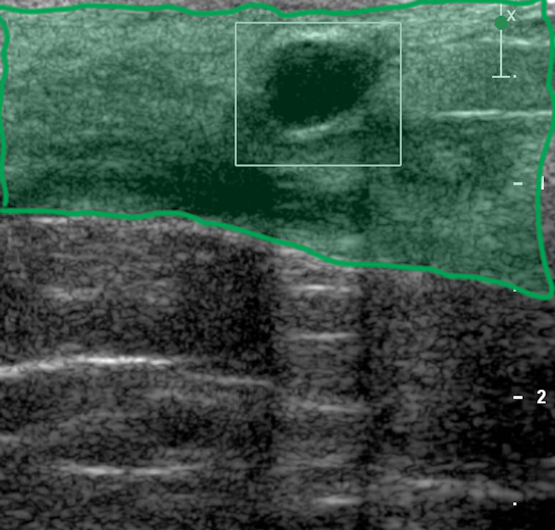
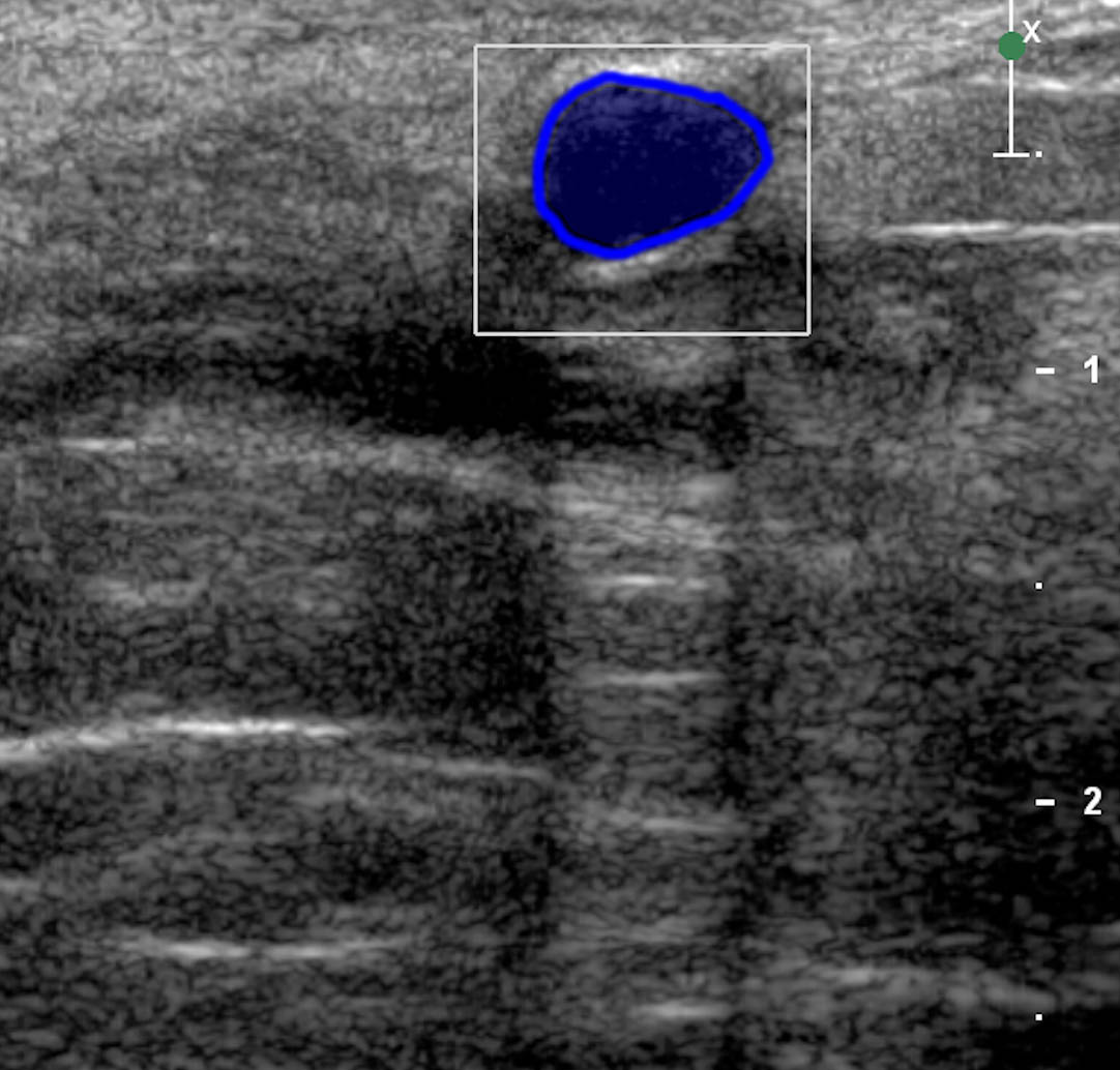
b) How would you describe the finding on this image?
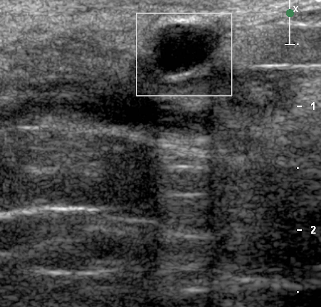
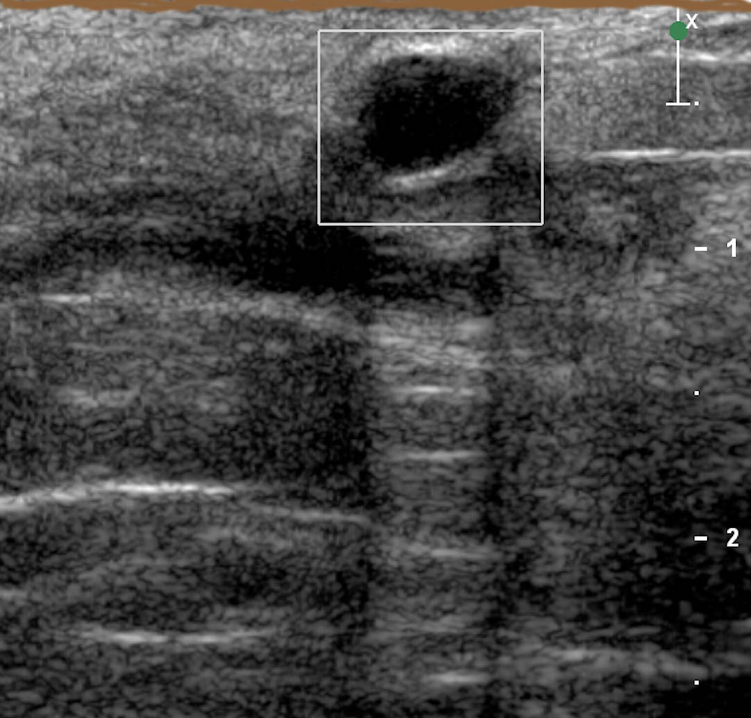
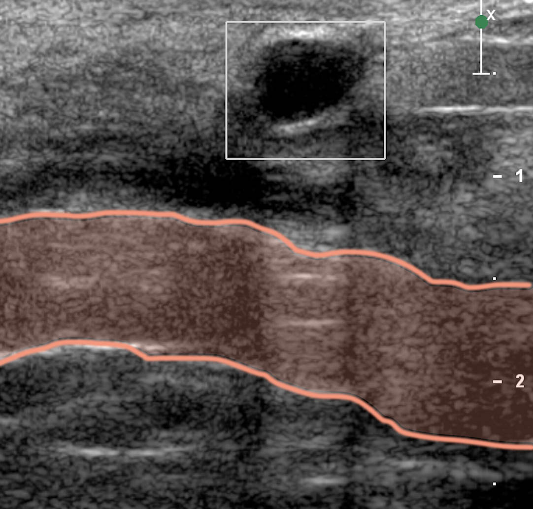
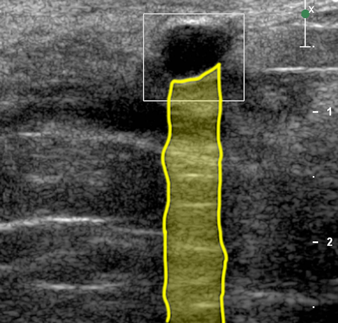
There is a round, smooth, well-marginated mass that is very black (anechoic, or without echos). There is a band of white extending behind the mass, away from the skin surface and transducer (which are always at the top of the image).
c) What is the white band?
The white band is an ARTIFACT, but one that we use to our advantage. When the sound beam encounters a watery structure, the sound is transmitted more easily than through other tissues (hence the very black appearance, with no echos within the lesion), and the beam that is beyond the lesion is stronger than expected, producing what is called 'posterior acoustic enhancement'. This white band tells us that the black lesion is likely a cyst.
d) What should be done next for this patient?
Since this is a simple cyst, nothing needs to be done. Cysts are very common in the breast and generally need no treatment. When very large and painful, they can be aspirated with a needle, but they often come back. This cyst is quite small and needs no treatment.
Case 1
How to hold the US probe (transducer)
Question 2:
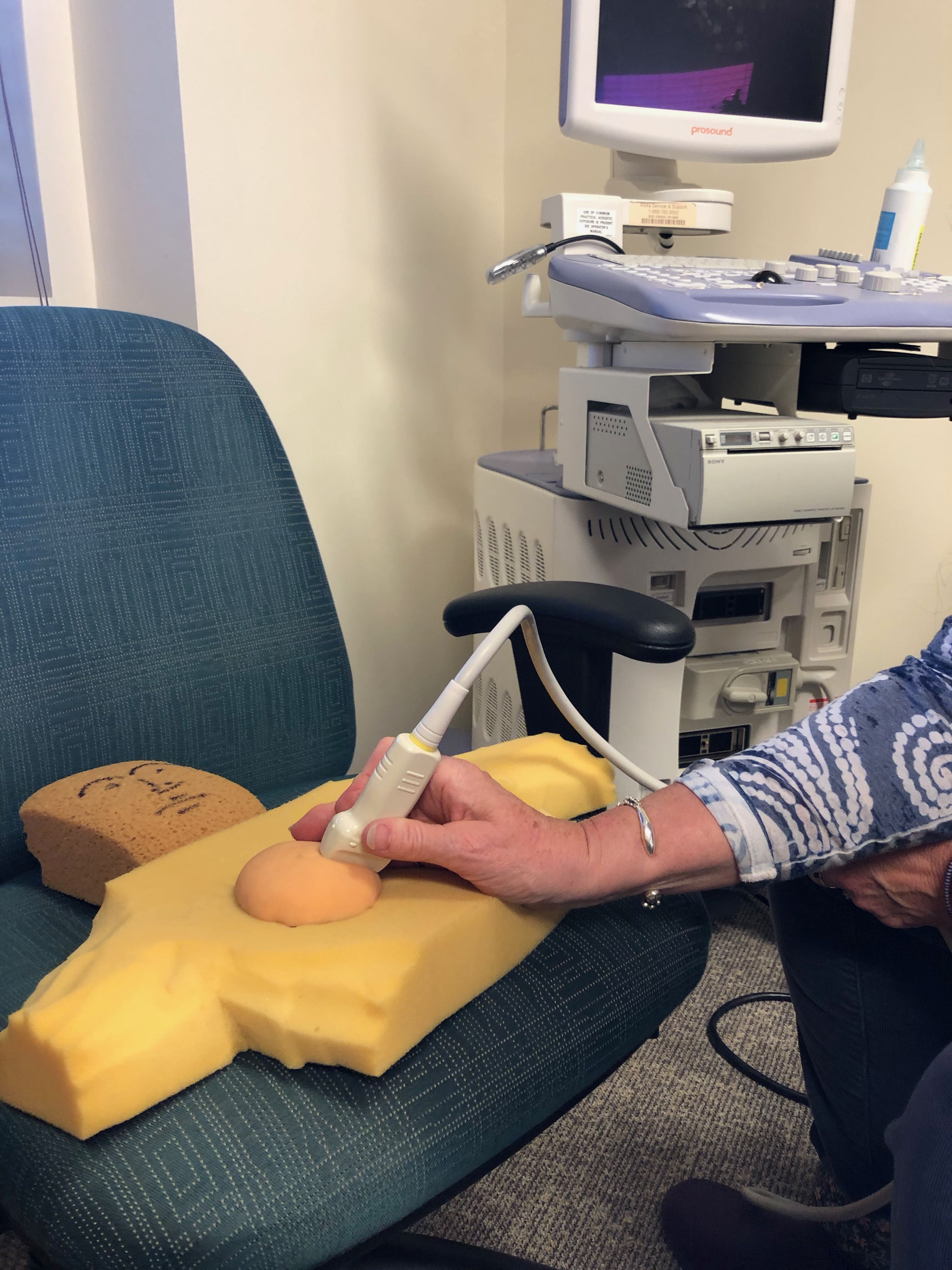
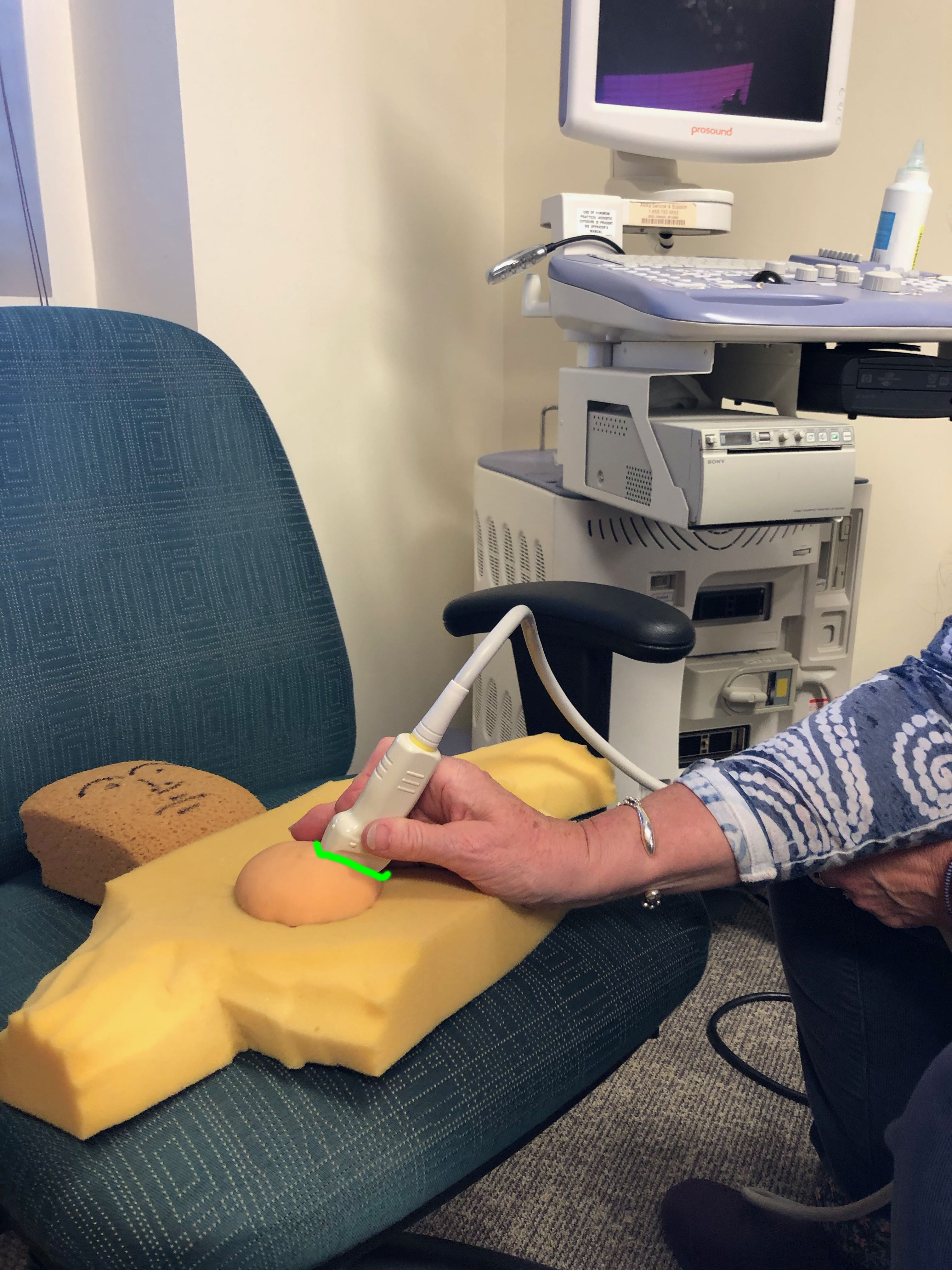
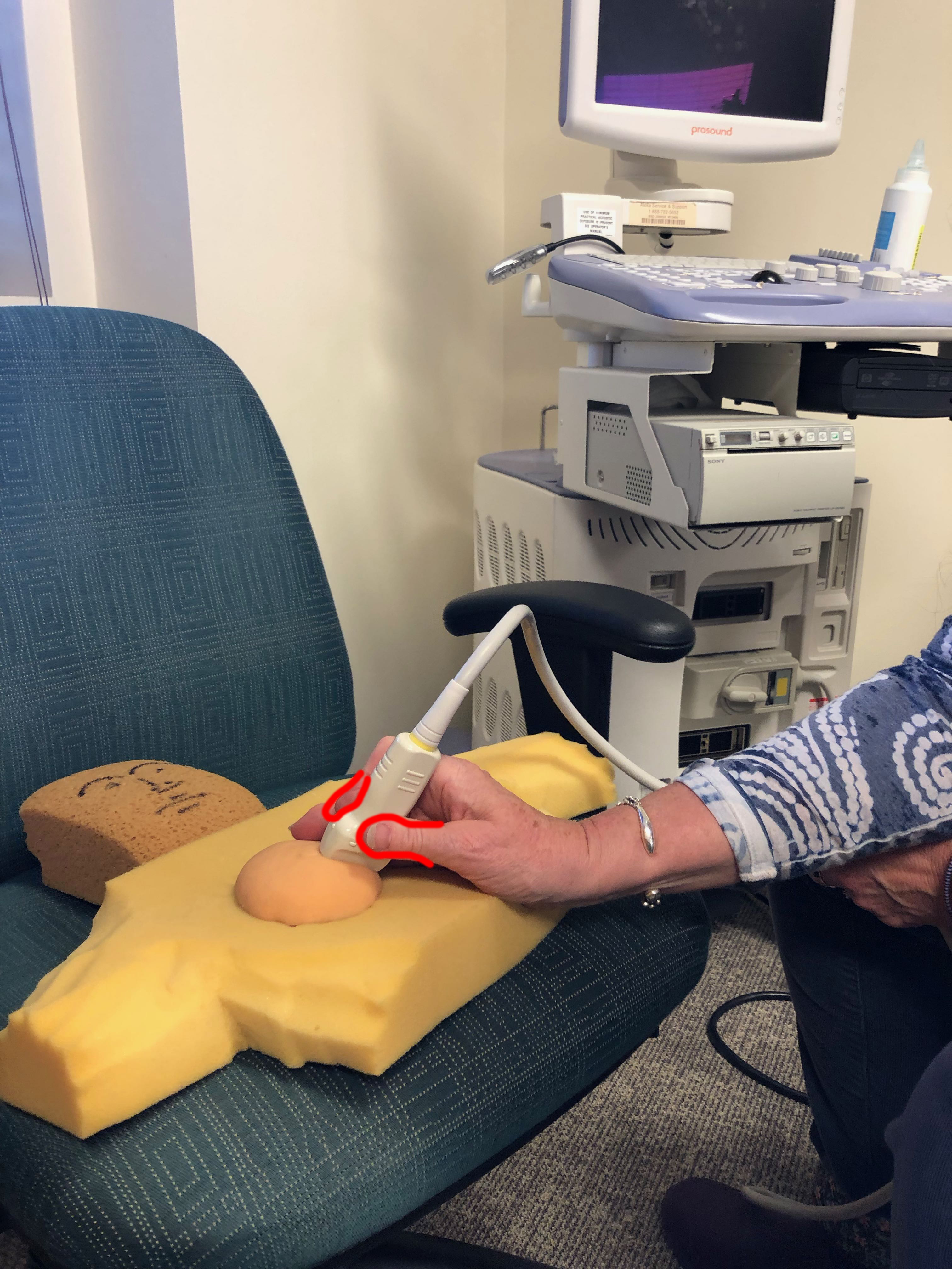
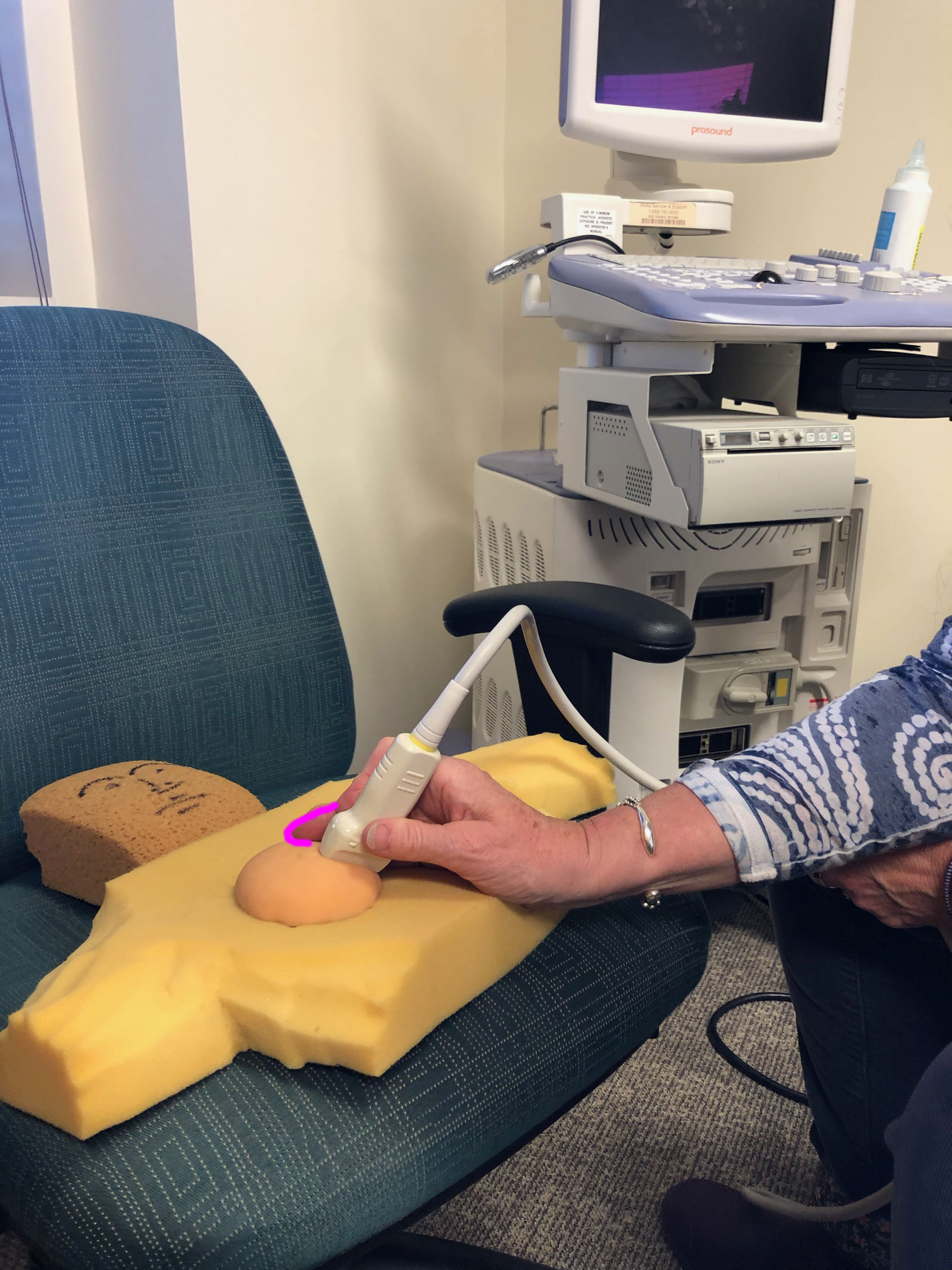
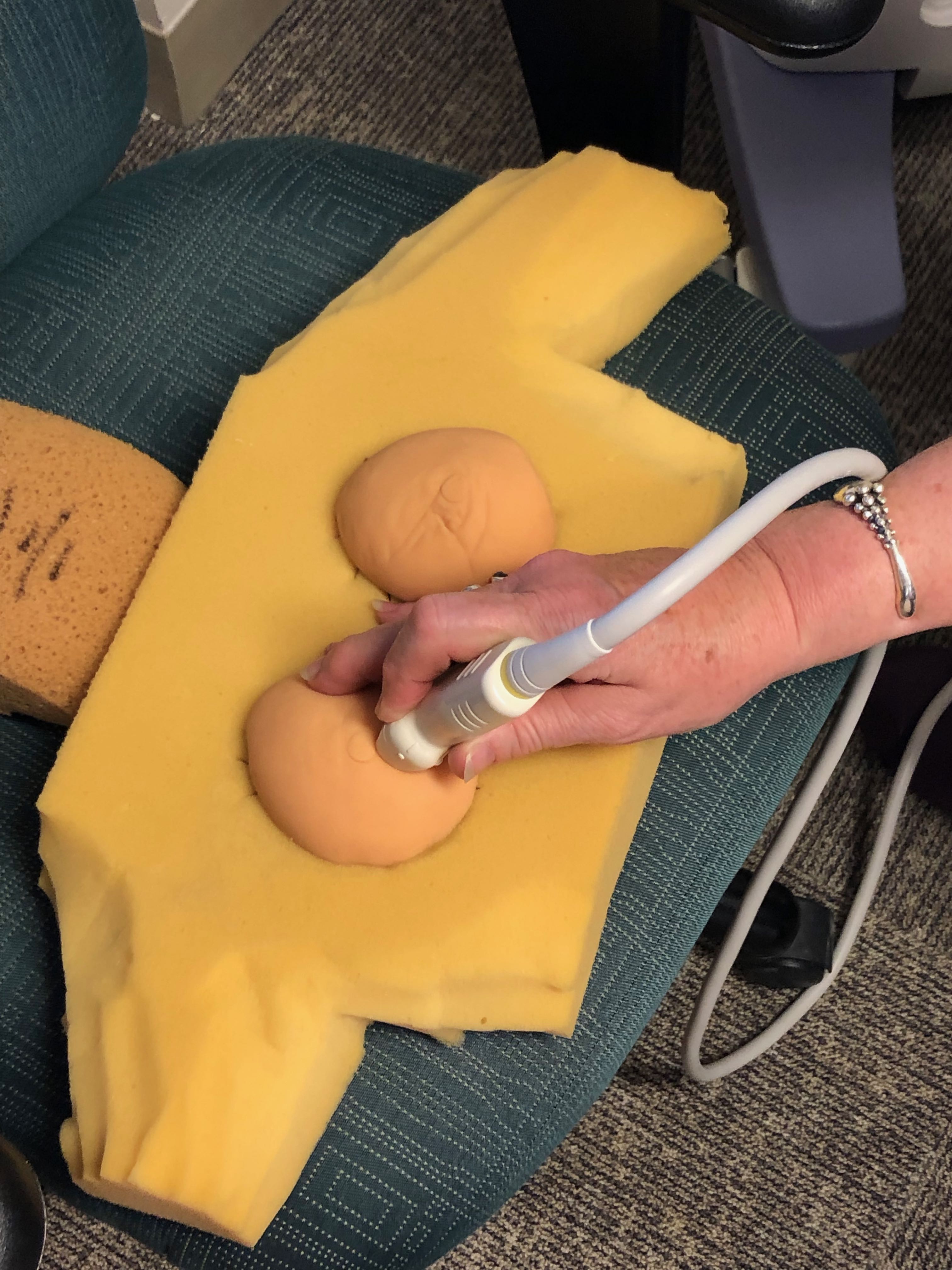
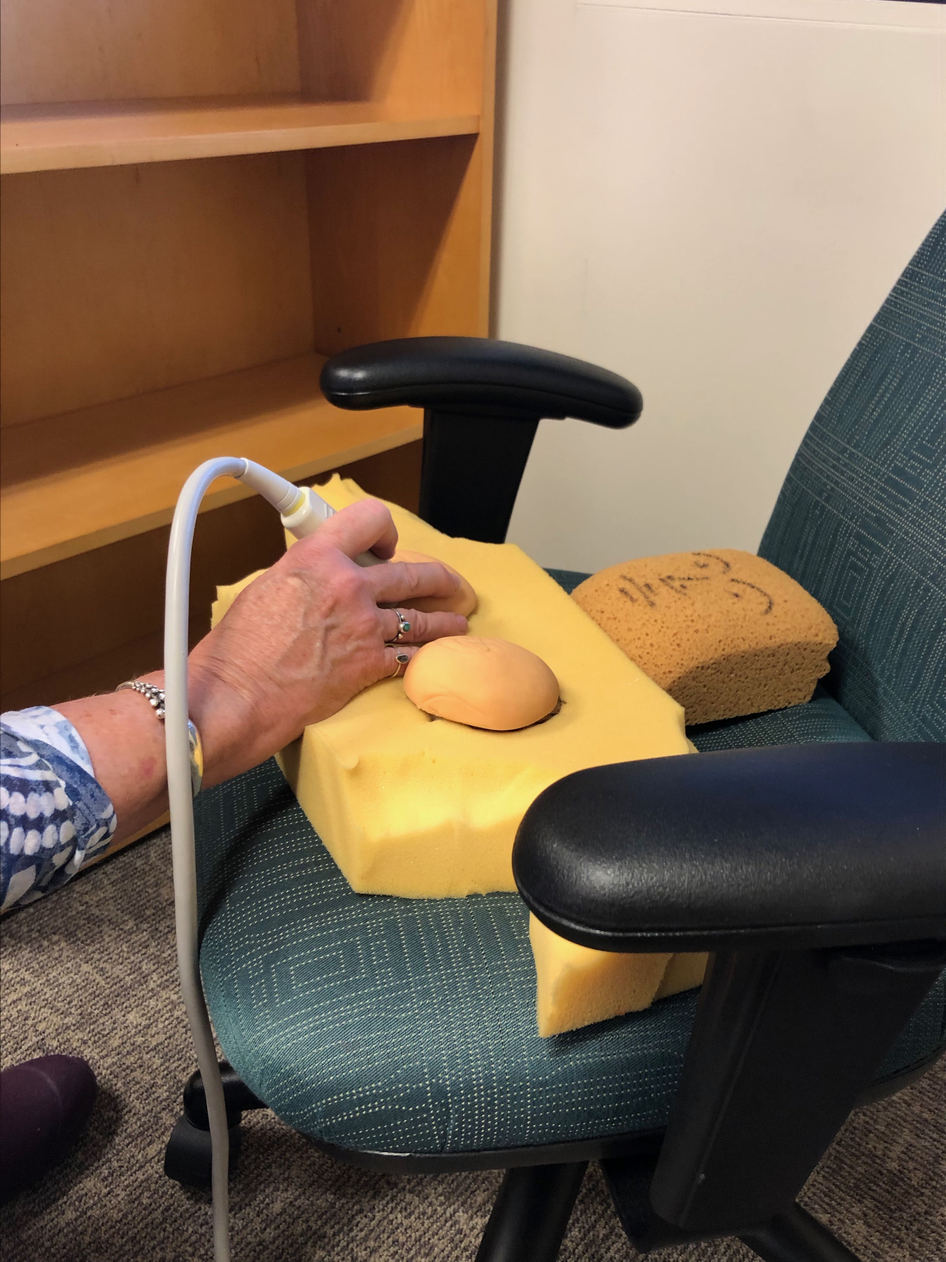
a) Where should you grasp the probe when performing a breast ultrasound?
You will have the best control (important on a slippery surface of a soft pliable organ like the breast) if you grasp the probe near the end that touches the patient, holding it lightly between thumb and index finger. This allows you to rest the other fingers on the patient, to steady your hand.
b) What do you need to put on the probe before placing it on the patient?
We use ultrasound GEL to provide acoustic coupling between the surface of the probe and the patient's skin. If any air is between the two, it will block the sound beam.
c) What tissues or materials block the sound beam of an ultrasound unit?
Air is the commonest thing that will block the sound beam. Sound travels well in air in the auditory range, but the much higher frequencies of sound used in ultrasound are blocked by air. Bone and other dense materials (like metal) will also block sound.
d) What type of tissue or material transmits ultrasound the best?
Water is the best transmitter of sound, and this is the basis of the 'increased through transmission' artifact that we see when we shine our beam through an area of water (like a breast cyst). The beam that emerges from the watery area is stronger than the other portions of the beam (that have been traveling through other types of tissue) so it appears whiter on the image.

Case 1
Radial coordinate system for localizing lesions in the breast
Question 3:
a) What is one potential problem with using the clock-face system to describe location of masses in the breast?
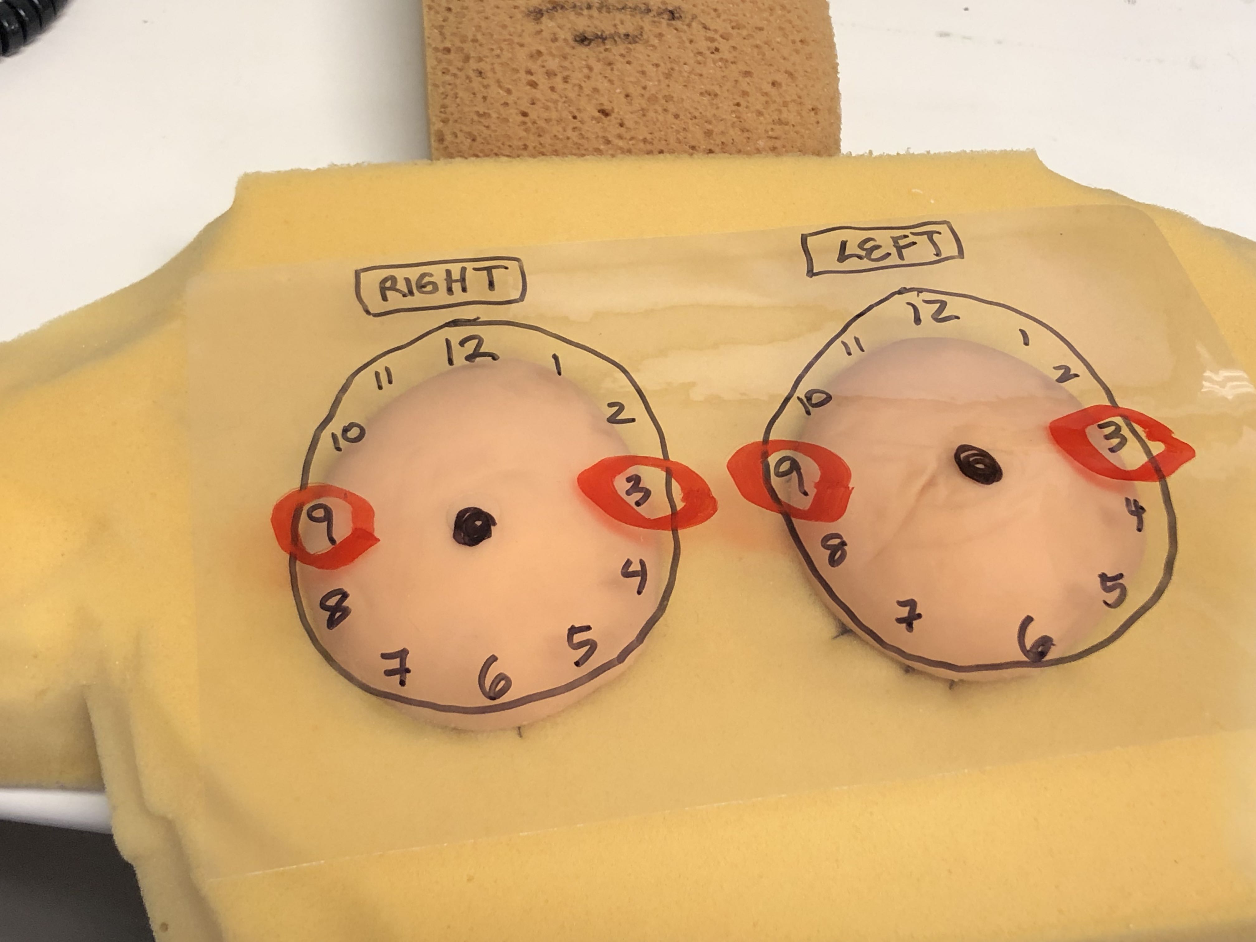
As you can see from the diagram, the 3:00 position on the right is INNER, while the 3:00 position on the left is OUTER! We use the clock face as if we are facing the patient when describing the location of a lesion, just as we would be oriented when examining the patient.
b) What is an alternative method to describe the location of lesions in the breast?
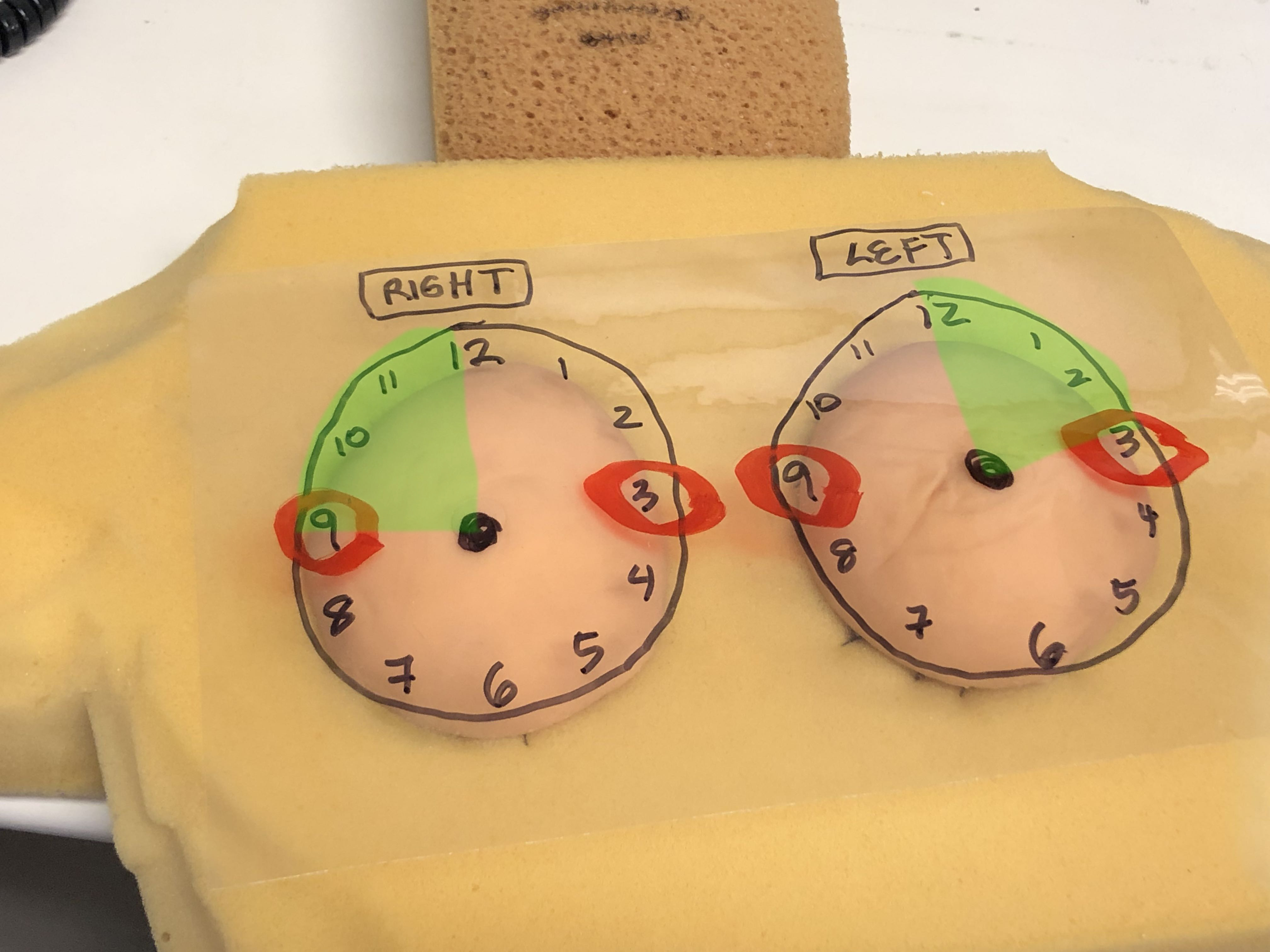
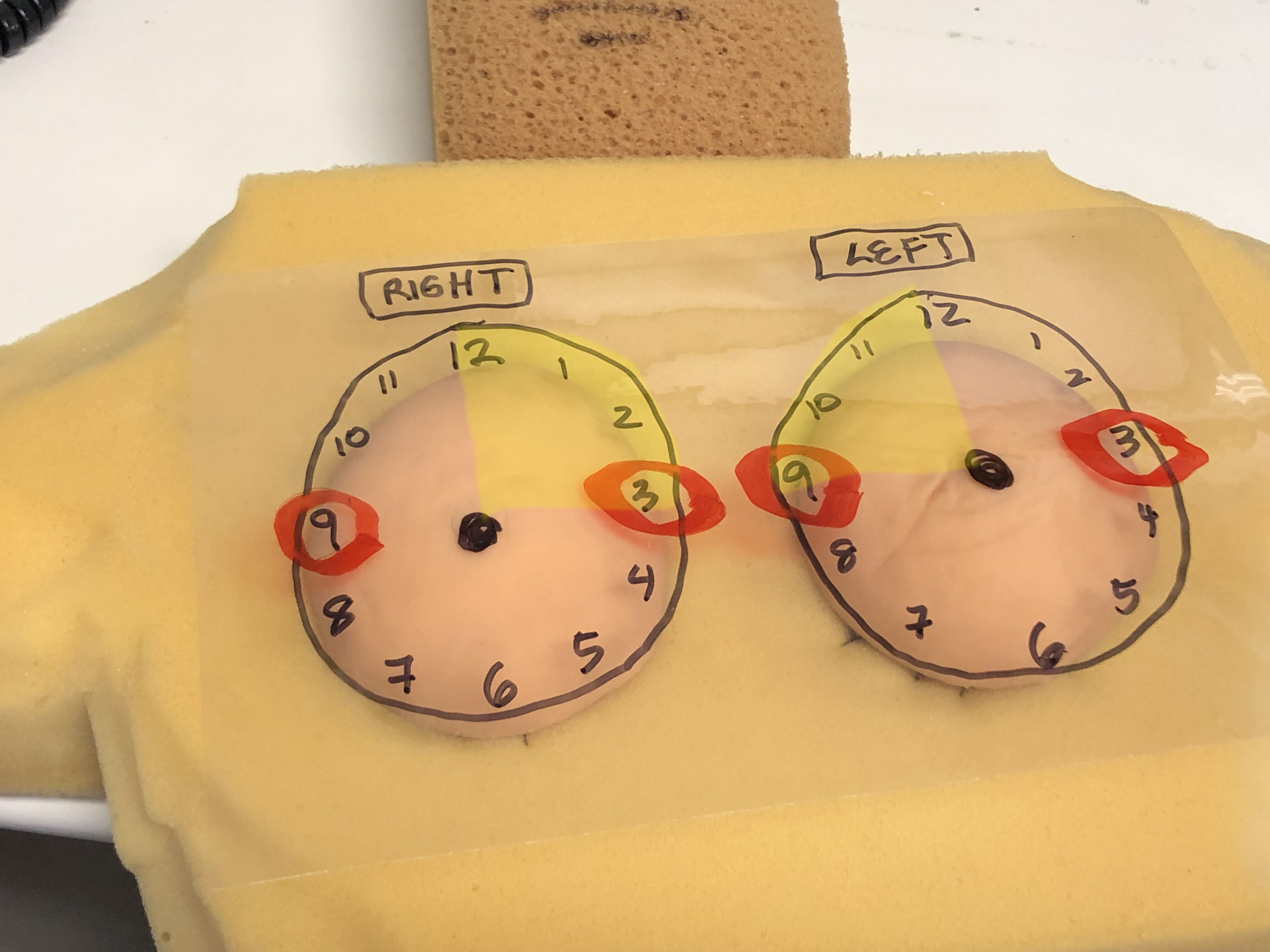
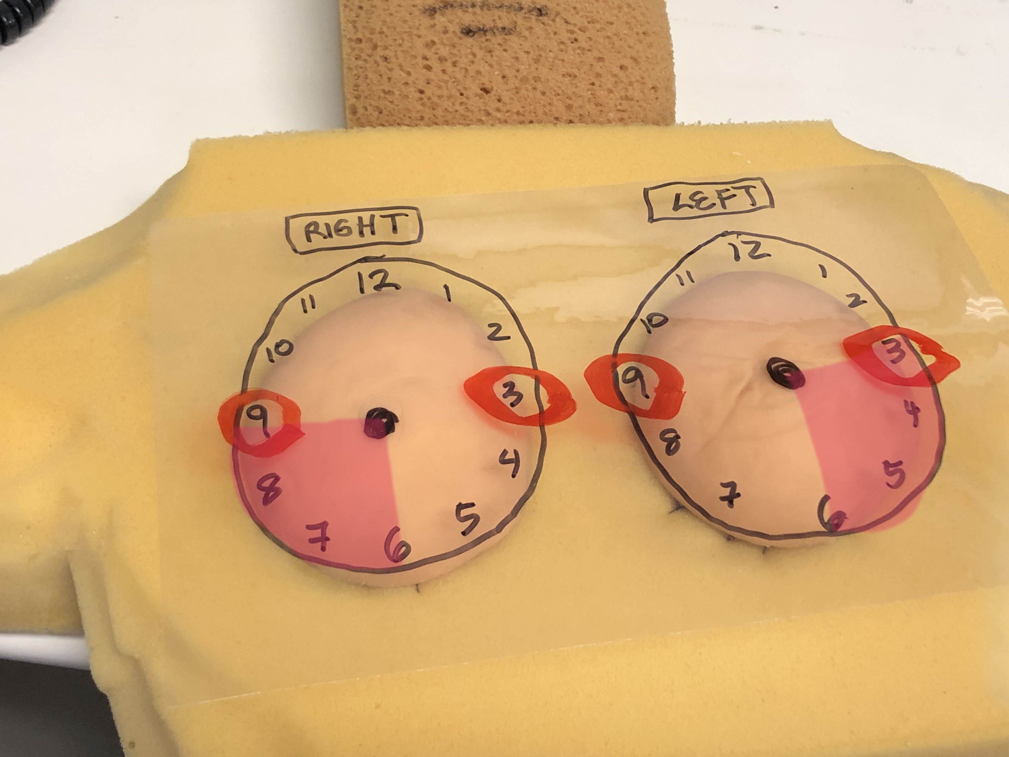
Instead of the clock-face, you can describe the location of lesions in terms of QUADRANTS, shown below. This is not as precise as the clock-face, but is less prone to error, since 'upper outer on the right' would not be as likely to be confused with 'upper inner on the left', both of which would be at 11:00 on the clock-face.
