

Case 2
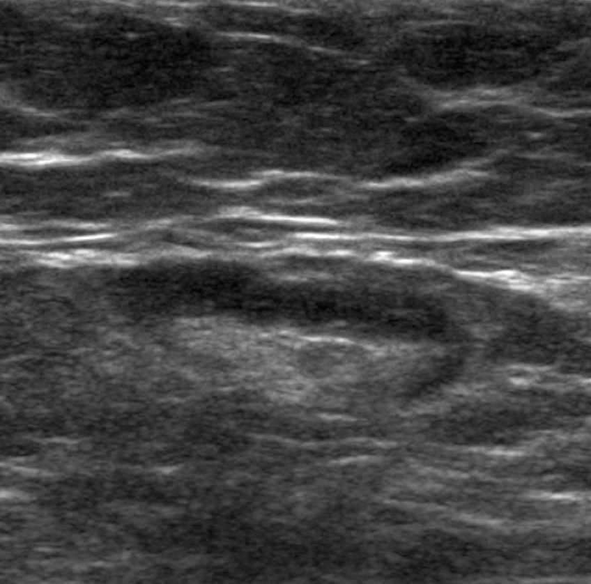
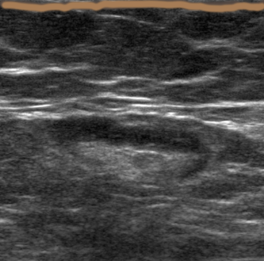
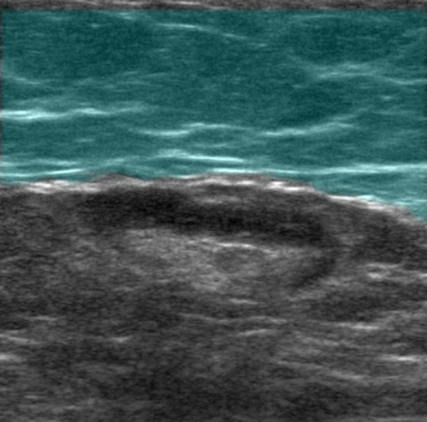
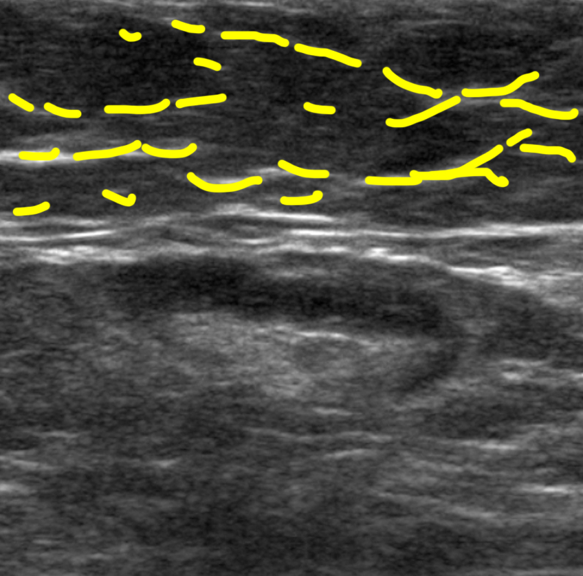
45 year old woman with left axillary mass, 1:00 position at a distance of 16 cm from the nipple
Question 1:
a) How do we determine if an axillary node is abnormal?
×
Answer:
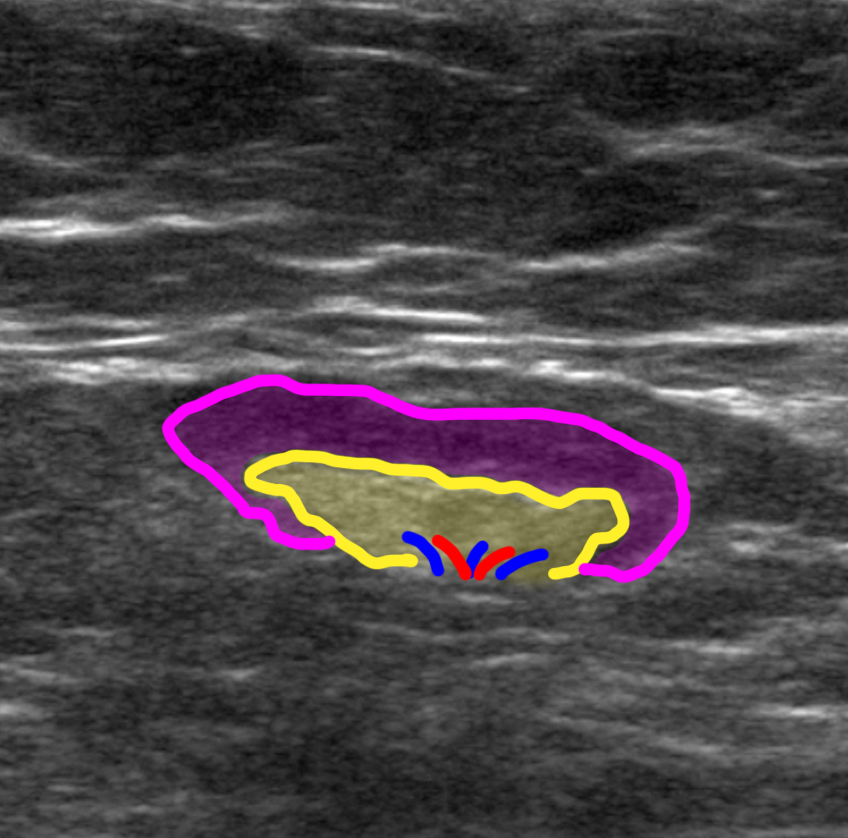
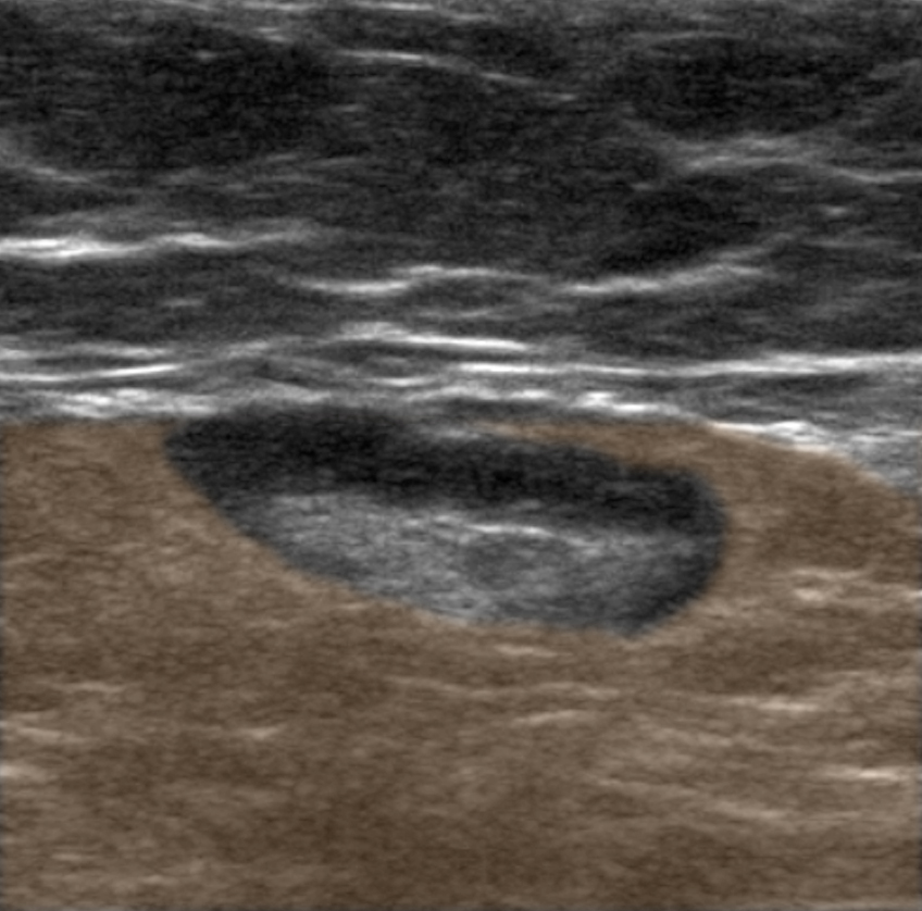
Axillary nodes vary widely in overall dimension, so we do not use that to determine if they are enlarged. Instead, we measure the THICKNESS of the cortex, and look at the overall morphology of the node. The cortex (purple on the labeled image below) should be smoothly draped around the echogenic fatty hilum (yellow on the labeled image below), where the blood vessels enter and exit. Vessels are often hard to see on routine grey scale ultrasound images, and are shown on the labeled image in red and blue for orientation only.
Axillary nodes vary widely in overall dimension, so we do not use that to determine if they are enlarged. Instead, we measure the THICKNESS of the cortex, and look at the overall morphology of the node. The cortex (purple on the labeled image below) should be smoothly draped around the echogenic fatty hilum (yellow on the labeled image below), where the blood vessels enter and exit. Vessels are often hard to see on routine grey scale ultrasound images, and are shown on the labeled image in red and blue for orientation only.
b) What is the mystery study indicated below
×
Answer:
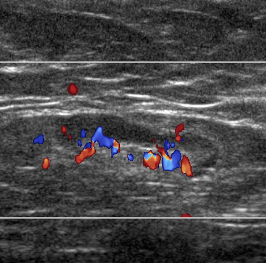
The mystery study is a color flow doppler ultrasound image of the node showing the extensive network of vessels in the region of the central fatty hilum. These vessels are generally not large enough to see clearly on the greyscale images, but show up well on doppler.
The mystery study is a color flow doppler ultrasound image of the node showing the extensive network of vessels in the region of the central fatty hilum. These vessels are generally not large enough to see clearly on the greyscale images, but show up well on doppler.
c) What is indicated by the red and blue colors on the mystery study below
×
Answer:
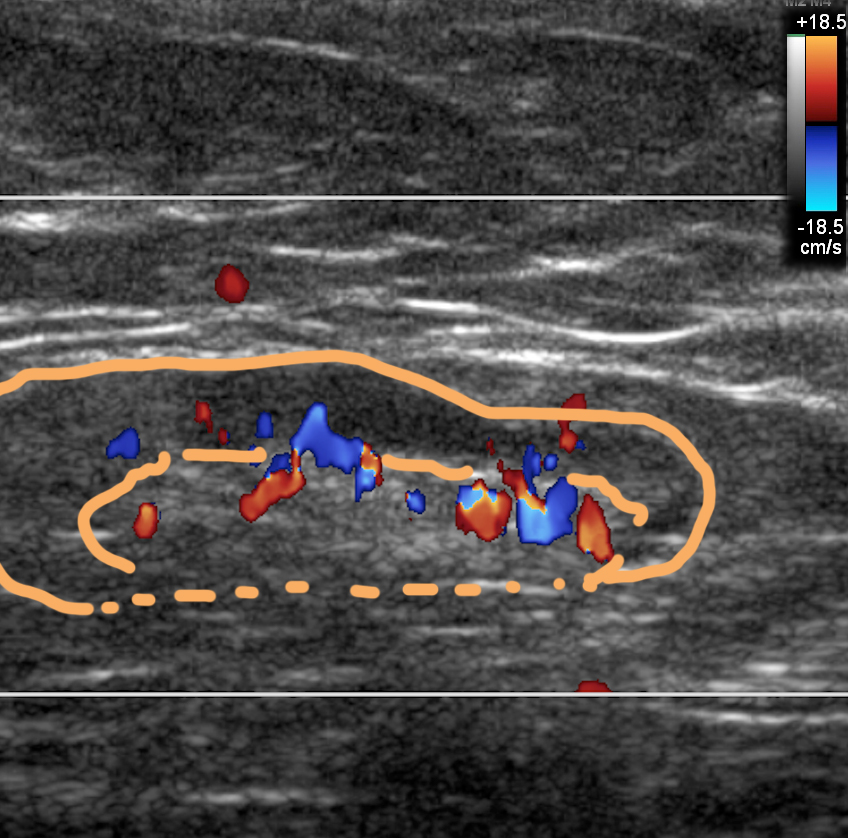
Red does NOT mean artery, and blue does NOT mean vein. The red-to-blue scale indicates whether the flow is TOWARD or AWAY from the transducer. There is usually a scale shown on the image , in the upper right hand corner on this particular image. The red at the top indicates that flow TOWARD the top (in this case, toward the skin) would be shown as red and flow AWAY from the top (in this case, deeper into the axilla) would be shown as blue.
Red does NOT mean artery, and blue does NOT mean vein. The red-to-blue scale indicates whether the flow is TOWARD or AWAY from the transducer. There is usually a scale shown on the image , in the upper right hand corner on this particular image. The red at the top indicates that flow TOWARD the top (in this case, toward the skin) would be shown as red and flow AWAY from the top (in this case, deeper into the axilla) would be shown as blue.
d) What is normal cortical thickness for an axillary lymph node
×
Answer:
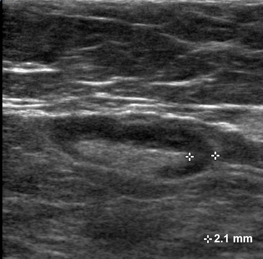
A normal axillary node, as noted above, should have a smooth cortex (hypo echoic) draped around a central oval or round fatty hilum (hyperechoic), and the maximum thickness of the cortex should be no more than 3 mm, measured as shown below by the cross-shaped calipers. This measurement could be made anywhere around the rim of cortex, usually at the place where it appears thickest.
A normal axillary node, as noted above, should have a smooth cortex (hypo echoic) draped around a central oval or round fatty hilum (hyperechoic), and the maximum thickness of the cortex should be no more than 3 mm, measured as shown below by the cross-shaped calipers. This measurement could be made anywhere around the rim of cortex, usually at the place where it appears thickest.
