

Case 3
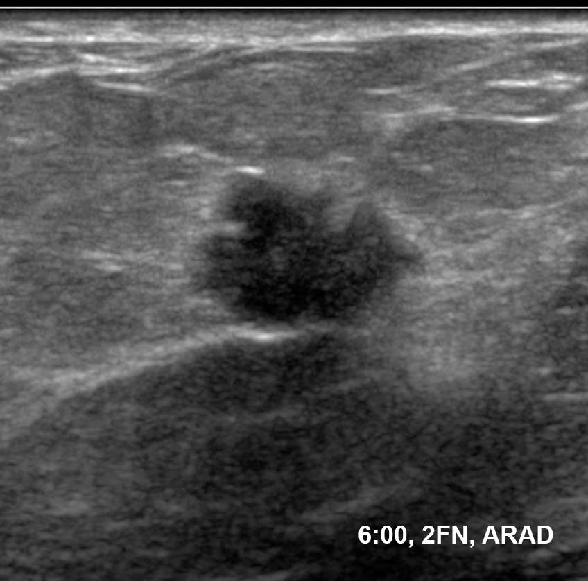
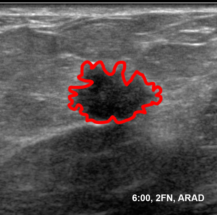
35 year old female with right breast mass, 6:00 position at a distance of 2 cm from the nipple
Question 1:
a) What does the label on the image below mean?
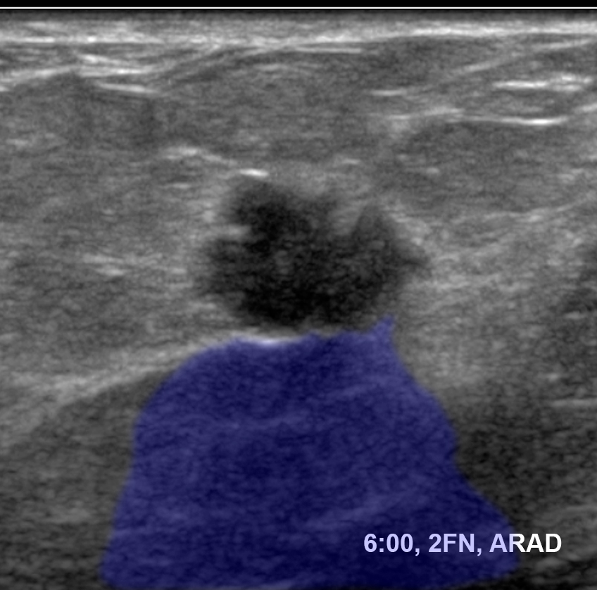
6:00 refers to the lower central breast, and '2FN' means that the lesion is 2 cm away from the nipple, measuring out in the radial direction. 'RAD' means that the transducer was positioned along that radial axis.
b) Click on the orientation image and see where you would place the mass on the model.
Click on the labeled orientation image to see where the mass would be. Additional images show you how the transducer is oriented for a RAD and an ARAD view of the mass.
c) Why do you need perpendicular views of the lesion?
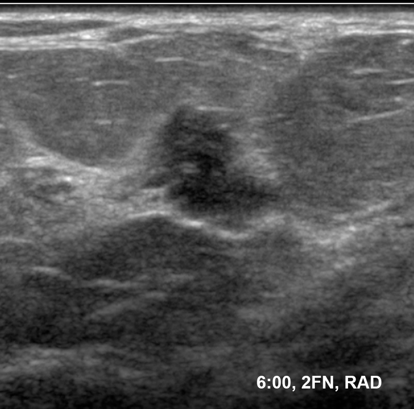
It is very easy on ultrasound to make normal tissue look like a mass. If you see what you think is a mass, if you can TURN your transducer around by 90 degrees and the lesion is still visible, then it is much more likely that it is a real finding. Turning on a lesion (without moving off of it or losing it) is a useful skill to practice when learning to do ultrasound.


Case 3
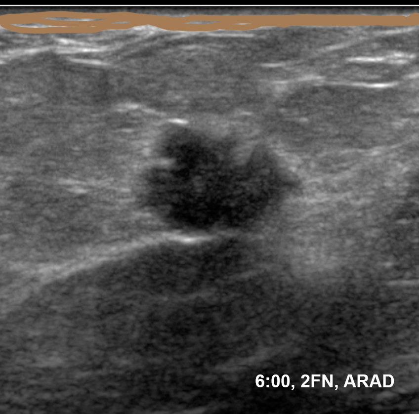
ARAD image from the same patient
Question 2:
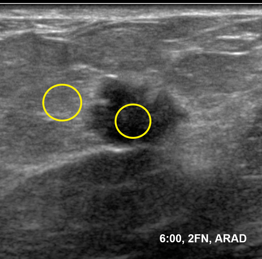
a) How would you describe the echogenicity of this mass on ultrasound?
When describing the echogenicity of a lesion, you can decide first if it is homogeneous or heterogeneous, and then compare the average shade of grey of the mass to surrounding normal breast tissue. If it is BLACKER than surrounding breast, it is hypoechoic. If it is EQUAL to surrounding breast, it is isoechoic. If it is WHITER than surrounding breast, it is hyperechoic or echogenic. Most breast cancers are hypoechoic, and slightly heterogeneous, as is seen in this example.
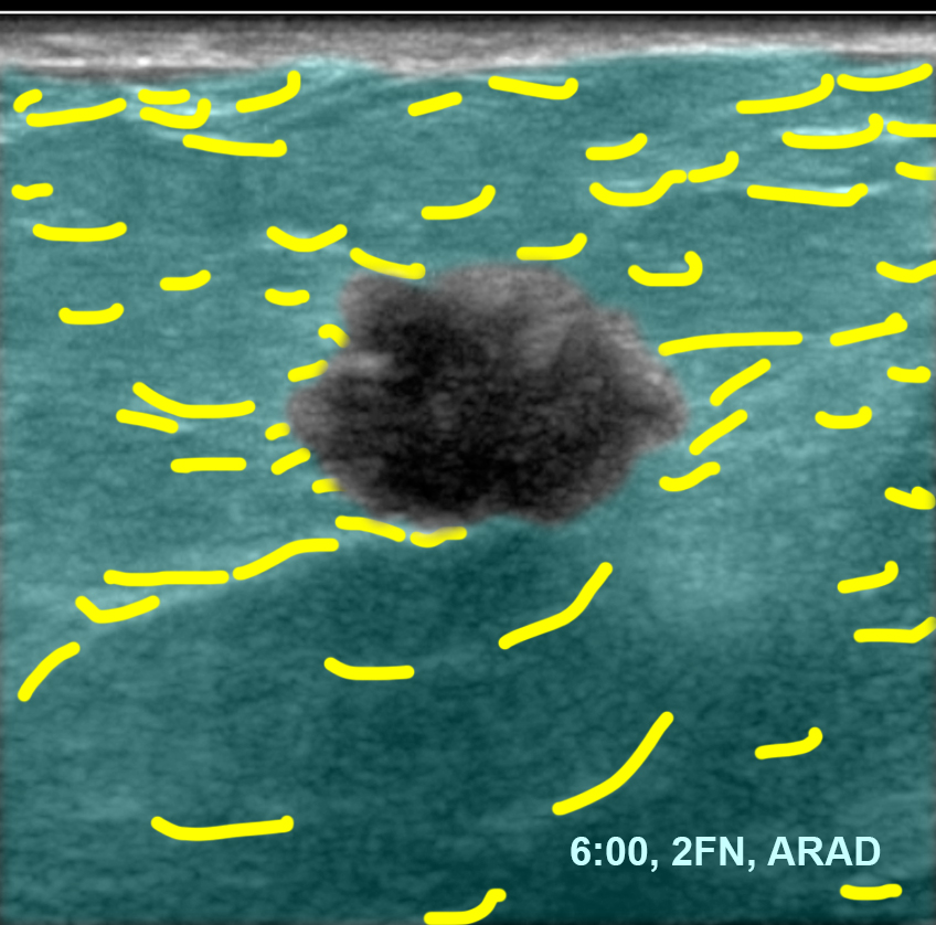
b) How would you describe the margin of this mass on ultrasound?
This mass has a very irregular margin, with small projections in every direction. Smooth margins are more often associated with benign processes, such as cysts or fibroadenomas. An irregular margin like this is worrisome for malignancy.
c) Are there any useful artifacts to help characterize the mass?
Artifacts that we use to characterize tissue on ultrasound include increased through transmission (which was shown in case 1, for a breast cyst). This is most often seen in benign processes, either cysts or occasionally very uniform masses like some fibroadenomas. In this case, there is a faint shadow behind the mass (darker than elsewhere in the breast). This can be seen in malignancies but also in cases of calcifications. If the shadow is caused by calcification it is usually quite dark and there is an echogenic line at the point of interaction with the calcium, which is not seen here. This type of vague shadowing is worrisome for malignancy.
