


Session 1 summary
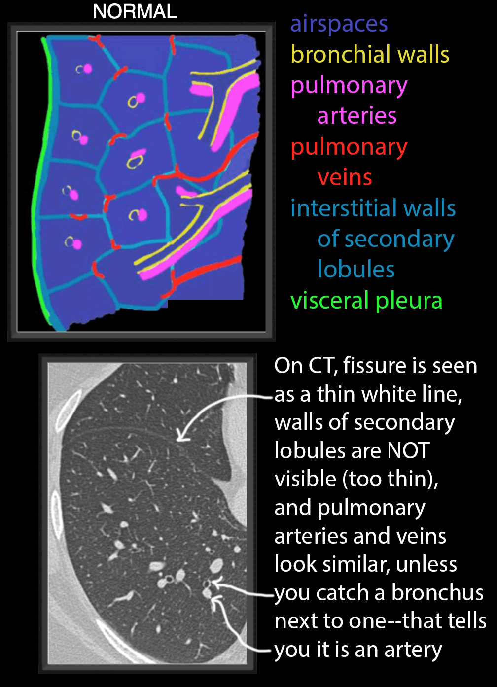
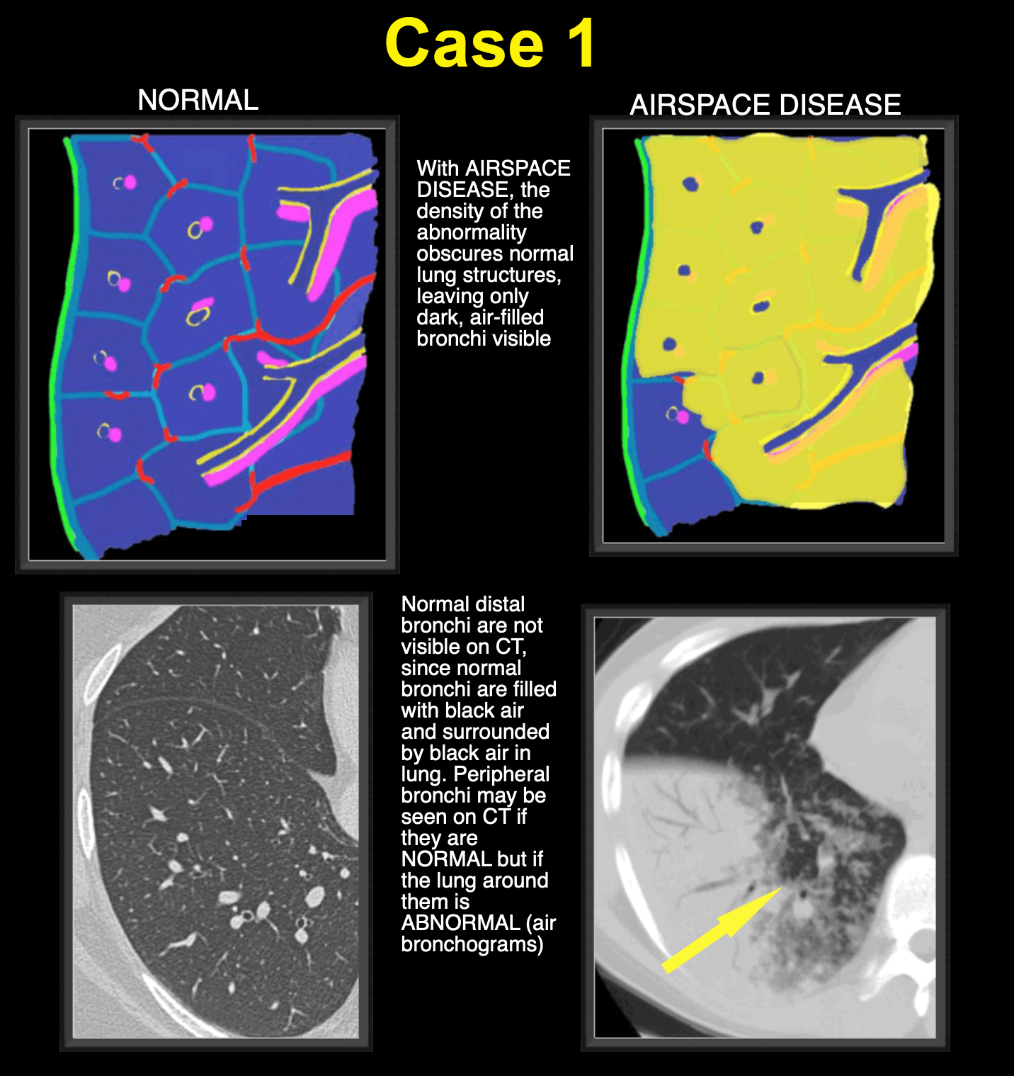
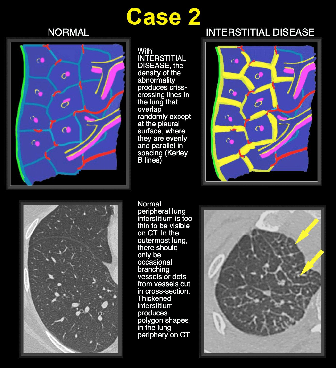
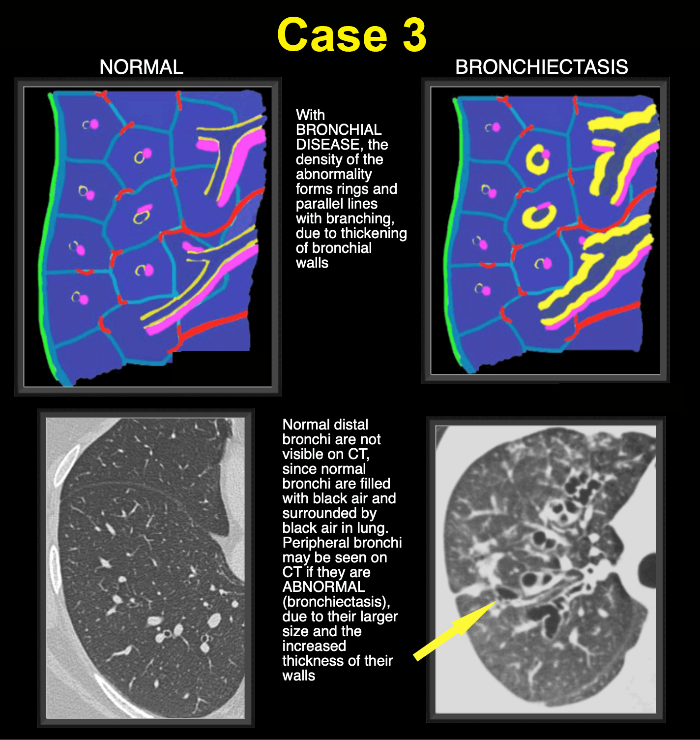
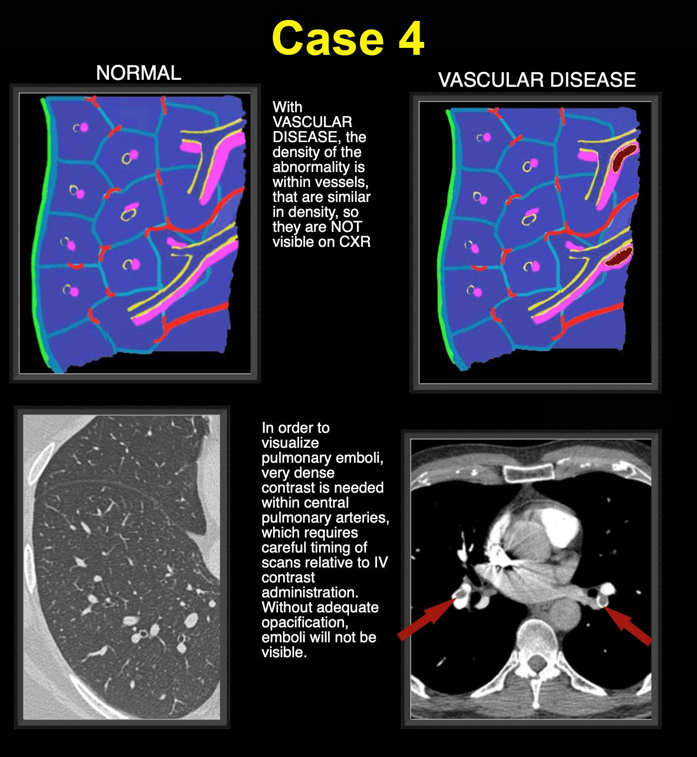
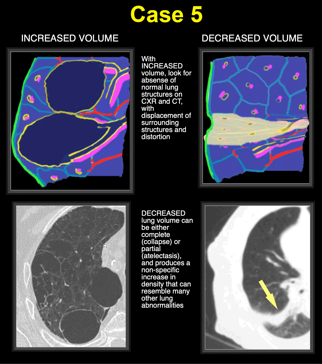
Normal diagram and CT slice are shown, with explanations of the colors used for different structural elements. Click the links below to see summaries for each case. Case 1 concerned airspace/alveolar disease, Case 2 concerned interstitial disorders, Case 3 showed bronchial disease, Case 4 was about vascular disease, and Case 5 reviewed volume changes in the lungs, both increased and decreased.
Further Explanation:
Further Explanation:
This concludes the pre-class review of imaging the various anatomic components of the chest.