

Case 5
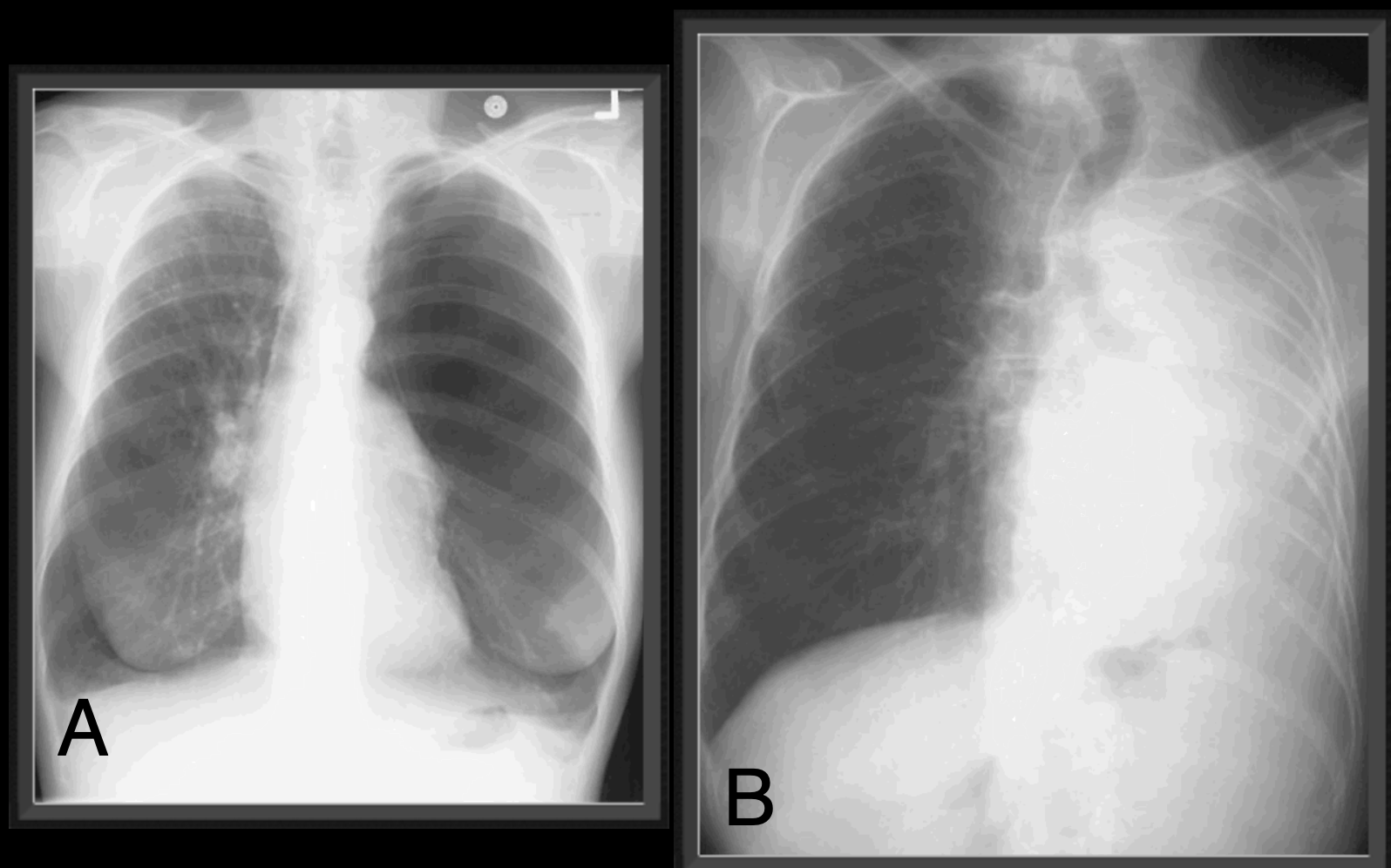
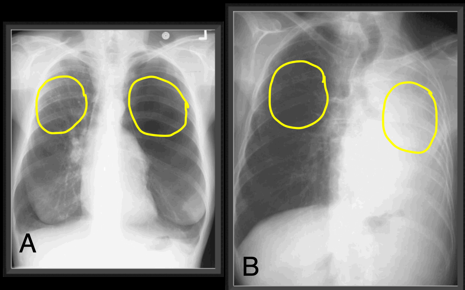
Case 5 focuses on changes in overall lung volume, also called 'aeration', rather than a specific anatomic region of the lung. Both patients are short of breath.
Question 1:
What other pertinent history might be present in both cases?
×
Answer:
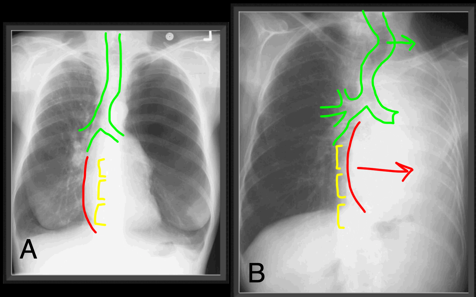
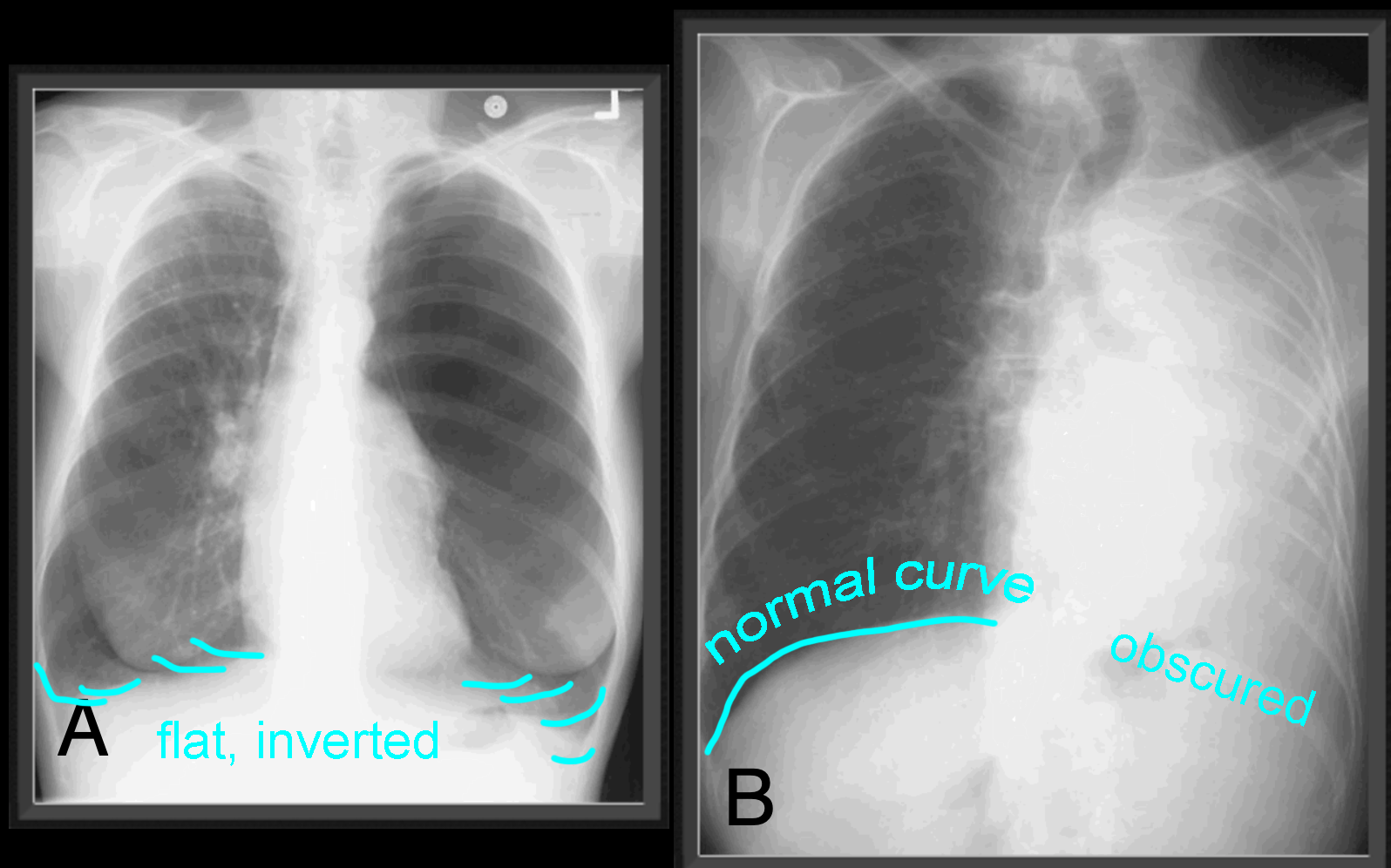
Patient A has very large lung volumes and is likely a smoker. There are very few markings on the left, which may represent emphysema, often better seen on CT. Note that the diaphragms appear flat or even slightly inverted, another sign of increased lung volumes. Patient B has no aerated lung on the left, which could be a result of surgery (removal of the left lung), but there are no changes in left ribs to suggest prior left thoracotomy). This could also represent an obstructing lesion blocking the left mainstream bronchus. Note how far to the left the heart is shifted (no heart is seen to the right of the spine), and how the trachea in the neck is also shifted to the left. These are signs of volume loss (atelectasis, collapse) on the left.
Patient A has very large lung volumes and is likely a smoker. There are very few markings on the left, which may represent emphysema, often better seen on CT. Note that the diaphragms appear flat or even slightly inverted, another sign of increased lung volumes. Patient B has no aerated lung on the left, which could be a result of surgery (removal of the left lung), but there are no changes in left ribs to suggest prior left thoracotomy). This could also represent an obstructing lesion blocking the left mainstream bronchus. Note how far to the left the heart is shifted (no heart is seen to the right of the spine), and how the trachea in the neck is also shifted to the left. These are signs of volume loss (atelectasis, collapse) on the left.
Case 5
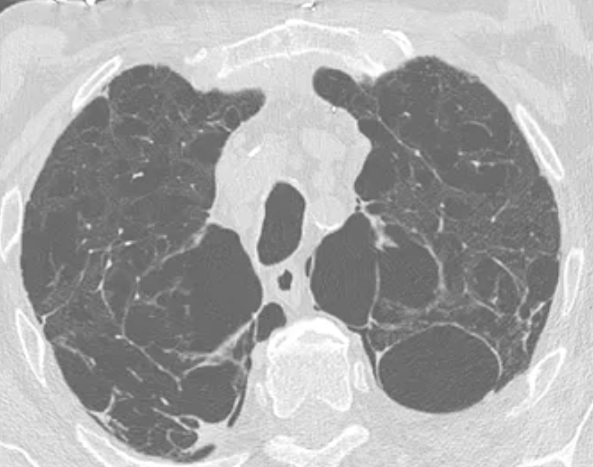
This is a study on a middle-aged patient with chronic shortness of breath.
Question 2:
What would you expect this patient's chest radiograph to look like, in particular their lung volumes?
×
Answer:
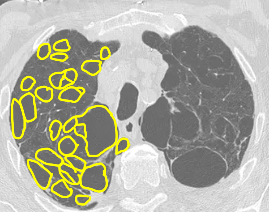
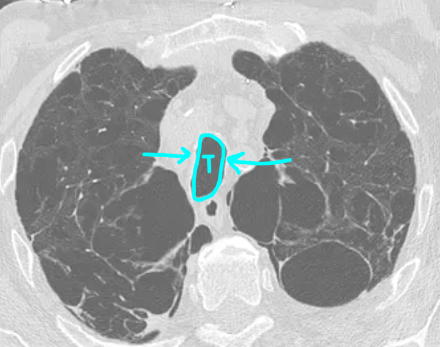
This lung window axial CT series shows extensive emphysema (large holes in the lungs) that is worst in the upper lung zones. This is an example of a condition that typically causes INCREASE in lung volumes, also called hyperinflation. When severe, this can cause flattening of the diaphragms or even inversion of diaphragms (curving downward instead of upward) on chest radiographs.
This lung window axial CT series shows extensive emphysema (large holes in the lungs) that is worst in the upper lung zones. This is an example of a condition that typically causes INCREASE in lung volumes, also called hyperinflation. When severe, this can cause flattening of the diaphragms or even inversion of diaphragms (curving downward instead of upward) on chest radiographs.
