

Back and Limbs Case 3
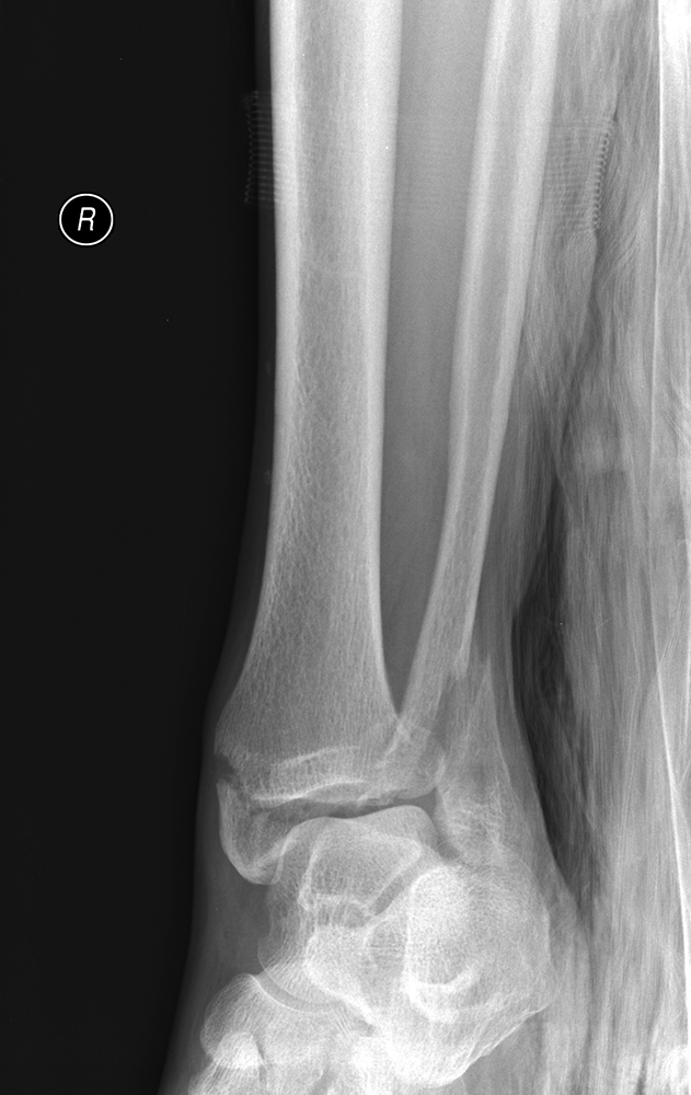
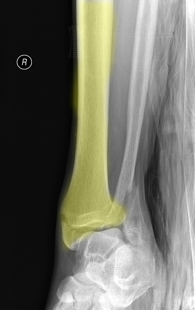
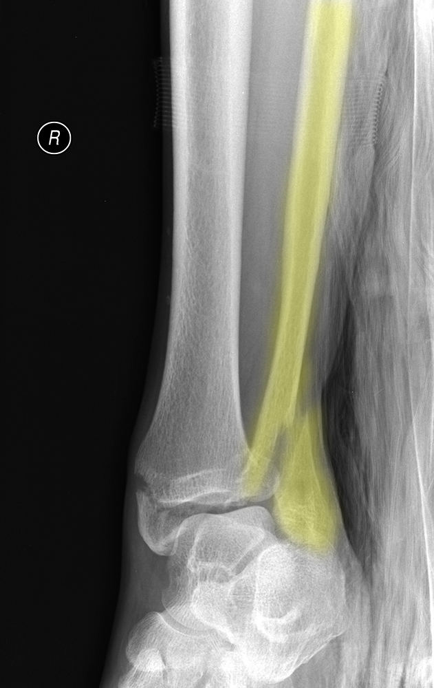
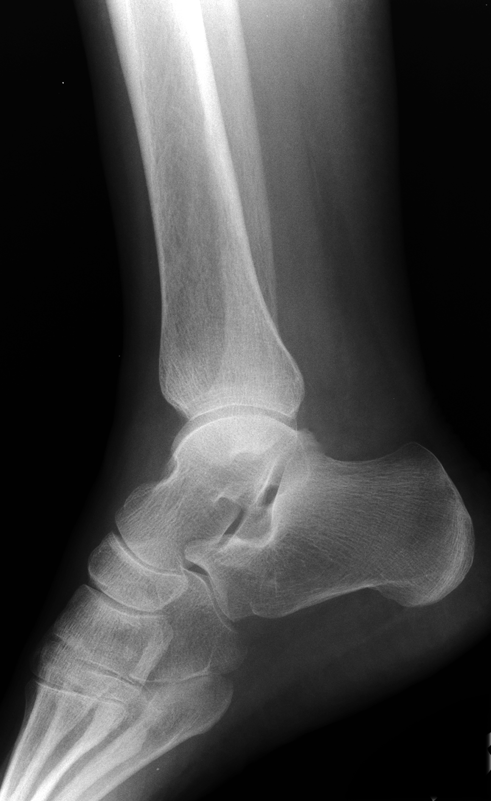
58 yo patient with painful ankle after fall
Question 1:
a) What are technical factors for this image?
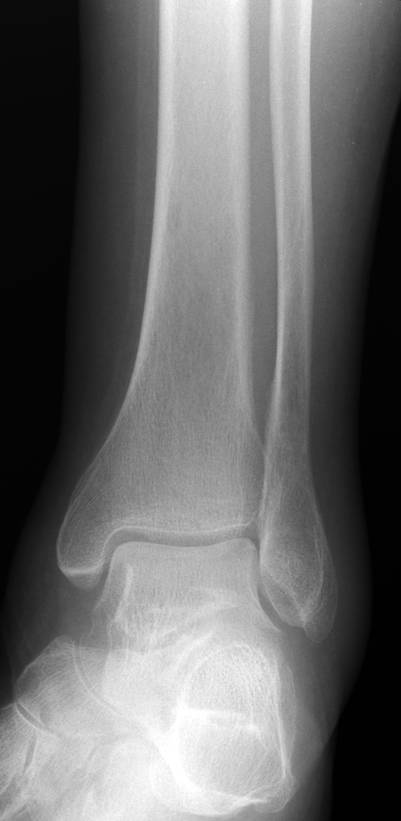
This is an oblique radiograph of the ankle. The standard views are AP and lateral for most extremities. This image is in between an AP and a lateral, and this may be due to inability to precisely position this patient, due to pain.
b) Is this radiograph an adequate examination of this patient's ankle?
No, we would like to see at least one more view, a lateral view. In general, for most bones we like to look at two 'orthogonal' views, meaning at 90 degrees from each other. This gives us a better idea of the 3D shape of a structure than a single radiograph can provide.

Back and Limbs Case 3
Another image on this same patient
Question 2:
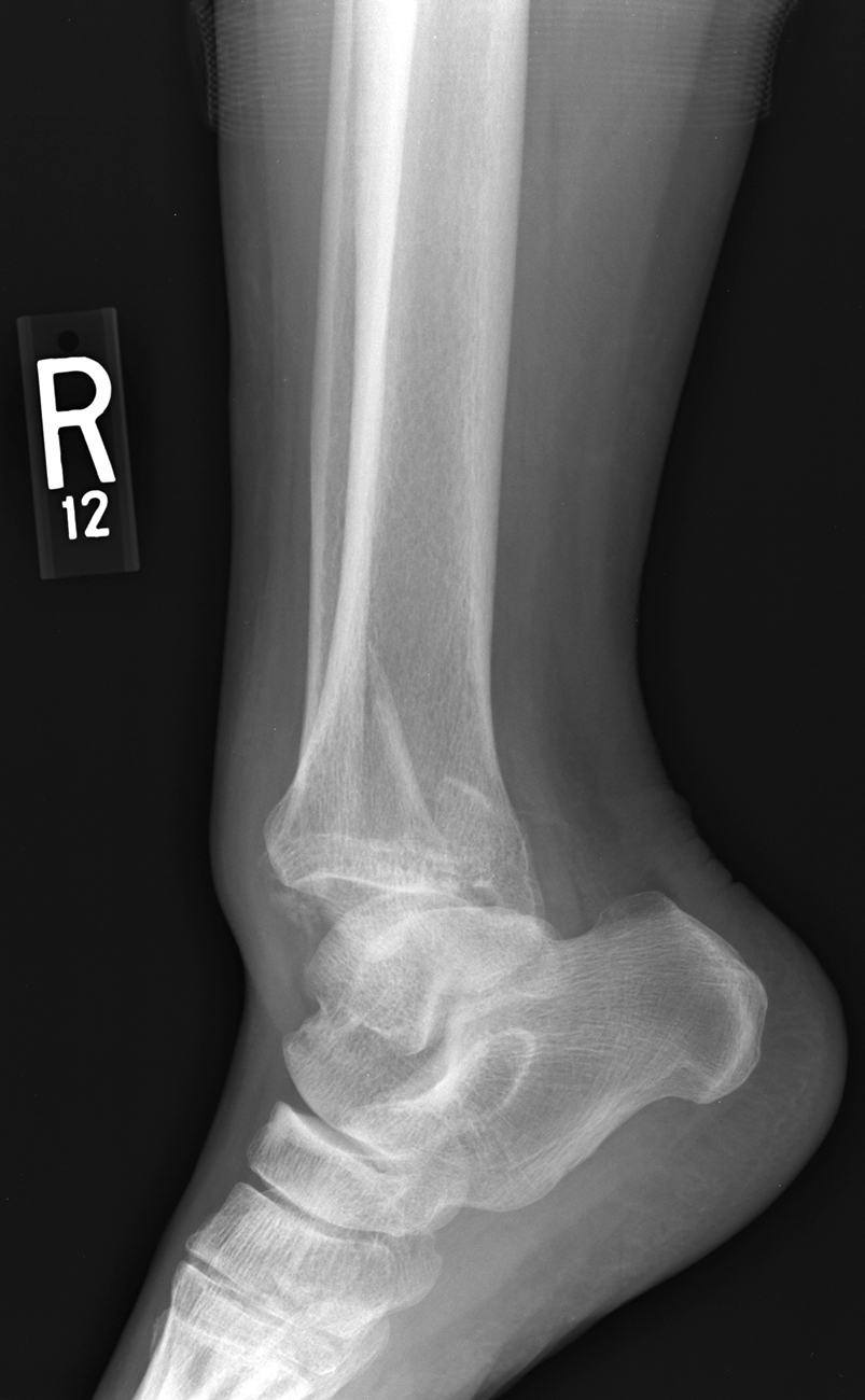
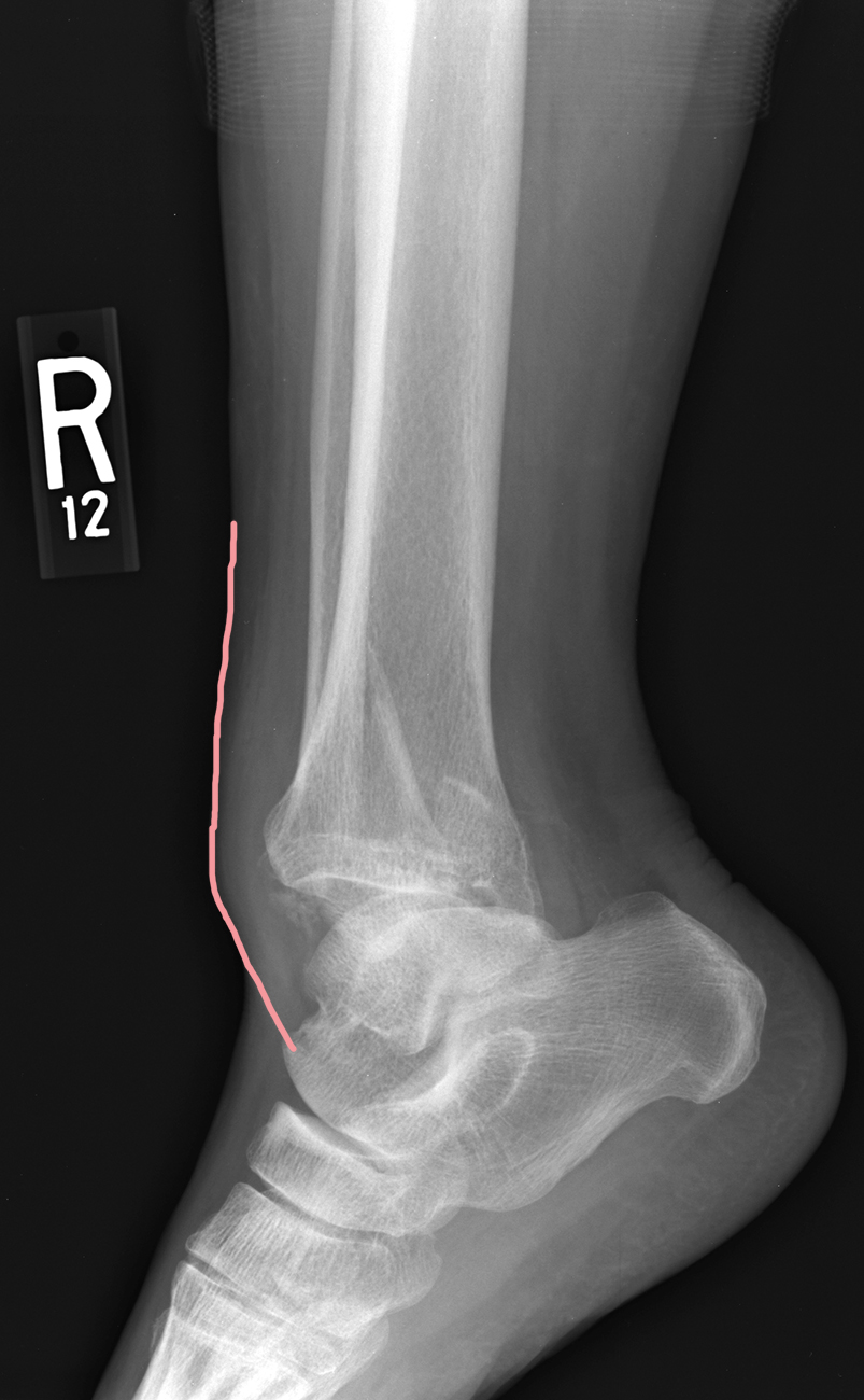
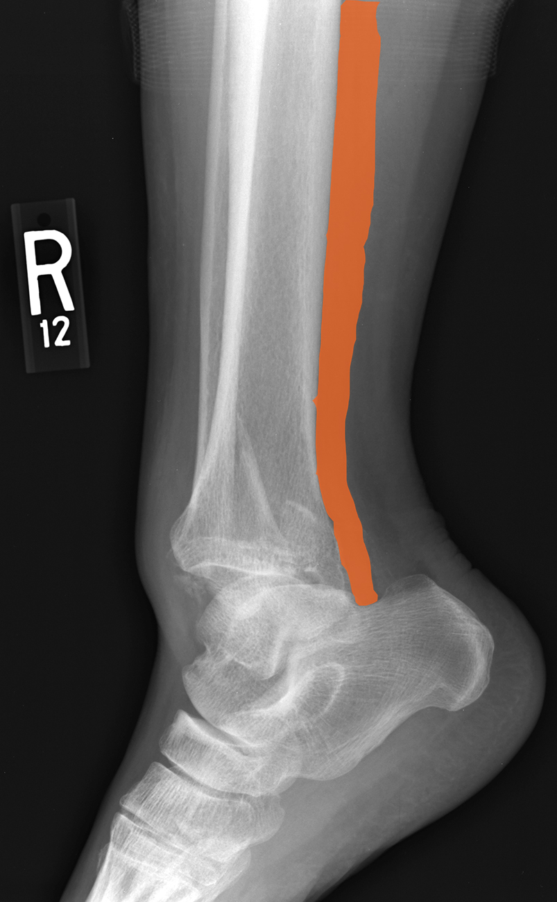
How would you describe the technical parameters of this image?
This is a lateral radiograph of the ankle. These are generally done in addition to an AP view, to provide two orthogonal views (at 90 degrees to each other). We do not generally do PA views of extremities, as there is not enough geometric distortion (limbs are not as thick as the chest) to make much of a difference between an AP and a PA view, and it is generally easier to position the limb for an AP view, particularly in someone in pain.
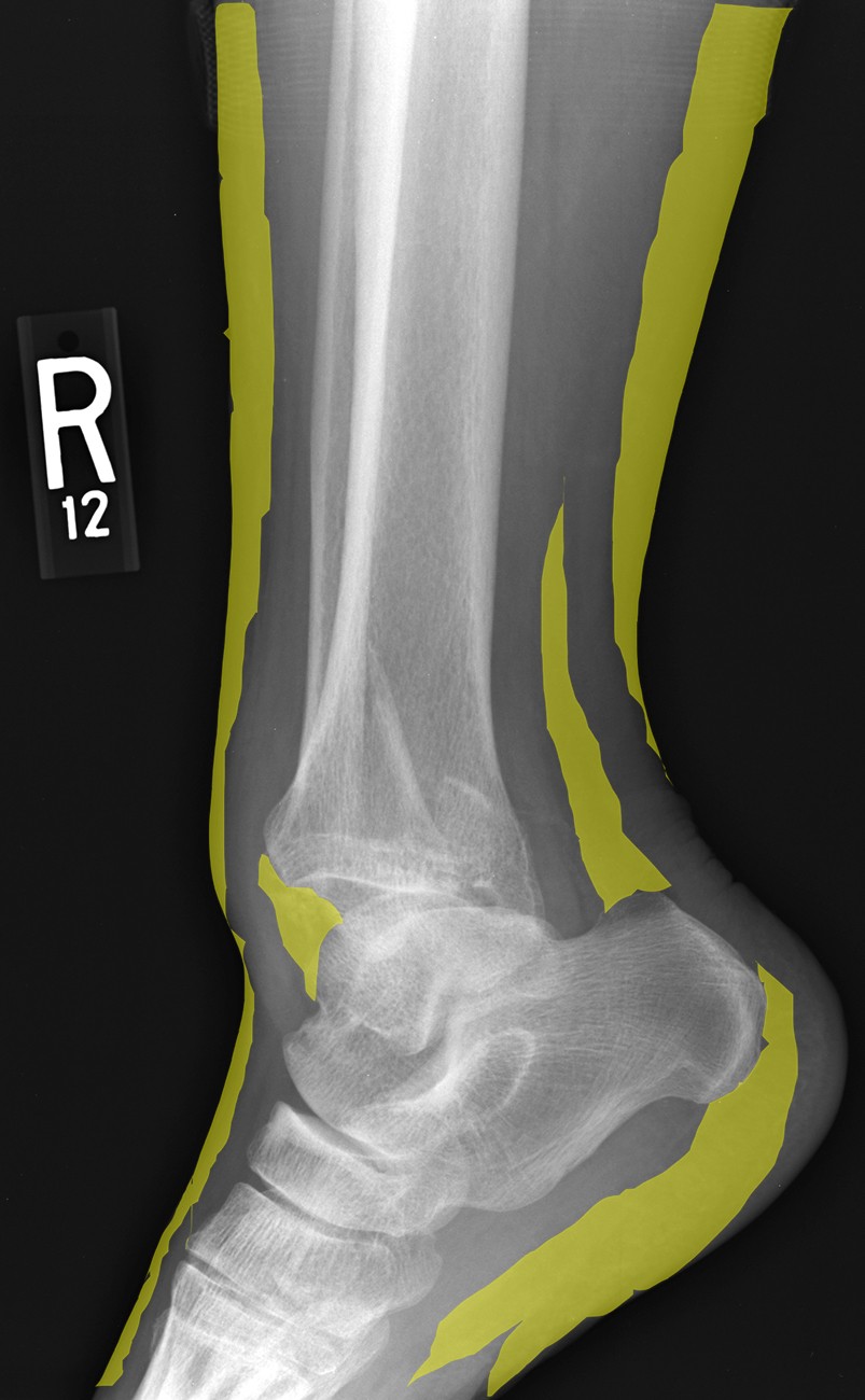
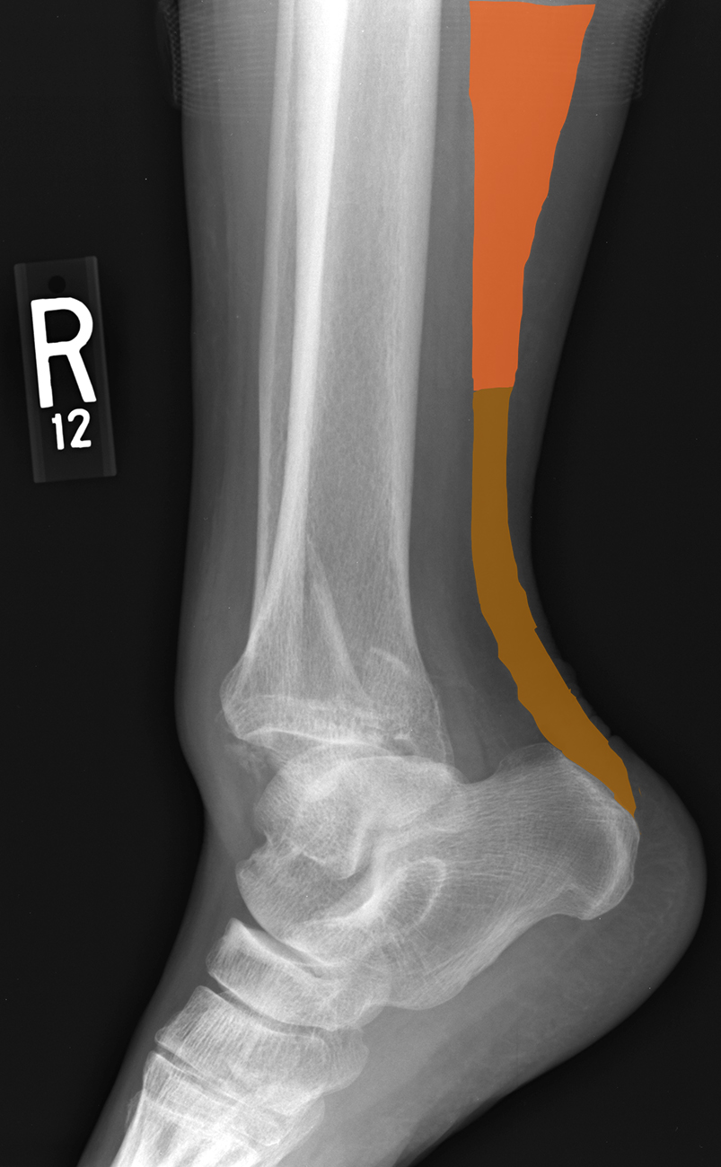
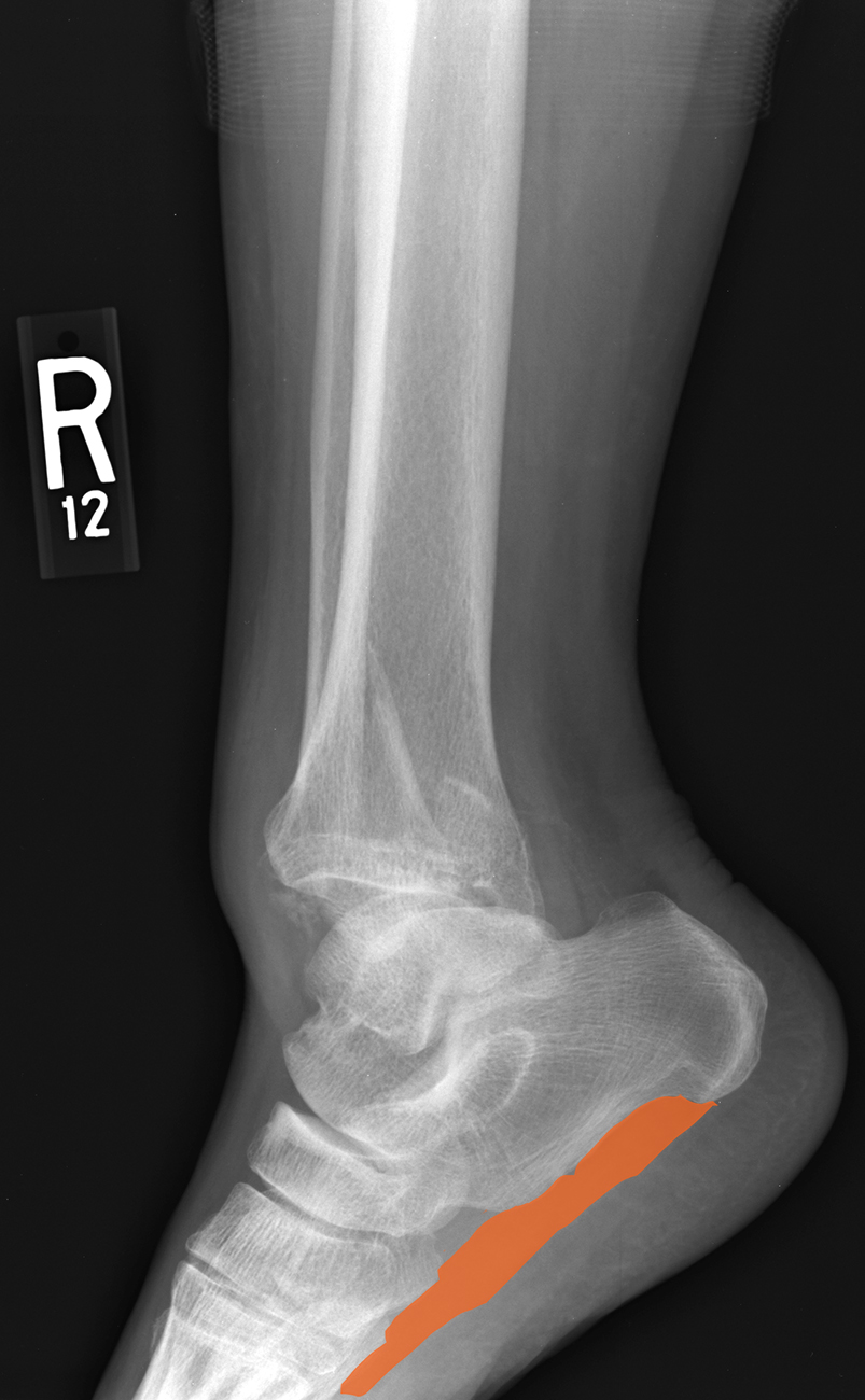
Try to identify structures on the image before clicking on the labels.
Back and Limbs Case 3
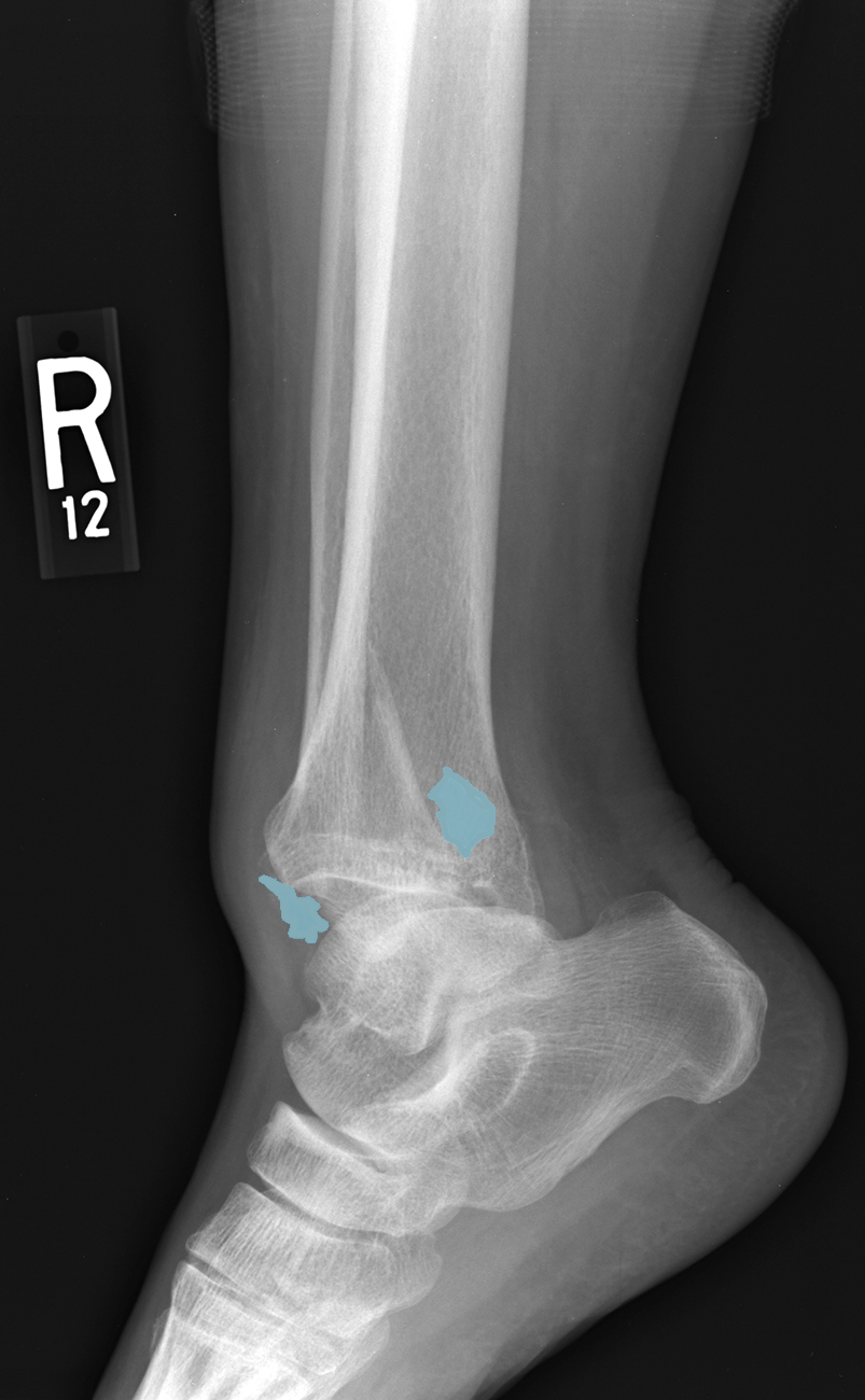
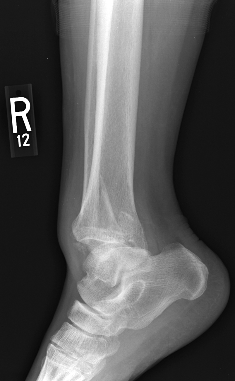
This is the same lateral image shown on the previous page.
Question 3:
a) What ligaments must be disrupted in this patient?
anterior tibiotalar and talofibular, and probably others. The most disruption appears to be at the anterior margin of the talotibial joint.
b) Do you think that casting is sufficient treatment for this injury?
This is a very severe fracture with dislocation, so simple casting will not result in a functional ankle. The patient needs surgical repair of this to provide enough stability for healing. The surgical procedure in general is called an open reduction and internal fixation, or ORIF for short.
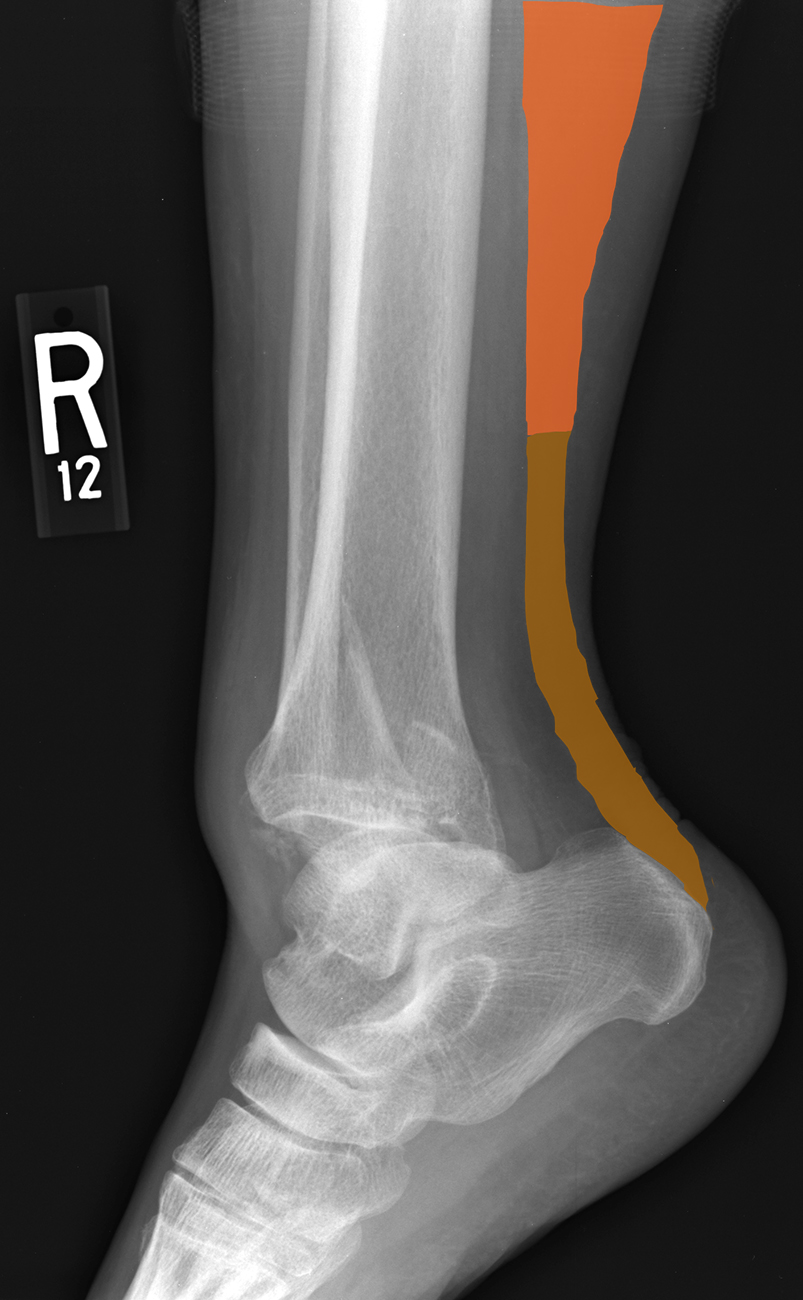
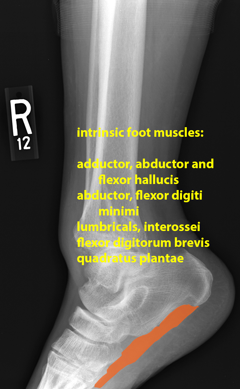
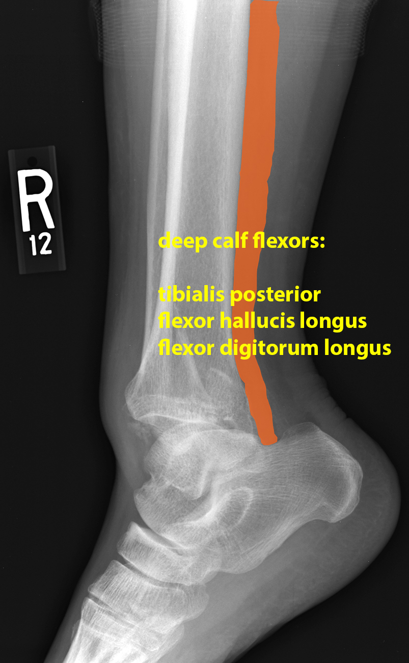
The muscles indicated on the prior page are shown here, with answers.
Back and Limbs Case 3
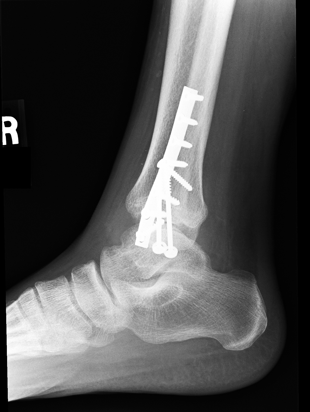
Followup image on this patient
Question 4:
a) What are the technical parameters of this image?
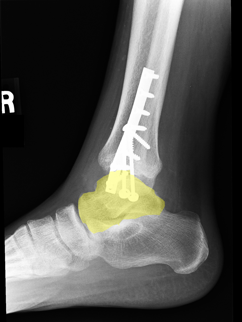
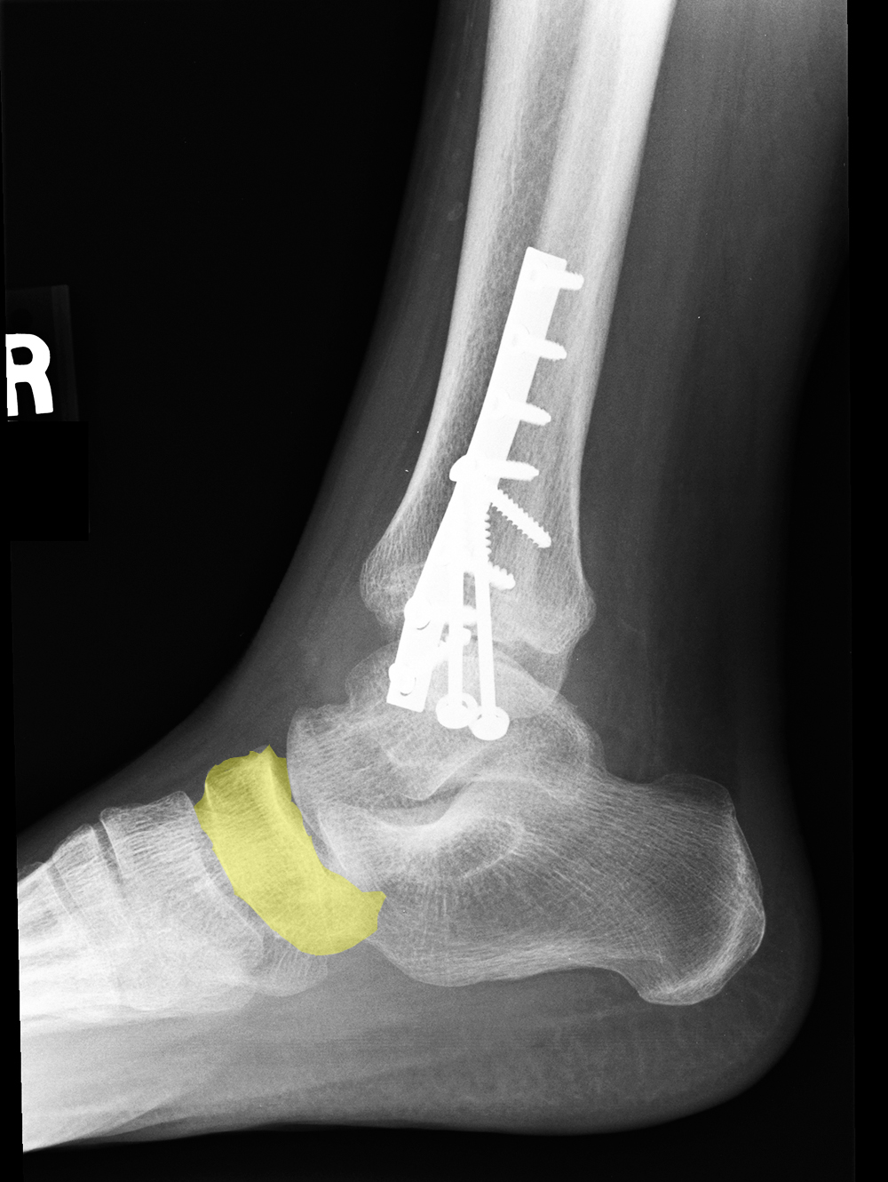
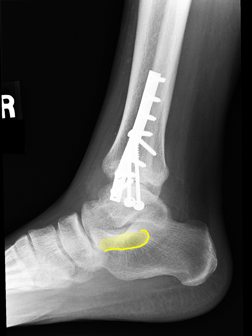
This is a lateral radiograph of the ankle.
b) What has been done?
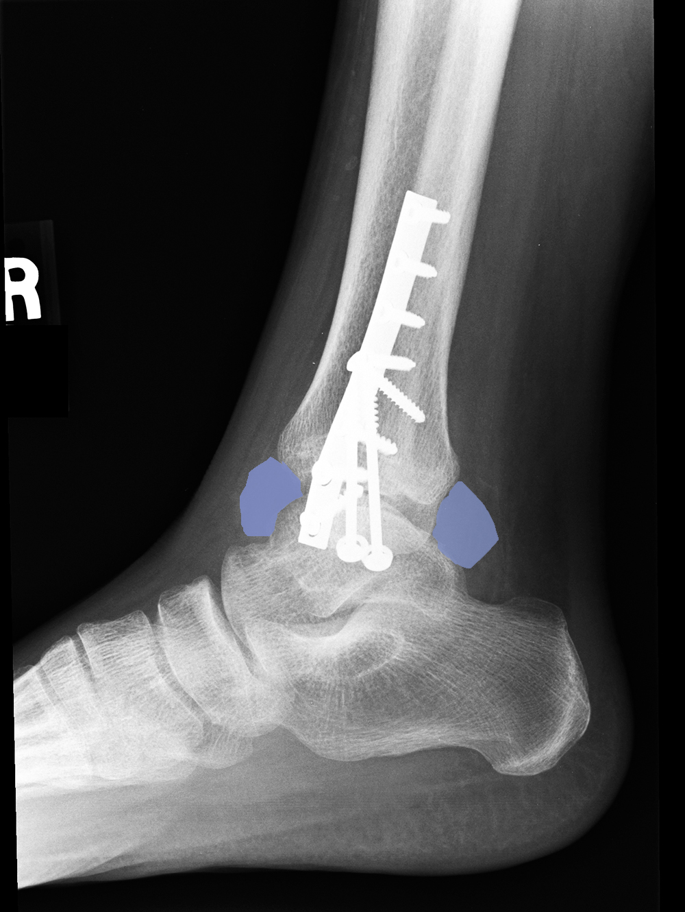
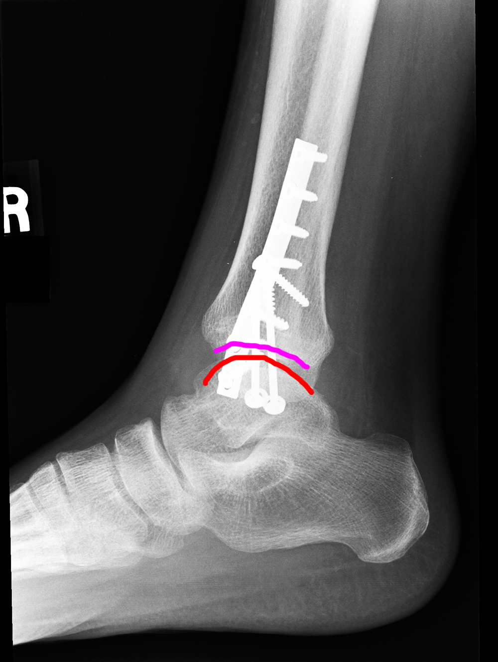
The patient has had surgery (open reduction and internal fixation, meaning that the dislocation was 'reduced' to anatomic alignment in the operating room, and that hardware was placed that is internal, or within the patient's body--as opposed to external fixation, which is a metal framework outside the patient with screws that hold it in place), or ORIF for short.
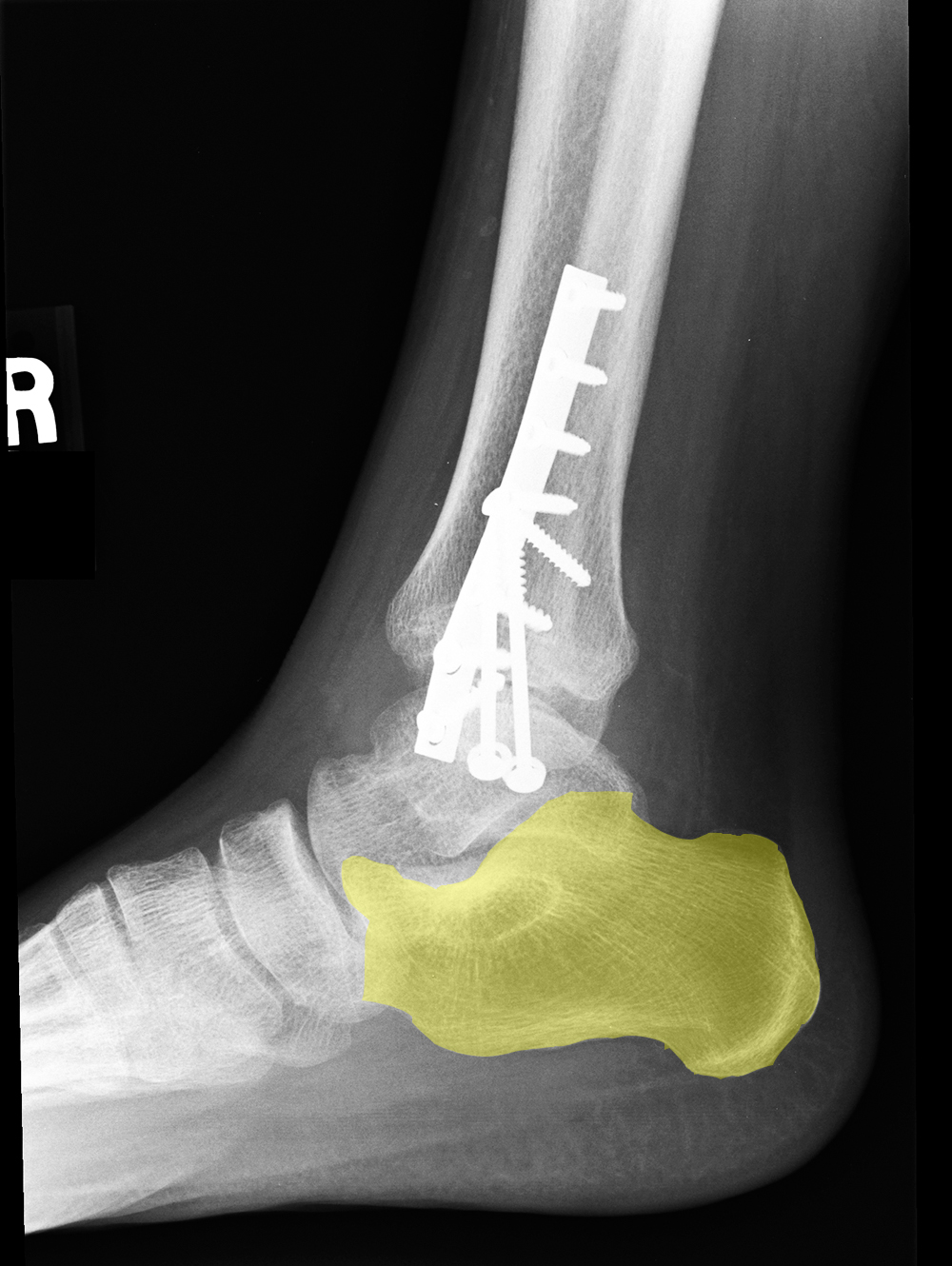
Several foot bones are outlined for you to practice identification. Try to figure out which bone is which before clicking on the label.
