

Back and Limbs Case 4
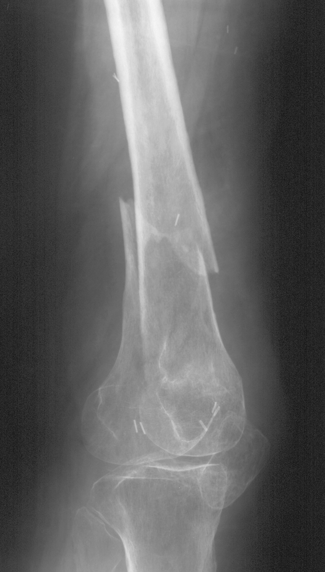
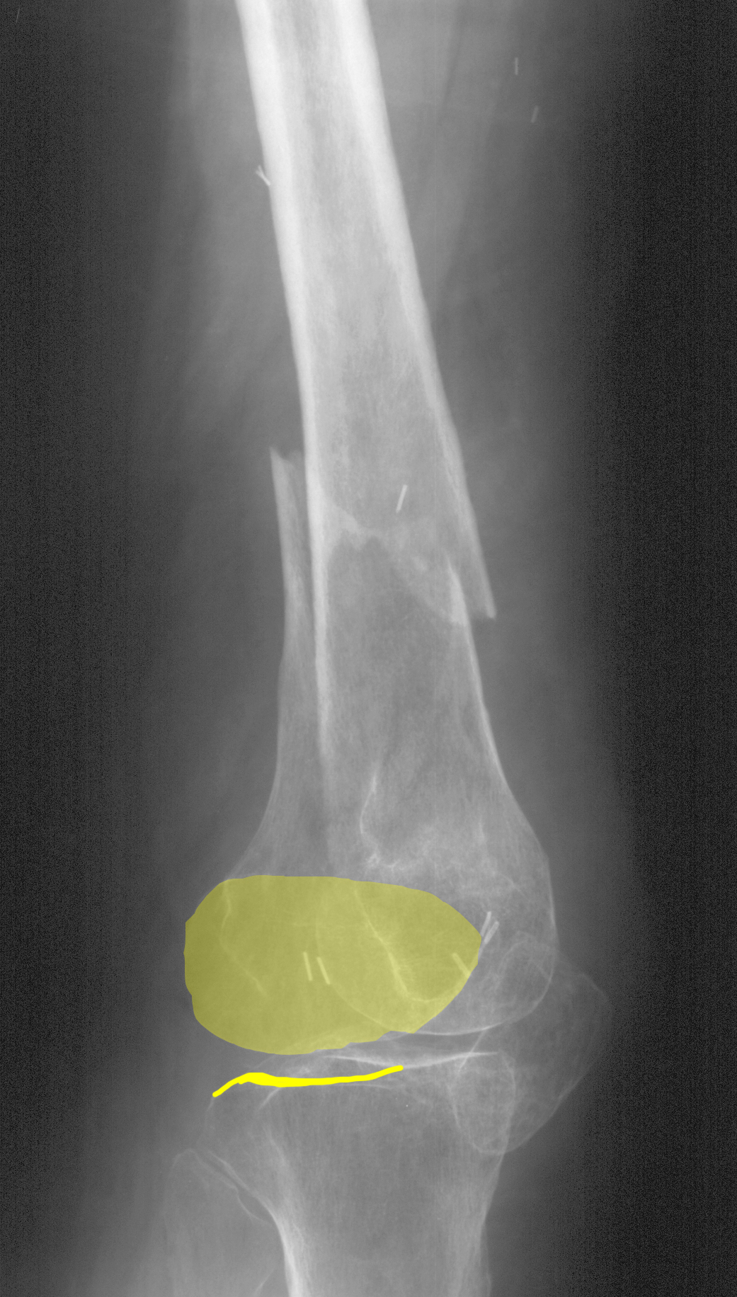
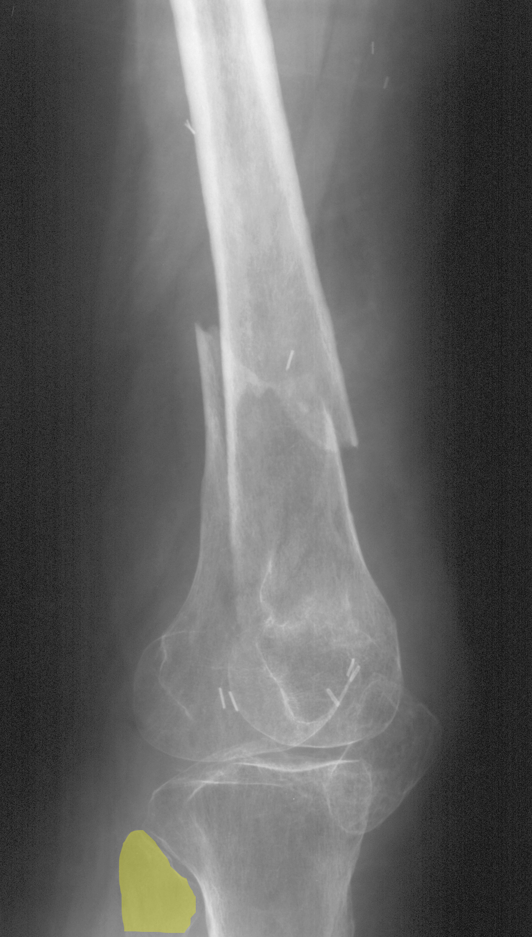
89 year old patient who cannot stand after a fall in a nursing home.
Question 1:
a) What are the technical parameters of this image?
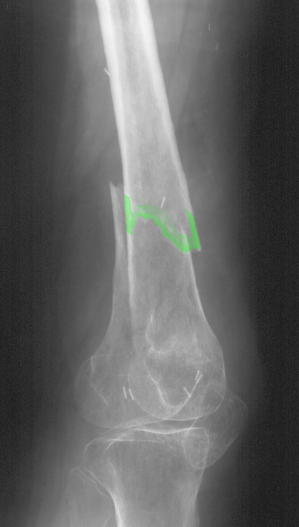
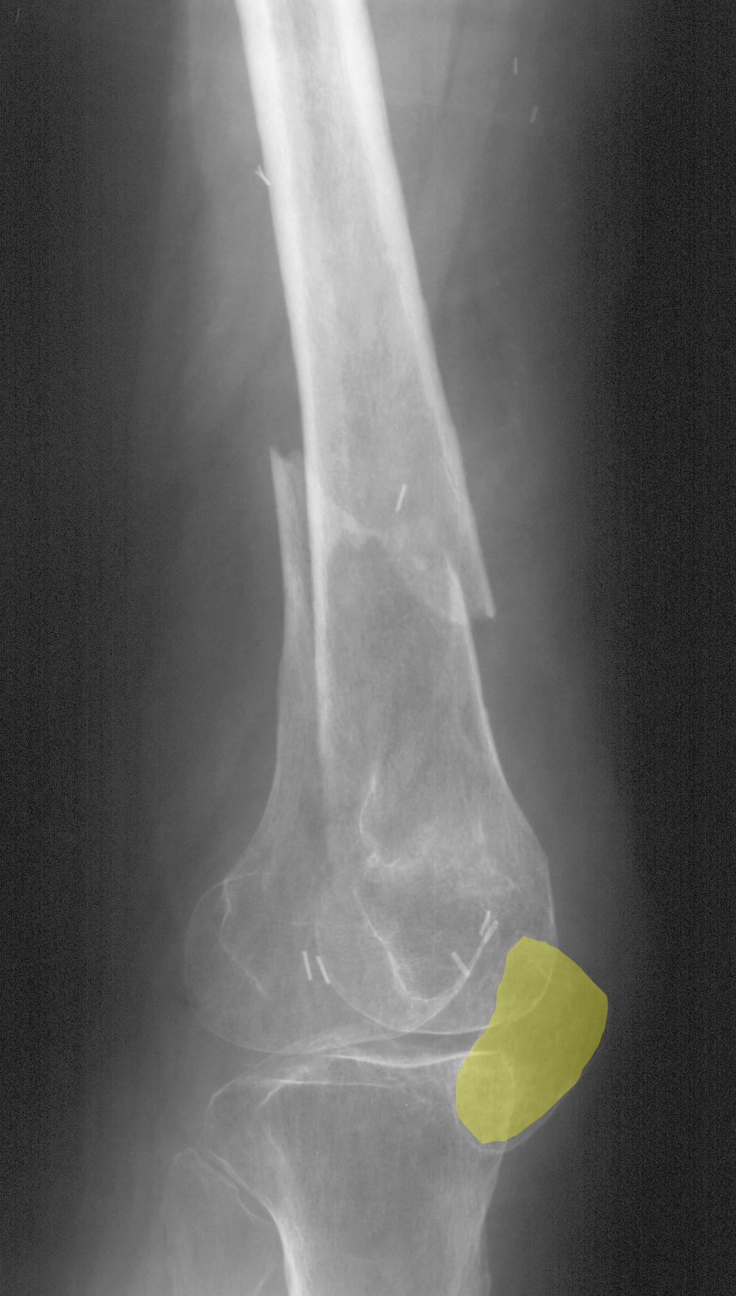
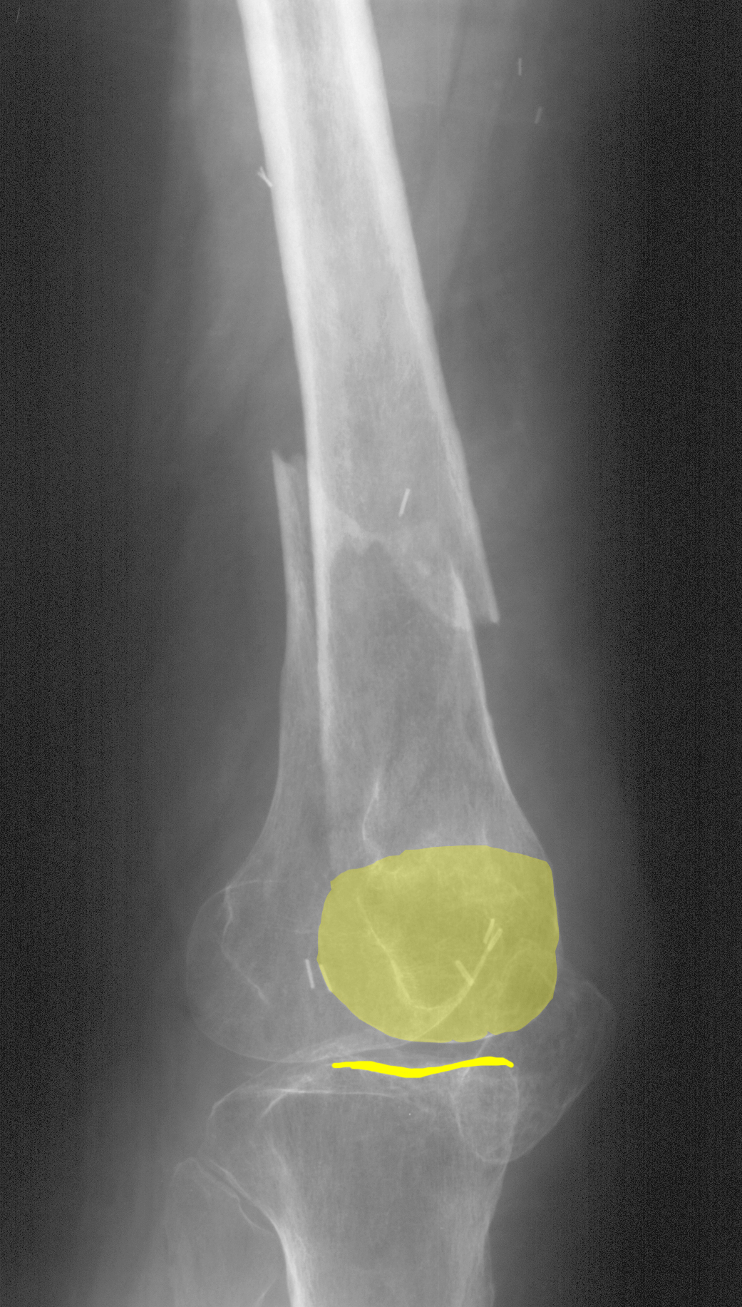
This is an oblique radiograph of the thigh and knee. Positioning is close to a lateral view but slightly rotated, probably because of limitations in positioning the patient due to pain.
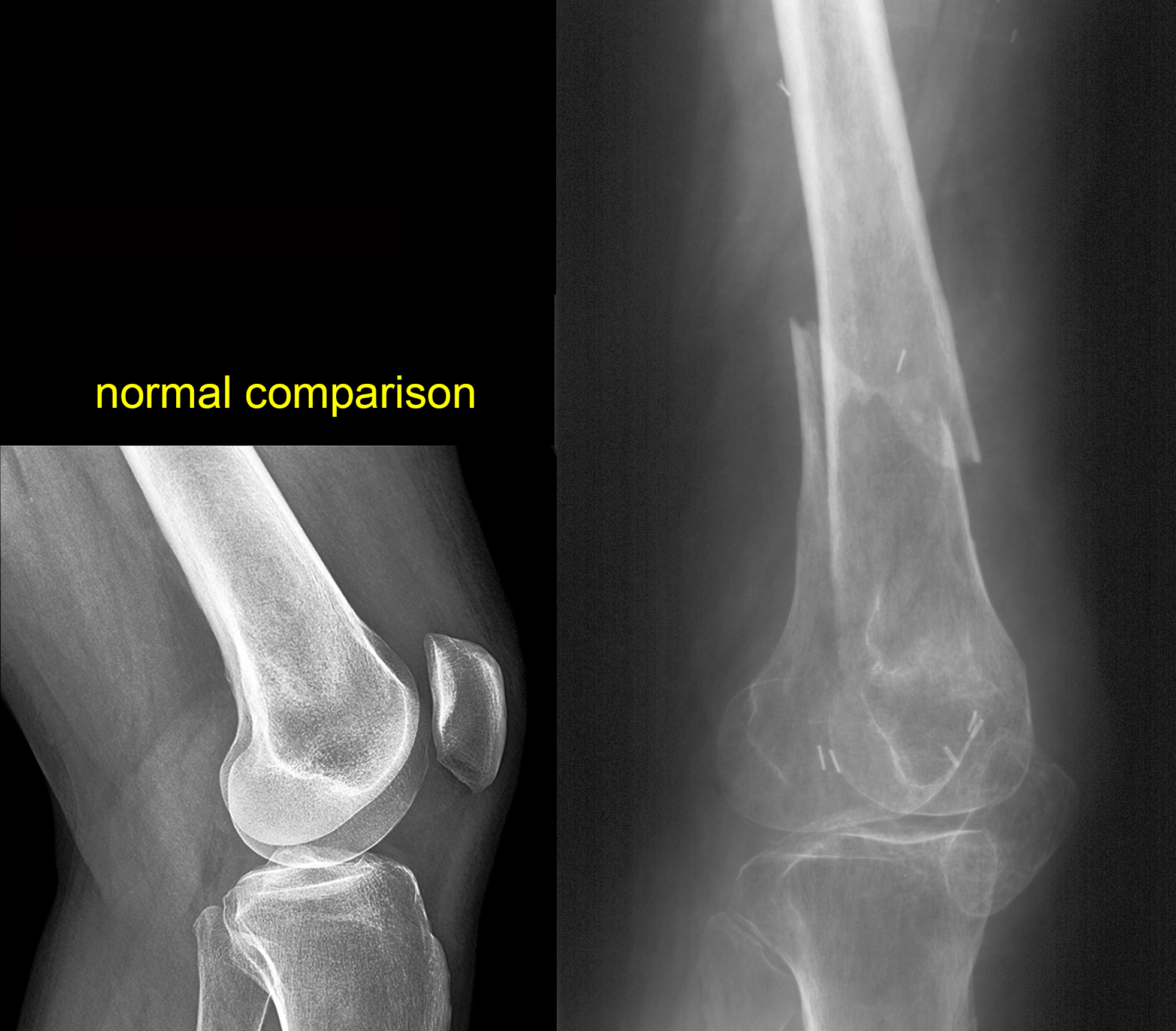
b) What do you think of the overall bony mineralization?
The cortical bone appears very thin. A comparison image in a young normal patient is shown below.

Back and Limbs Case 4
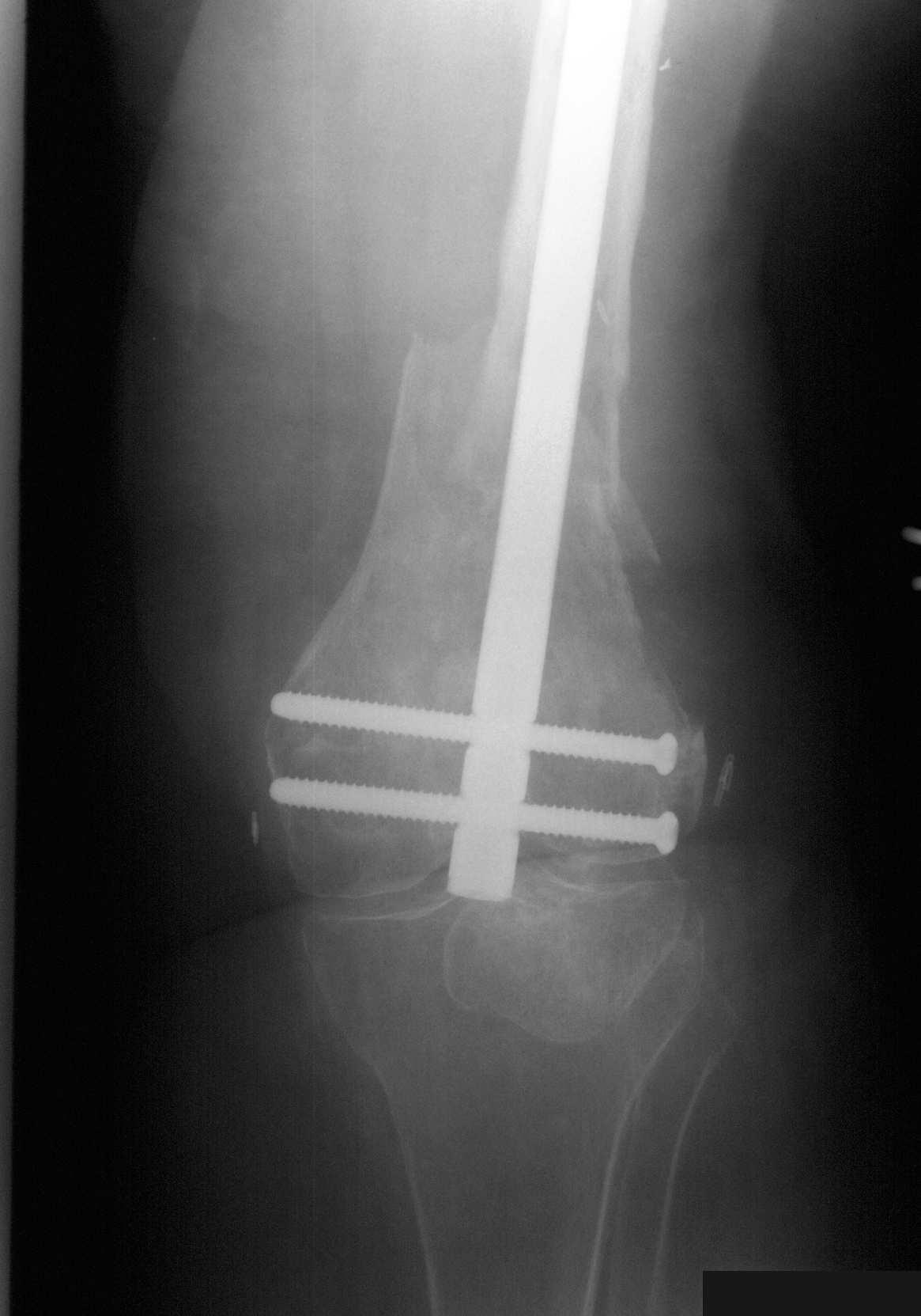
This is the next imaging study on this patient. The surgical clips on the previous image were related to prior venous surgery done several years ago.
Question 2:
a) What has been done to the patient?
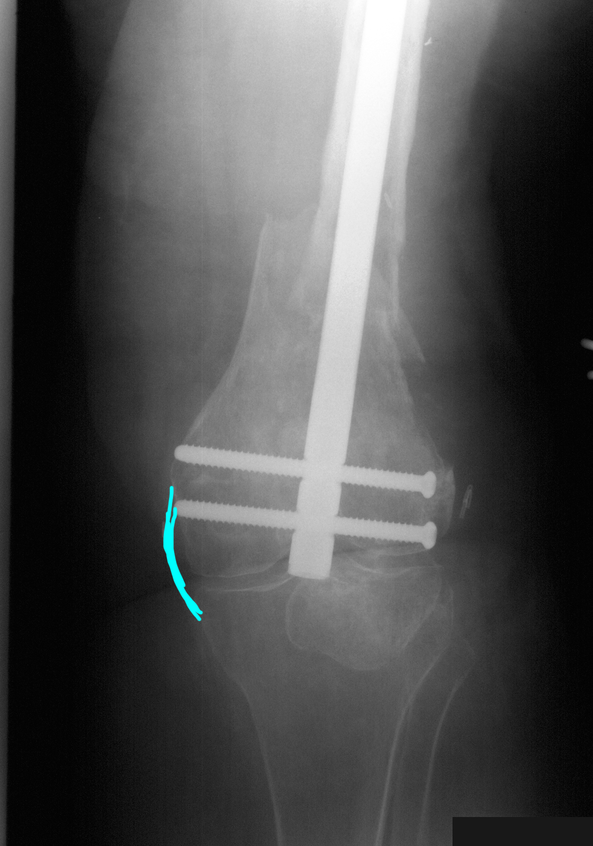
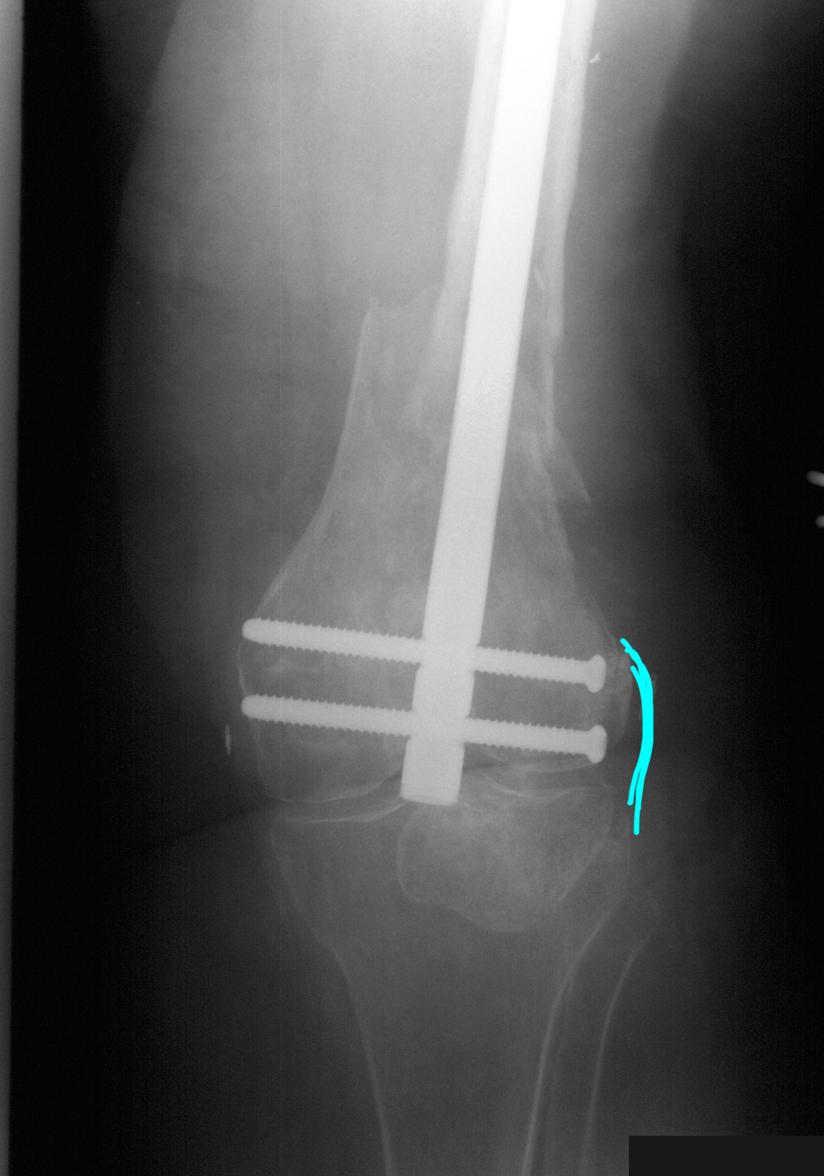
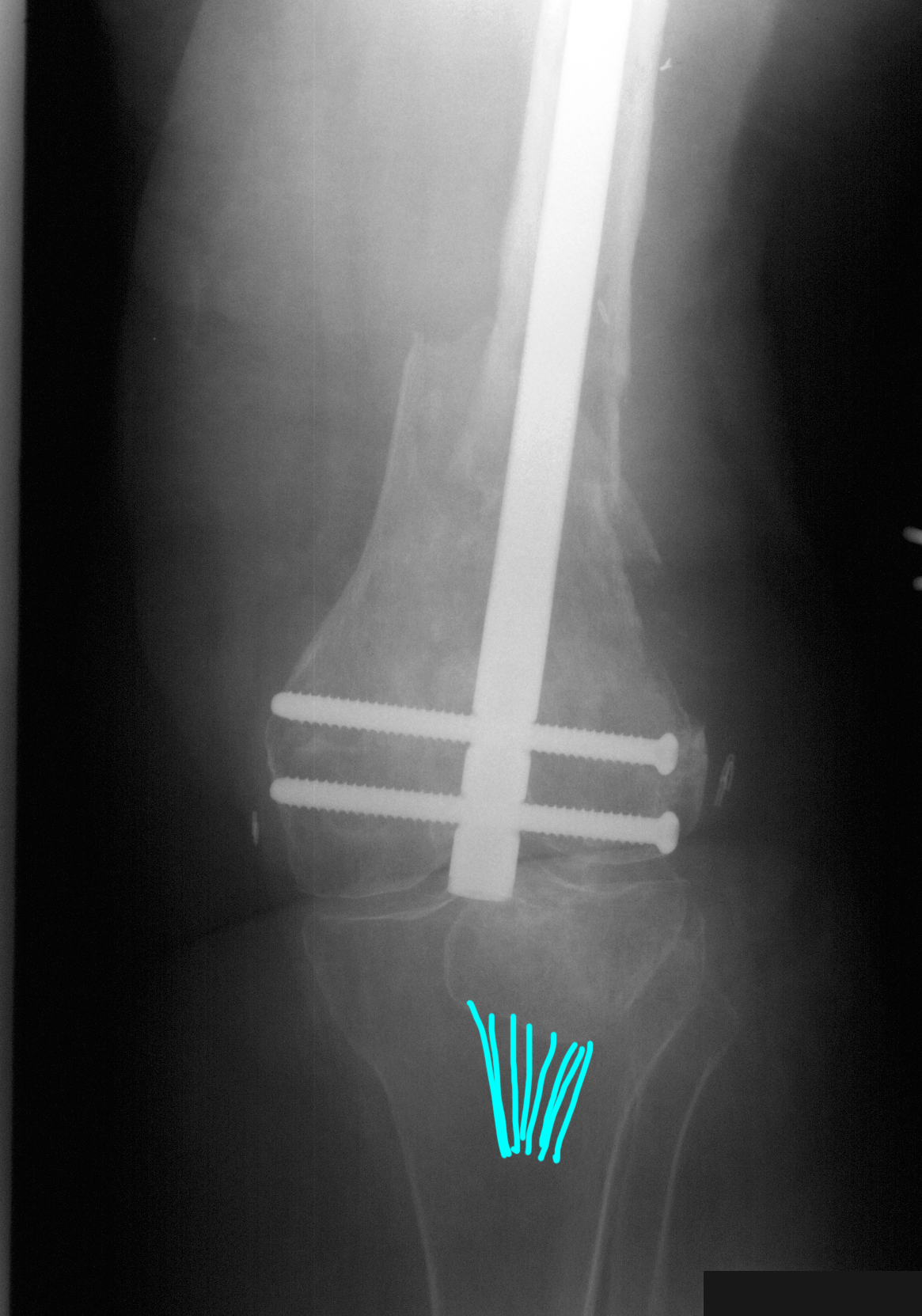
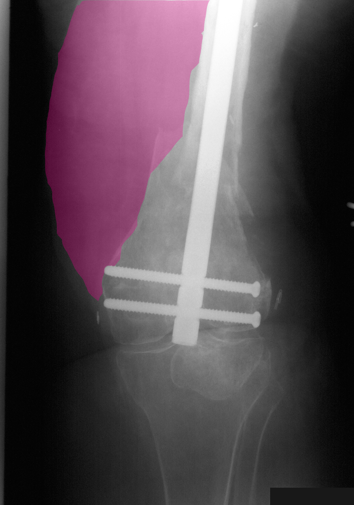
This is another example of an open reduction and internal fixation (ORIF). The reduction in this case is not a repositioning of a joint, but a repositioning of the shaft of the femur so that it is more anatomical in alignment.
b) What are technical parameters of this image?
This is a much more straight AP view of the distal femur and knee.
It is important when looking at extremity radiographs to remember to look at the soft tissues. The bones tend to catch and hold our attention, but the other parts of the image are also important.
Back and Limbs Case 4
Additional imaging on this same patient.
Question 3:
a) What are technical parameters of these images?
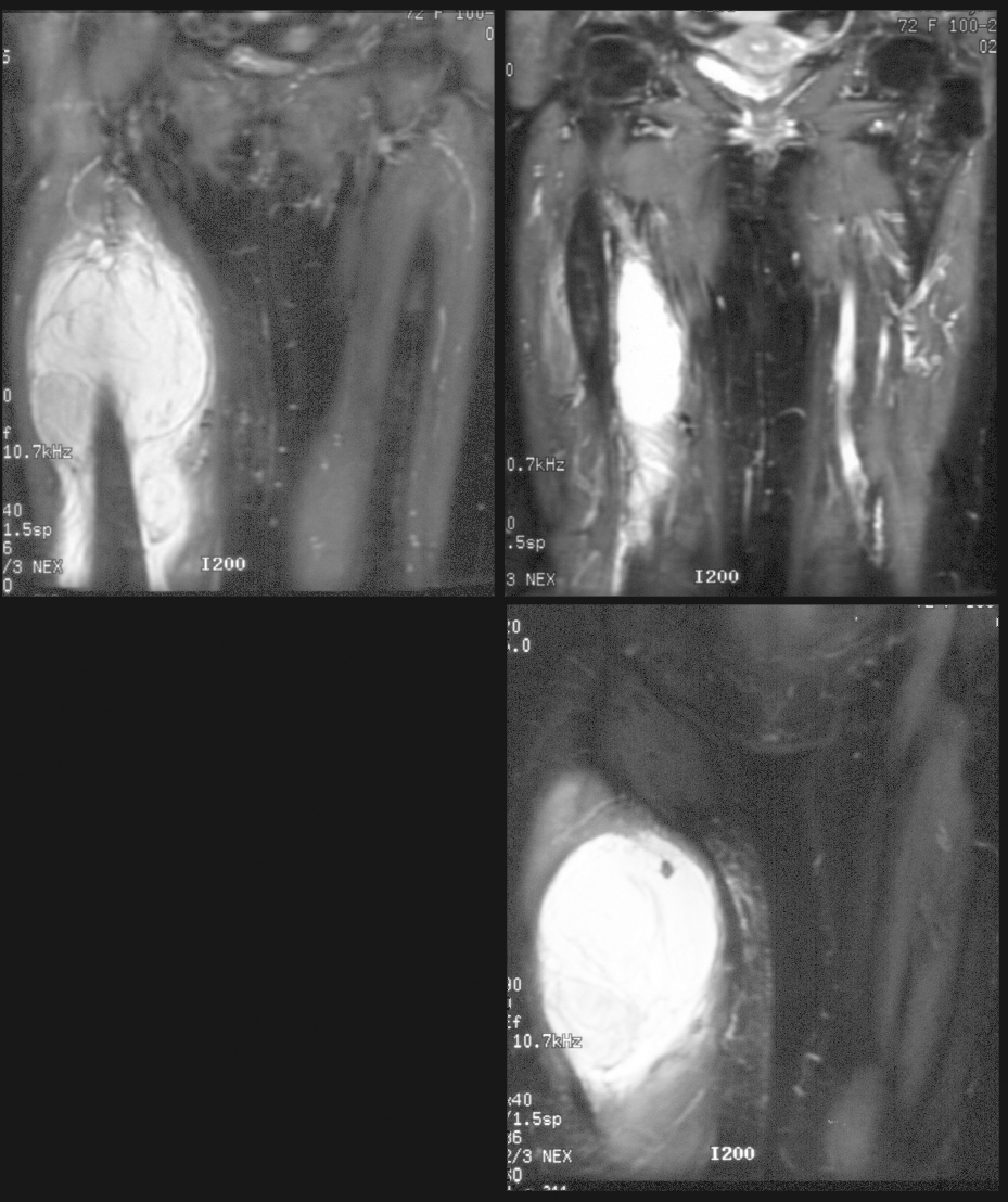
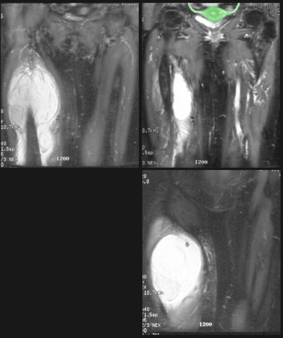
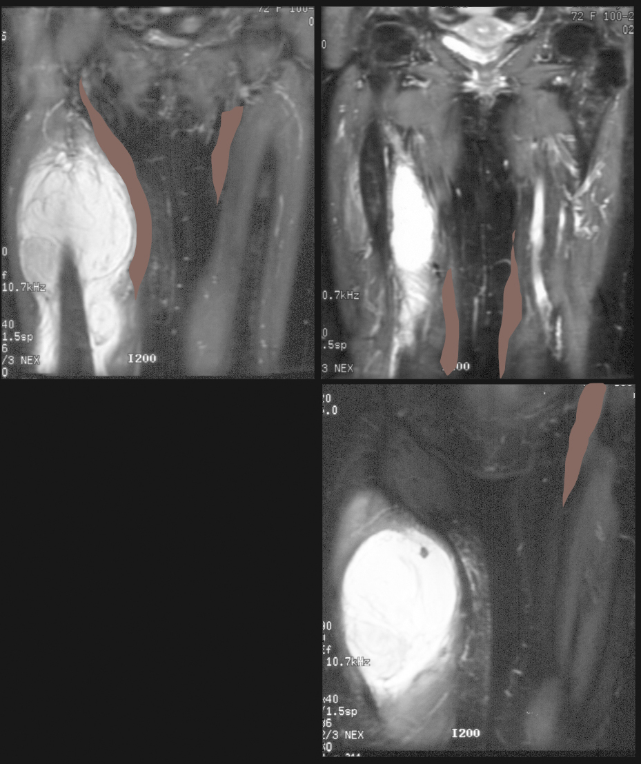
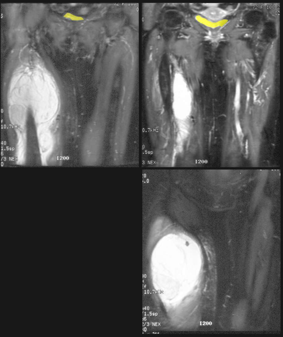
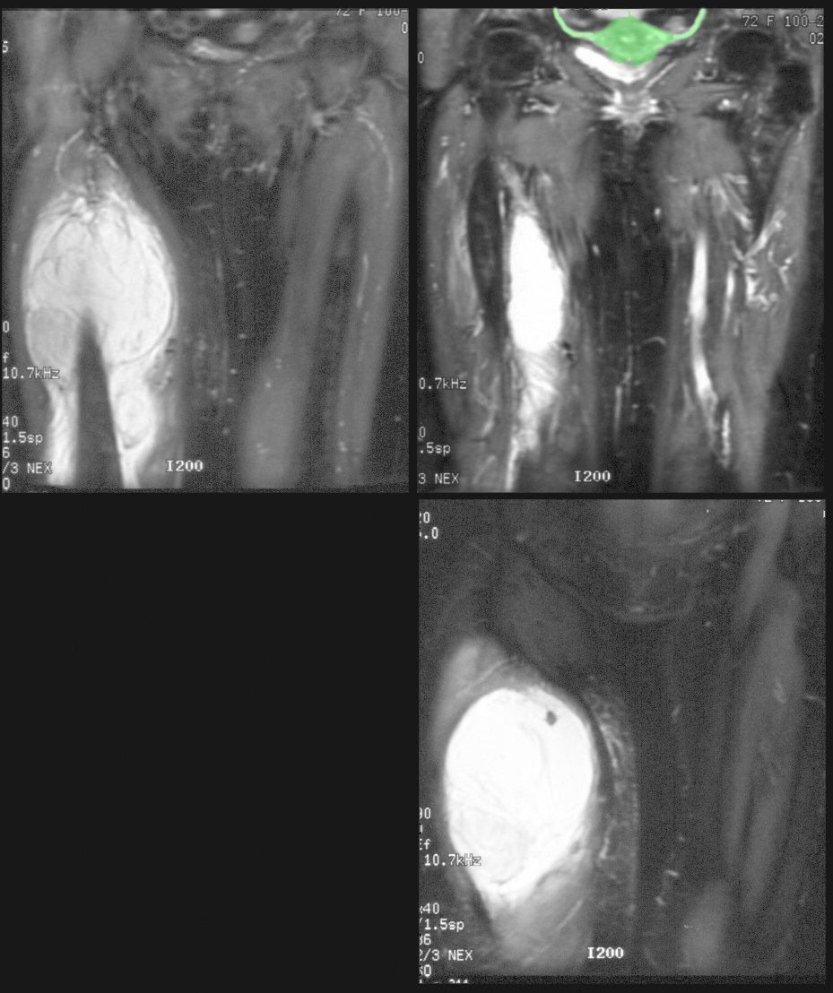
These are coronal MR images of the lower pelvis and thighs with fat suppression and T2 weighting.
b) What tissue is most important to assess when trying to identify whether a sectional image is a CT or an MR?
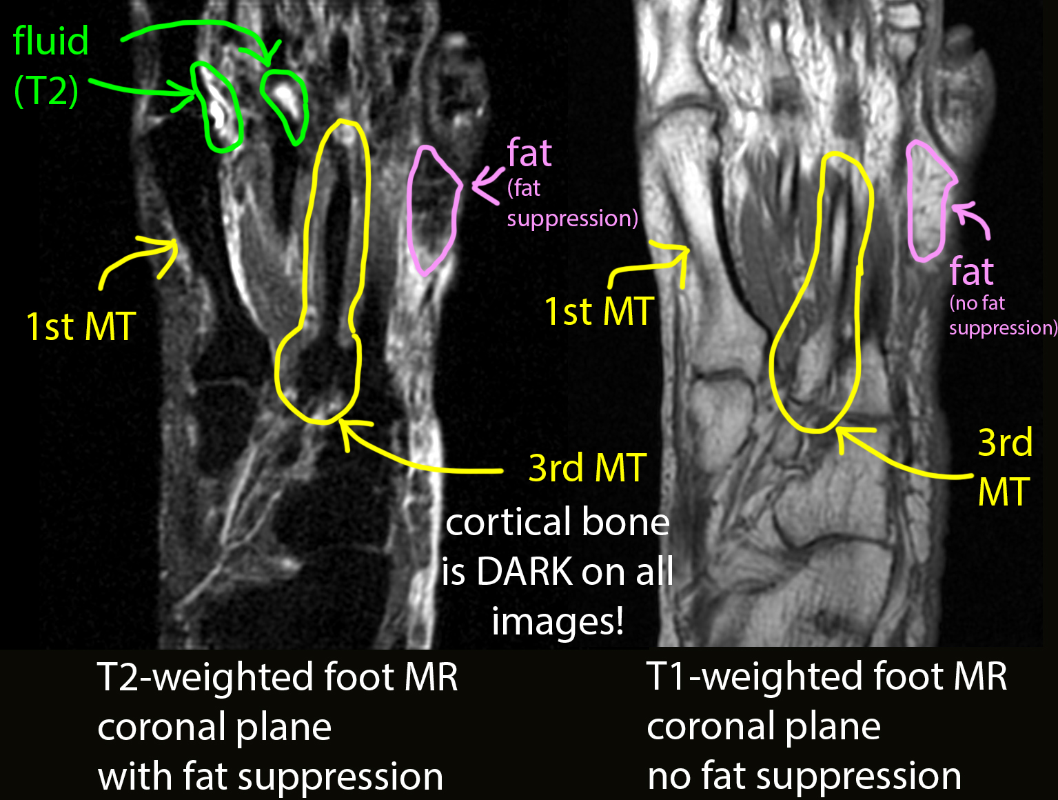
Cortical bone is key. Cortical bone (the outer margin of the bone) should be high attenuation (white) on all CT windows, and should be low signal (dark) on all MR sequences. Fat can sometimes be helpful as it is relatively low attenuation (dark grey) on CT images, but on MR it can be either low or high signal, depending on how the scan was acquired. In this sequence, fat suppression was used, so the signal from fat is low and the fat appears dark, similar to how it would look on a CT.
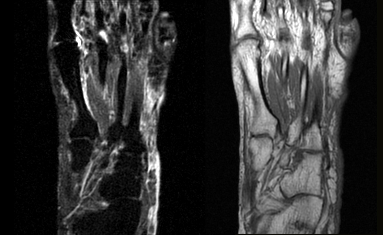
c) On the additional MR examples below, why does the medullary area of the bones appear white on one image and dark on the other?
We think of bone as being hard and mineralized, but it is important when looking at imaging of bones to recognize that the marrow space contains relatively little mineralized bone, but a large amount of other tissues. The blood-forming cellular structures are present in the marrow, embedded in fat. The amount of fat that is present varies with age. In young children, there is little marrow fat, but in adults, there is quite a bit. One radiographs, the medullary area will be darker than the outer cortex. And on MR sequences without fat suppression, the marrow space will tend to be quite bright (white) because of the fat that is present. But the outer rim of cortical bone should always be DARK on all MR sequences (unlike CT, where it is always WHITE). On MR images using fat suppression, the signal in the marrow will drop, just like the dark appearance of other areas containing a large amount of fat (breasts, subcutaneous regions).
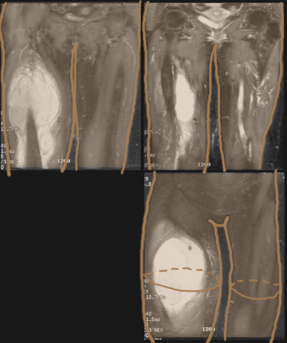
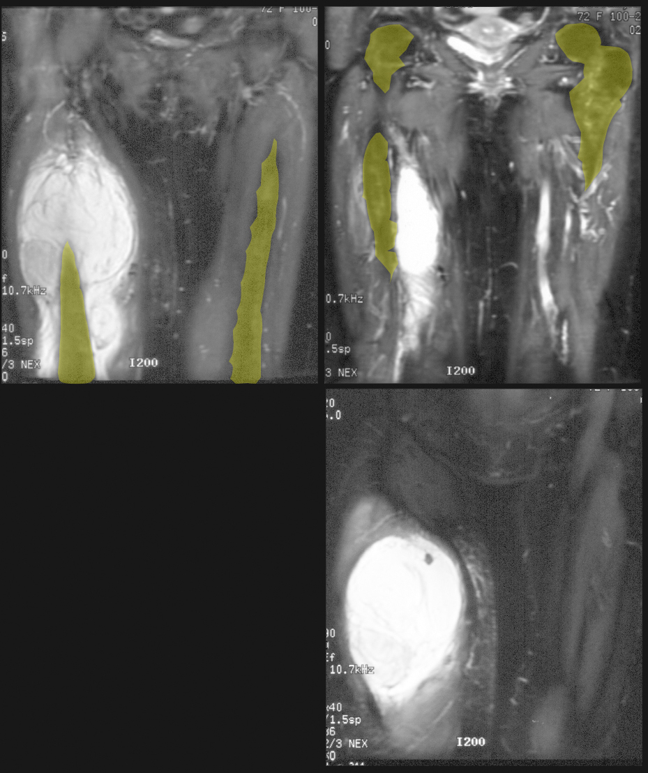
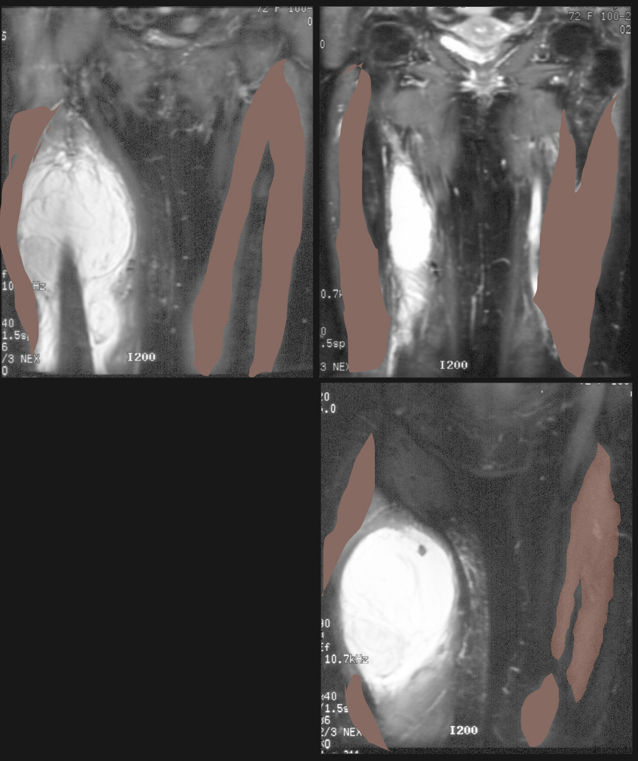
Again, try to identify listed structures before clicking on labels. For the two mystery structures, go to the next page for answers.
Back and Limbs Case 4
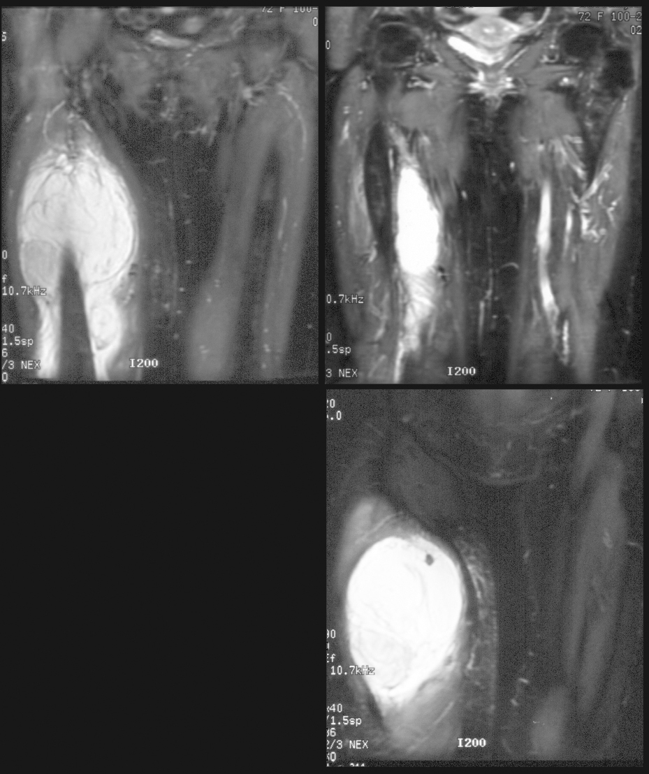
The patient's right thigh became massively enlarged after her surgery to stabilize her femoral fracture.
Question 4:
a) Why is bleeding into the thigh particularly dangerous?
The thigh is a large potential space where blood can freely collect, stretching and moving muscles, fascia and skin out of the way. The thigh can hold a very large amount of blood, so that patients may actually become hypotensive from bleeding in this region. There are not enough firm boundaries to stop the leakage of blood, which can accumulate to a very large collection as in this case.
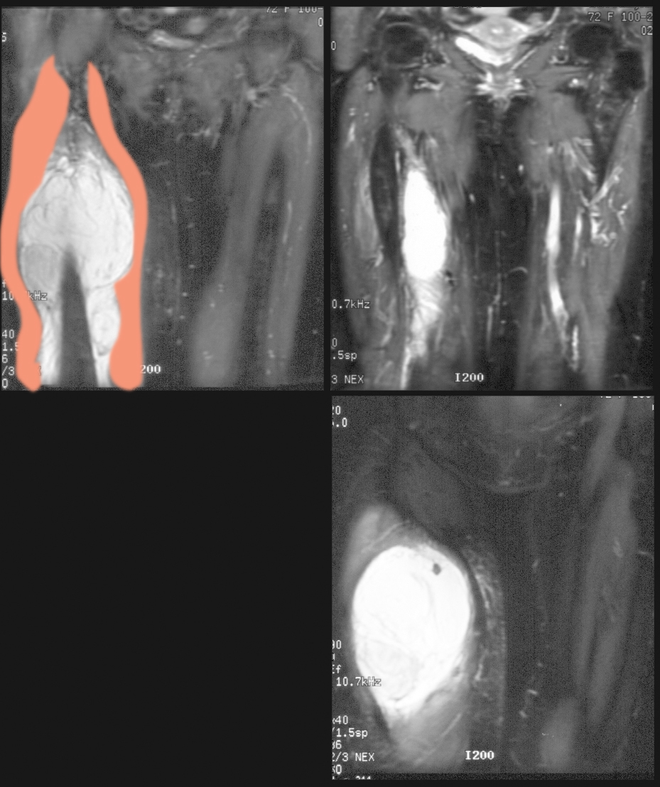
b) What muscles are being displaced by the right thigh hematoma (indicated by the label below)?
Since the femoral vein runs medial to the majority of the anterior thigh muscles, this hematoma is likely pushing the rectus femoris and vastus lateralis to the lateral side, and lifting or displacing sartorius and vastus medalis to the medial side. Vastus intermedius, lying directly anterior to the femur is probably deep to the plane of the hematoma.
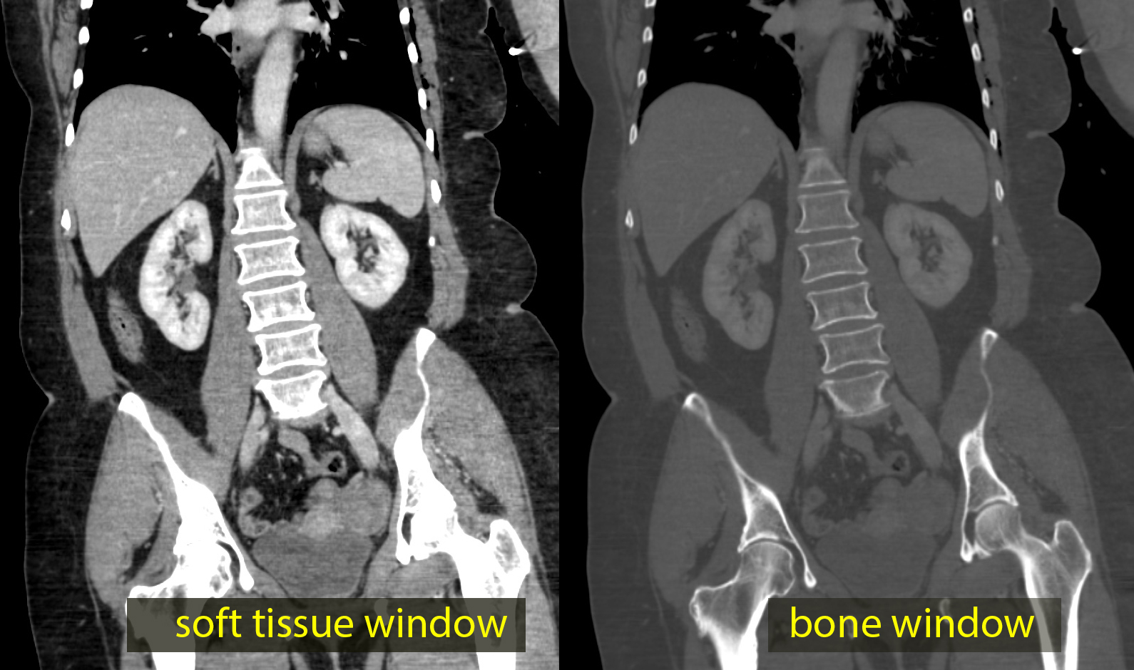
c) What modality is shown below as 'comparison study', in a different patient?
The comparison study is a coronal CT scan, shown in soft tissue and bone windows. Notice that the cortical bone is high attenuation (white) on both images, which are the same data displayed with different brightness and contrast settings. This patient does NOT have a thigh hematoma.
The patient required repair of damage to the femoral vein at the time of her surgery, and required transfusions due to the large amount of blood that accumulated in her thigh.
